N201 Wk2
 Call Kai
 Learn
Learn Practice Test
 Spaced Repetition
 Match
Match Flashcards
 Knowt Play
1/149

There's no tags or description
Looks like no tags are added yet.
Name | Mastery | Learn | Test | Matching | Spaced | Call with Kai | Chat |
|---|
No analytics yet
Send a link to your students to track their progress
150 Terms
Function of the heart
pumps oxygenated blood and nutrients to the rest of the body
receives deoxygenated blood and pumps away unwanted waste products
maintains heart rate and blood pressure
Point of maximal impulse
aka apical pulse
5th intercostal space, midclavicular line
Layers of the heart
endocardium
myocardium
epicardium
Endocardium
lining of heart and heart valves
inner layer
Myocardium
muscle for pumping
middle layer
Epicardium
serous layer; protects the heart
outer layer
Pericardium
sac surrounding the heart
Flow of blood through the heart
Deoxygenated blood flows through inferior/superior vena cava into right atrium
Right atrium delivers blood to right ventricle through tricuspid valve
Right ventricle delivers blood to pulmonary arteries through pulmonary valve into lungs
Lungs receive blood and oxygenate them
Oxygenated blood returns to the heart through pulmonary veins and into the left atrium
Left atrium delivers blood to left ventricle through mitral valve
Left ventricle delivers blood to aorta through aortic valve
Aorta transports the blood to rest of the body
Arteries
high pressure blood is transported from the heart to arteries and arterioles
delivers blood away from the heart
Veins
acts as a reservoir of blood and carries low-pressure blood to the heart from venules
delivers blood to the heart
Capillaries
permits gas exchange, transfer of nutrients, and removal of waste between blood and fluid of tissues
Sinoatrial (SA) node
cardiac impulses start at SA node
located in right atrium
considered heart’s natural pacemaker because it sets the rate and rhythm of heart
Atrioventricular (AV) node
impulse received by AV node from SA node
slight delay of impulse in AV node allows atria to fully contract and empty into ventricles
Heart’s conduction system
SA node
Impulse travels through atria (causing it to contract) and then travels to AV node
AV node has slight delay of impulse to allow atria to fully contract
Impulse travels to the Bundle of His
Impulse continues along Purkinjie fibers reaching terminal point and ventricular contraction
Systole
contraction phase
“lub”
Diastole
relaxation phase
“dub”
Cardiac output
total amount of blood ejected from one ventricle of the heart in L/min
to calculate: Stroke volume x Heart rate
Normal cardiac output
5-6 L/min
Factors that affect cardiac output
vasoconstriction
compliance of arteries
arterial pressure
amount of volume of blood entering the heart from veins
exercise
Stroke volume
volume of blood ejected by one ventricle per heartbeat
Three factors determining stroke volume
preload
afterload
contractility
Preload
volume of blood in the ventricle at the end of diastole
volume of blood is highest in the ventricle at end of diastole (meaning stretch of muscle fibers is greatest)
Afterload
resistance directly related to arterial blood pressure and diameter of the blood
the resistance the heart must overcome to push blood out
Contractility
can be affected by multiple factors
sympathetic stimulation or positive inotropic medications may cause contractions to increase
hypoxia or negative inotropic medications may cause contractions to decrease
Atrioventricular valves
tricuspid
mitral
Semilunar valves
aortic
pulmonic
S1
first heart sound heard indicating the closure of atrioventricular valves (mitral and tricuspid)
“Lub”
S2
second heart sound signifies closure of semilunar valves (aortic and pulmonic)
“Dub”
S3 and S4
abnormal heart sounds
best heard with bell of stethoscope
can be normal in children and adults up to age 40
S3
in adults older than 40, could indicate heart failure and decreased ventricular compliance
ventricular gallop
heard as a loud “dub” following S2
S4
due to decreased ventricular compliance caused by hypertension, aortic stenosis, coronary artery disease or cardiomyopathy
atrial gallop
heard as a loud “lub” immediately before S1
Murmurs
indicate turbulent blood flow through normal or abnormal valves
whooshing sound
classified according to timing in cardiac cycle
Systolic murmurs
occurs between S1 and S2
Diastolic murmurs
occurs between S2 and S1
Pericardial friction rub
abnormal heart sound that is typically heard over left sternal
grating sound
occurs due to inflammation, infection, or infiltration in the pericardial sac
Electrocardiogram
records cardiac electrical activity
Depolarization
transmission of electrical impulse that activates contraction of the heart
Repolarization
occurs when the heart relaxes and prepares to receive the next electrical impulse
allows chambers to refill with blood

P wave
atrial depolarization (contraction)
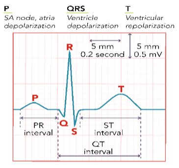
PR interval
related to the rate of cardiac impulse transmitted from the SA node to the AV node
should be between 0.12-0.20 seconds

QRS complex
ventricular depolarization (contraction) and atrial repolarization (relaxation)
should be less than 0.12 seconds

ST segment
follows ventricular depolarization and occurs prior to start of ventricular repolarization
elevation indicates myocardial ischemia (MI)

T wave
ventricular repolarization (relaxation) is occurring

QT interval
time for ventricular repolarization to complete

ECG strip
ECG grid is used to measure components of the ECG impulses
typical strip is 6 seconds in length and would have total of 30 large blocks
one large square is equal to 0.20 seconds
5 small squares located within one large square
1 small square is equal to 0.04 seconds
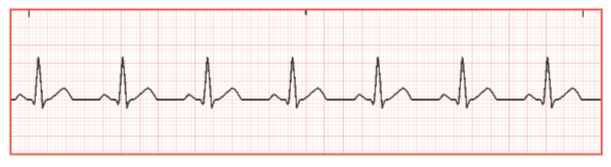
Identify the heart rhythm on ECG strip
normal sinus rhythm

Identify the heart rhythm on ECG strip
sinus bradycardia
Sinus bradycardia
occurs when SA node sends an electrical impulse slower than 60 bpm
Causes for sinus bradycardia
genetics
sleep apnea
increased intracranial pressure
decrease in metabolic needs
increased exercise tolerance
hypothyroidism
medications
vagus nerve stimulation
normal aging
prolonged hypoxia
Medications that can cause sinus bradycardia
parasympathomimetics (acetylcholine)
beta blockers (metoprolol)
digitalis glycosides (digoxin)
calcium channel blockers (diltiazem)
antiarrhythmics (amiodarone)
chemotherapy agents
lithium
Sinus bradycardia signs and symptoms
may be asymptomatic or symptomatic
symptoms include
fatigue
increased SOB
dizziness
Sinus bradycardia (role of the nurse)
fall precautions for symptomatic bradycardia
identify cause of bradycardia
instruct client on lifestyle changes that decrease potential for injury
Sinus bradycardia treatment
observe asymptomatic clients
unstable clients
IV atropine 1 mg repeat every 3-5 minutes; not to exceed total of 3 mg
monitor for changes in HR
temporary transcutaneous pacemaker if client continues to remain unstable and symptomatic

Identify the heart rhythm on ECG strip
sinus tachycardia
Sinus tachycardia
occurs when the SA node sends a faster electrical impulse, greater than 100 BPM
excessive excitability of the sympathetic nervous system and under stimulation of parasympathetic nervous system can trigger increased electrical impulses fired from SA node
prolonged tachycardia can lead to decrease in cardiac output which affects amount of oxygenated blood being pumped throughout body
Sinus tachycardia causes
fluid volume loss
fluid volume excess
pain
fever
shock
hyperthyroidism
stress
medications
Medications that can cause sinus tachycardia
atropine
catecholamines
theophylline
illicit drugs (cocaine, amphetamines)
caffeine
nicotine
Sinus tachycardia signs and symptoms
may be asymptomatic or symptomatic
symptoms include
palpitations
dizziness
lightheadedness
elevated temperature
chest pain
difficulty breathing
Sinus tachycardia (role of the nurse)
fall precautions
identify precipitating factors (infection or illness, chronic health condition, stress, pain, anxiety, medications)
client education
stress management
discontinue or reduce substances that can increase heart rate
change positions slowly
decrease physical intensity or activities
Sinus tachycardia treatment
interventions to decrease fever, pain, stress, fear, or anxiety
administer medications as ordered (adenosine, beta blockers)
catheter ablation
Catheter ablation
performed to destroy abnormally excited cardiac cells responsible for increased heart rate
considered minimally invasive and is performed using sedation
cardiac catheter tube is inserted into femoral vein or artery, and tube is advanced to heart. areas identified as causing excitability are then heated or frozen to eliminate blood supply to those cells
continuous cardiac monitoring detects early arrhythmias post-ablation and assessing pulses ensure adequate circulation and early identification of vascular complications
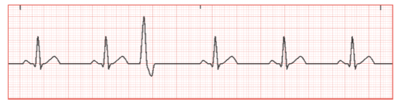
Identify the heart rhythm on ECG strip
Premature ventricular contraction (PVCs)
Premature ventricular contractions (PVCs)
occur when early electrical impulses, originating from irritated ventricular cells, are transmitted before the next normal impulse is expected from the SA node
the QRS complexes identified as PVCs occur early in cardiac cycle and appear much wider than QRS complexes seen in normal sinus
can be normal and may occur often in healthy clients
PVCs that occur without identification of abnormal heart structures are considered benign and medically insignificant
Premature ventricular contraction (PVCs) causes
may result from
cardiac muscle damage
chronic lung conditions
electrolyte imbalance (hypokalemia)
caffeine or alcohol consumption
Bigeminy
every normal heartbeat is followed by a premature beat
Trigeminy
every two normal heartbeats is followed by a premature beat
Premature ventricular contractions risk factors
inactivity
poor sleep
tobacco use
obesity
lipid levels
hypertension
diabetes
stress
hyperlipidemia
depression
joint pain
gout
osteoarthritis
cancer
Premature ventricular contractions signs and symptoms
may be asymptomatic or symptomatic
symptoms include
palpitations
lightheadedness
chest pain
shortness of breath
Premature ventricular contractions (role of the nurse)
assess for contributing factors that increase likelihood and frequency of PVCs
ask client to provide timeline of manifestations of PVCs
obtain a list of medications and chronic health conditions
analyze ECG (identify frequency and pattern of PVCs)
client teaching to minimize or prevent PVCs
smoking and alcohol cessation
eliminate illicit drug use
reduce caffeine intake
Premature ventricular contractions treatment
medications
beta blockers (atenolol, bisoprolol, carvedilol, metoprolol, propranolol)
antiarrhythmic medications (flecainide, propafenone, amiodarone)
catheter ablation
if medications aren’t effective after 30 days

Identify the heart rhythm on the ECG strip
premature atrial contractions
Premature atrial contractions
are benign cardiac rhythm occurrences that cause the heart’s electrical conduction system to activate an early heartbeat or impulse from one of the two atrial chambers
occurs when irritated atrial tissue fires an early electrical impulse before the next normal impulse is expected from the SA node
seen most often in infants and older individuals
frequently asymptomatic and do not pose a safety risk to client
Premature atrial contractions (role of the nurse)
identify lifestyle conditions that are known to precipitate development of PACs
provide reassurance to client
lifestyle modifications
contact health care provider if palpitations or SOB interfere with activities of daily living
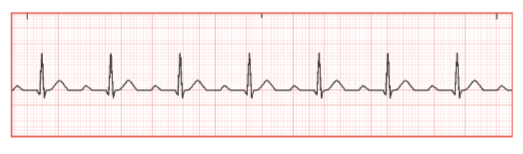
Identify the heart rhythm on the ECG strip
First-degree heart block
First-degree heart block
occurs when the cardiac conduction system is delayed in transmitting an electrical signal through the right atrium to the AV node
signal is not actually blocked from being sent but the process is slowed down
when conduction through the AV node is abnormally slow or prolonged, this results in a PR interval greater than 0.20 seconds
Causes of slow AV node conduction
increasing of age
history of cardiac disease (MI)
electrolyte imbalances (hypokalemia, hypomagnesemia)
antiarrhythmic medications
infection
athletes
male over 60
First-degree heart block (role of the nurse)
identify factors that contribute to first degree heart block
diets high in sodium, cholesterol or triglycerides
fall precautions
assess vital signs and ECG
provide education on lifestyle modifications
eliminate or limit smoking and alcohol consujmption
eat a healthy low-cholesterol diet
avoid excessive fatigue

Identify the heart rhythm on the ECG strip
Atrial fibrillation
Atrial fibrillation
common cardiac arrhythmia that occurs when the normal pacemaker of the heart SA node is not firing appropriately; electrical stimuli are being sent from multiple areas in the atria and are transmitting electrical impulses that are rapid, chaotic, and irregular
irregular impulses do not allow the atria to effectively contract and send blood into the ventricles
cardiac output decreased due to ineffective and irregular contractions of the heart
blood clots can form due to irregularity of the blood flow out of ventricles and can cause damage to the brain, lungs, kidneys, or other body organs
Atrial fibrillation risk factors
history of uncontrolled blood glucose levels
hyperthyroidism
obstructive sleep apnea
smoking
excessive alcohol intake
sedentary lifestyle
age over 60
obesity
chronic health conditions (hypertension, chronic heart failure, diabetes, COPD, kidney disease)
Atrial fibrillation signs and symptoms
may be symptomatic or asymptomatic
symptoms include
irregular pulse
hypotension
heart palpitations
increased heart rate
chest discomfort
shortness of breath (at rest or activity)
exertional fatigue
anxiety
dizziness
lightheadedness
syncope
weight gain
decreased urination
Atrial fibrillation ECG interpretation
ECG is key diagnostic test to confirm A-fib
irregular rhythm, HR 100-175 BPM (or even higher)
P waves replaced by atrial activities between QRS complexes
p waves not present before QRS
Atrial fibrillation (role of the nurse)
Bleeding precautions for clients on anticoagulant therapy
falls
shaving (electric razor)
avoid vigorous teeth brushing and flossing (use of soft bristle toothbrush)
avoid participation in contact sports
Client education
report any manifestations of A-fib to provider
take medications as prescribed
healthy lifestyle modifications
avoid stimulants
avoid herbal supplements
Atrial fibrillation treatment
electrocardioversion
catheter ablation
anticoagulants
diet modification
Electrocardioversion
procedure used to return an abnormal heartbeat to a normal rhythm
delivers a timed, low-energy electrical shock to the heart to help restore normal rhythm

Identify the heart rhythm on the ECG strip
Atrial flutter
Atrial flutter
common supraventricular (occurring before the ventricles) cardiac arrhythmia that occurs when the atria are beating in a regular rhythm and increased rate
less common than AF
atrial rate can range from 240-400 BPM which is caused by multiple rapid electrical impulses being sent from the atrial too quickly for the AV node to process before transmission to ventricles
P waves are replaced (no P waves) by multiple sawtooth or flutter waves between QRS complexes
Atrial flutter risk factors
recent MI or postoperative cardiac surgery
antiarrhythmic medications (beta blockers, calcium channel blockers, amiodarone, dioginx)
hypertension
heart failure
diabetes
COPD
obesity
thyroid disease
kidney disease
stroke
Atrial flutter signs and symptoms
lightheadedness
palpitations
hypotension
dizziness
chest discomfort
shortness of breath

Identify the heart rhythm on the ECG strip
supraventricular tachycardia
Supraventricular tachycardia
is in a category of cardiac dysrhythmias also known as narrow complex tachycardia
also called AV nodal reentrant tachycardia and occurs without precipitating factors
often occurs abruptly and suddenly ends without warning
originates in AV node and can cause excessive excitability of the atrial tissue resulting in an increased heart rate
Supraventricular tachycardia ECG interpretation
regular rhythm
heart rate of 100-220 BPM
narrow QRS complex less than 0.12 seconds
undiscernible P wave
Supraventricular tachycardia risk factors
increased stress levels
smoking or alcohol intake
caffeine and other stimulants
history of heart disease
Wolff-Parkinson-white syndrome
pregnancy
chronic lung disease
coronary artery disease
hypertension
diabetes
cardiomyopathy
Supraventricular tachycardia signs and symptoms
symptoms may present suddenly
dizziness
lightheadedness
syncopal episodes
fainting
hypotension
shortness of breath
palpitations
Supraventricular tachycardia treatment
vagal maneuvers
IV adenosine
calcium channel blockers (IV diltiazem)
beta blockers (IV esmolol, IV metoprolol)
Vagal maneuvers
simple, non-invasive physical techniques that help stimulate the vagus nerve to slow down abnormally fast heart rates
types include:
Valsalva maneuver
modified Valsalva maneuver
cold water immersion
coughing
carotid sinus massage
Coronary artery disease
occurs with a build up of plaque within the coronary arteries
can cause
stable or unstable angina
ST elevation myocardial infarction (STEMI)
Non-ST elevated myocardial infarction (NSTEMI)
Atherosclerosis
causes narrowing or occlusion of arteries
narrowed or occluded arteries cause decreased blood from which leads to decreased oxygenation and perfusion to myocardium
Angina pectoris
term used to describe chest pain and often occurs when the heart has decreased perfusion that can be caused by narrowing of the blood vessels (atherosclerosis)
Types of anginas
stable (exertional)
unstable (preinfarction)
variant (Prinzmetal/vasoplastic)