Cytoskeleton and the cell divison
1/53
There's no tags or description
Looks like no tags are added yet.
Name | Mastery | Learn | Test | Matching | Spaced | Call with Kai |
|---|
No analytics yet
Send a link to your students to track their progress
54 Terms
What are the 3 types of cytoskeletal filaments?
Actin filaments, microtubules, intermediate filaments.
Subunit of actin filaments?
Actin monomers (G-actin).
Subunit of microtubules?
α/β-tubulin heterodimers.
Subunit of intermediate filaments?
alpha helices
subunits wrap around each other
Arrangement of actin filaments?
Two-stranded helix, polarized (plus/minus ends).
Arrangement of microtubules?
13 protofilaments form a hollow tube.
Arrangement of intermediate filaments?
Rope-like, made of staggered tetramers, non-polar.
Which end of a microtubule grows faster?
Plus end (β-tubulin exposed).
How are microtubules polar?
α-tubulin at minus end, β-tubulin at plus end.
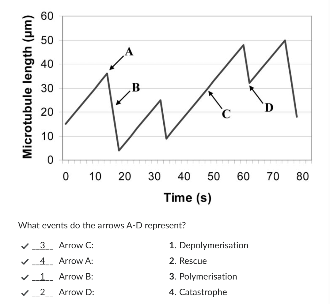
Define dynamic instability.
Random switching between growth and shrinkage of microtubules.
Catastrophe: Switch from growth to rapid shrinkage.
Rescue: Switch from shrinkage back to growth.
What stabilizes a growing microtubule?
A GTP cap on the plus end.
What causes catastrophe?
Loss of the GTP cap → protofilaments peel away.
Where do microtubules nucleate?
At MTOCs (e.g., centrosome) which is anchored in the minus end (alpha tubulin)
LONG: What molecular mechanism underlies dynamic instability?
Tubulin dimers bind GTP.
GTP on α-tubulin is non-exchangeable; GTP on β-tubulin is hydrolyzed after incorporation.
A stabilizing GTP cap exists at the growing plus end.
Loss of the cap causes protofilaments to peel outward → rapid depolymerization (“catastrophe”).
Regaining a GTP cap allows growth to resume (“rescue”).
How do α- and β-tubulin differ in GTP binding and hydrolysis?
Both bind GTP, but α-tubulin’s GTP is never hydrolyzed, while β-tubulin hydrolyzes GTP → GDP after incorporation into the microtubule.
What is the GTP cap, and why is it important?
A stabilizing region of GTP-tubulin at the microtubule plus end; it prevents depolymerization by strengthening subunit interactions.
What causes catastrophe, and what happens during it?
Loss of the GTP cap exposes GDP-tubulin at the end → protofilaments peel outward → rapid shrinkage (catastrophe).
The hydrolysis of GTP to GDP of tubulin subunits at the plus end of the microtubule has occurred before new tubulin subunits were added. Consequently, the GTP-cap disappeared.
What is rescue, and how can it be triggered?
Switch from shrinkage back to growth, when GTP-tubulin is added faster than hydrolysis or when rescue proteins stabilize the end.
How are tubulin dimers recycled?
Shrinking microtubules/microtubules which depolymerize release GDP-tubulin, which exchanges GDP for GTP and is reused.
What is the role of katanin and how is it activated?
Severing enzyme that chops microtubules, creating many unstable GDP-tubulin ends for rapid depolymerization.
It gets activated at the onset of rapid microtubule reorganization (e.g., spindle assembly).
What is the structural arrangement of an actin filament?
two protofilaments of serially linked actin monomers twisted into a helix; ~7 nm wide.
What nucleotide does actin monomer bind, and what happens after incorporation?
Each actin monomer carries an ATP molecule when free in the cytoplasm. After the monomer is incorporated into a growing actin filament, this ATP is hydrolyzed to ADP, which reduces the strength of the bond between subunits.
This difference in stability between ATP-actin at the filament’s “plus end” and ADP-actin in the older filament body drives treadmilling: a dynamic process where monomers are added at the plus end while simultaneously being removed from the minus end.
How does nucleotide hydrolysis affect filament stability?
Hydrolysis from ATP → ADP weakens subunit interactions, making the filament prone to shrinkage.
How is actin filament dynamics similar to microtubules?
Both undergo growth and shrinkage controlled by nucleotide hydrolysis at filament ends (ATP for actin, GTP for tubulin).
What happens during G1 phase?
Cell grows, performs normal metabolic functions, organelles replicate, protein synthesis is active. DNA is not yet replicated.
What occurs during S phase?
DNA replication → each chromosome forms two sister chromatids. Centrosomes duplicate. Cell prepares for mitosis.
What is the purpose of G2 phase?
Cell grows further, produces proteins needed for mitosis, checks DNA for errors. Microtubules begin reorganizing for spindle formation.
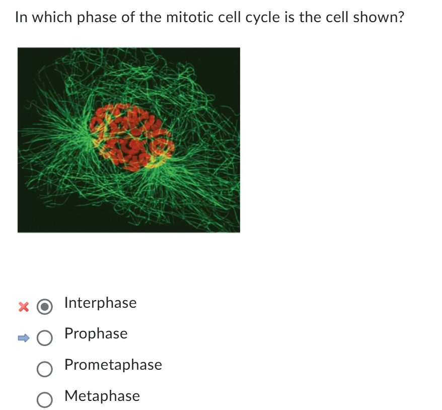
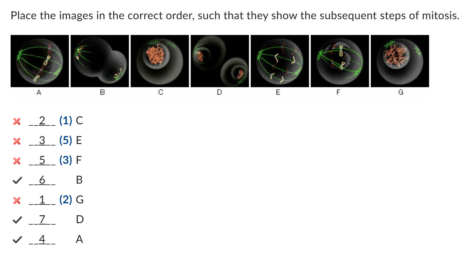
What key events occur in prophase?
Chromatin condenses into chromosomes; centrosomes migrate to poles; mitotic spindle begins forming; nuclear envelope still intact; DNA replication does not occur.
What happens during prometaphase?
Nuclear envelope breaks down; kinetochores assemble at centromeres; spindle microtubules attach to kinetochores; chromosomes begin moving toward metaphase plate.
What defines metaphase?
Chromosomes fully align at metaphase plate; sister chromatids bi-oriented and attached to microtubules from opposite spindle poles; spindle checkpoint ensures proper attachment.
Describe anaphase.
Cohesins cleaved → sister chromatids separate; kinetochore microtubules depolymerize to pull chromatids to poles; interpolar microtubules polymerize to push poles apart.
What occurs during telophase?
Chromosomes reach poles; nuclear envelope reforms; chromosomes de-condense; spindle disassembles.
What happens in cytokinesis?
Cytoplasm divides via actin-myosin contractile ring → cleavage furrow forms → two genetically identical daughter cells result.
How do microtubules function during mitosis?
Form mitotic spindle; kinetochore microtubules pull chromatids; interpolar microtubules maintain spindle and push poles apart; dynamic instability allows polymerization/depolymerization for chromosome movement.
Cell division in order
Interphase – Cell prepares for division
G1 phase (Gap 1):
Cell grows and performs normal metabolic functions.
Organelles replicate, protein synthesis is active.
No DNA replication occurs yet.
S phase (Synthesis):
DNA replication occurs → each chromosome forms two sister chromatids connected at the centromere.
Centrosome duplication occurs.
G2 phase (Gap 2):
Cell continues to grow and produce proteins required for mitosis.
DNA repair mechanisms check for replication errors.
Microtubules begin reorganizing in preparation for mitotic spindle formation.
2. Prophase – Chromosomes condense, spindle begins forming
Chromatin condenses into visible chromosomes (each with two sister chromatids).
Centrosomes begin migrating to opposite poles of the cell.
Mitotic spindle begins to form (microtubules start to assemble).
Nuclear envelope is still intact at the start.
Note: DNA replication is already completed in S phase; it does not occur in prophase.
3. Prometaphase – Nuclear envelope breakdown
Nuclear envelope fragments → spindle microtubules can access chromosomes.
Kinetochore proteins assemble at centromeres.
Microtubules attach to kinetochores of each sister chromatid.
Chromosomes start moving toward the equatorial plane.
4. Metaphase – Chromosomes align
Chromosomes fully align at the metaphase plate (cell equator).
Each sister chromatid is attached to microtubules from opposite spindle poles (bi-orientation).
Checkpoints ensure all kinetochores are properly attached before anaphase begins.
5. Anaphase – Sister chromatids separate
Cohesin proteins holding sister chromatids are cleaved.
Sister chromatids are pulled toward opposite spindle poles.
Microtubule dynamics during anaphase:
Kinetochore microtubules depolymerize at the plus end → pull chromosomes toward poles.
Interpolar microtubules polymerize → push poles apart.
6. Telophase – Chromosomes reach poles, nuclei reform
Separated chromosomes arrive at spindle poles.
Nuclear envelope re-forms around each chromosome set.
Chromosomes begin de-condensing back into chromatin.
Mitotic spindle disassembles.
7. Cytokinesis – Cytoplasm divides
Division of cytoplasm into two daughter cells.
Actin-myosin contractile ring forms at the cell equator → cleavage furrow forms.
Result: Two genetically identical daughter cells.
How does cytoplasmic streaming occur in plant cells?
Organelles bind to myosin, which walks along actin filaments toward the plus end using ATP hydrolysis.
Which motor proteins mediate organelle transport in animal cells, and in which directions?
Kinesins (plus-end, outward) and dyneins (minus-end, inward) move cargo along microtubules.
What is the structural arrangement of cilia and flagella?
Axoneme: 9 outer doublet microtubules + 2 central singlets (9+2 arrangement).
How does dynein cause bending of cilia and flagella?
Dynein “walks” toward the minus end of adjacent microtubules using ATP; nexin links convert sliding into bending.
Compare movement of cilia vs. flagella.
Cilia: short, wave-like beating; move fluid or cell.
Flagella: long, whip-like beating; propulsion (e.g., sperm).
Which enzyme synthesizes cellulose microfibrils, and how?
Cellulose synthase complexes (CSCs) in the plasma membrane polymerize glucose into cellulose chains, which are excreted outside the cell.

How do cellulose synthase complexes move through the plasma membrane?
CSCs track along cortical microtubules beneath the plasma membrane, leaving trails of cellulose microfibrils.
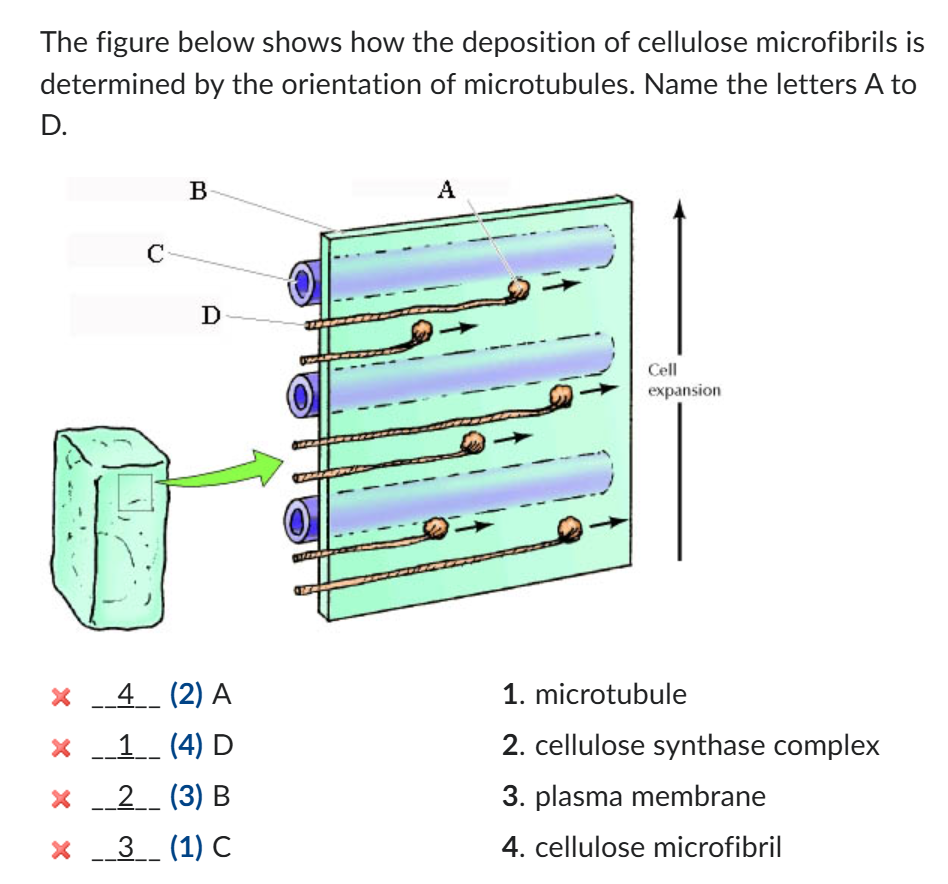
How does microtubule organization affect cell wall structure?
Microtubule orientation dictates the alignment of cellulose microfibrils, which determines the direction of cell wall growth.

Why does plant cell expansion occur perpendicular to cellulose microfibril orientation?
Microfibrils are rigid and resist stretching along their length, so growth can only occur in the perpendicular direction.

What is the building block of cellulose microfibrils?
Glucose molecule (linked into long β-1,4-glucan chains).
Can one Cdk bind multiple cyclins?
Yes; the cyclin determines which downstream proteins are activated or deactivated.
What regulates M-Cdk activity?
Cyclic changes in M-cyclin levels; when cyclin accumulates, M-Cdk is active → triggers mitosis.
How do checkpoints maintain cell cycle fidelity?
They pause the cycle when conditions are unfavorable (e.g., DNA damage, incomplete replication).
How do cancer cells bypass normal cell cycle control?
They evade checkpoints, allowing uncontrolled division → tumor formation.
How does cyclin-Cdk complexes G1-Cdk regulate the cell cycle and their functions.
phosphorylates proteins that prepare the cell for DNA replication and covmmitment to divide.
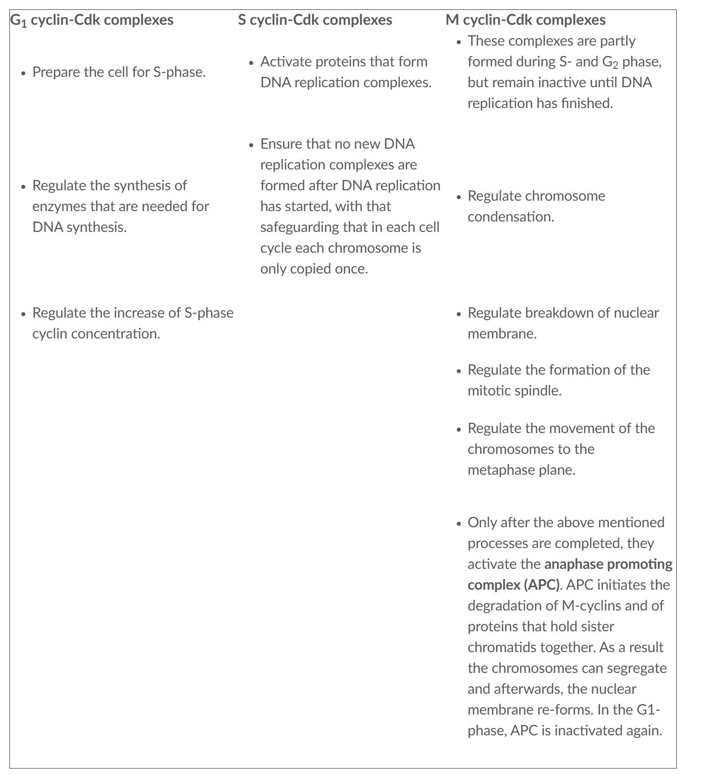
How does cyclin-Cdk complexes S-Cdk regulate the cell cycle and their functions.
Activate proteins that form DNA replication complexes.
Ensure that no new DNA replication complexes are formed after DNA replication has started, with that safeguarding that in each cell cycle each chromosome is only copied once.

How does cyclin-Cdk complexes M-Cdk(Cyclin B-Cdk1) regulate the cell cycle and their functions.
triggers entry into mitosis; promotes chromosome condensation, spindle assembly, and nuclear envelope breakdown.

What happens in G2 and G2 checkpoint?
the cell grows and all preparations for the cell division are completed. In humans, this phase takes approximately 4-6 hours.
At this checkpoint it is determined whether or not the environmental circumstances are sufficiently good to continue the cell cycle.
What are kinases?
proteins that activate or inactivate other proteins by their phosphorylation.
Kinases that play a role in the progression of the cell cycle are only active when they are coupled to cyclin.