Oral Potentially Malignant Disorders
 Call Kai
 Learn
Learn Practice Test
 Spaced Repetition
 Match
Match Flashcards
 Knowt Play
1/97

There's no tags or description
Looks like no tags are added yet.
Name | Mastery | Learn | Test | Matching | Spaced | Call with Kai | Chat |
|---|
No analytics yet
Send a link to your students to track their progress
98 Terms
Oral Potentially Malignant Disorders (OPMD): WHO Definition
Heterogeneous group of clinically defined conditions with a variable risk of progression to oral squamous cell carcinoma. Most produce clinically visible lesions.
OPMD: 2 Major Precursor Lesion Types
LEUKOPLAKIA and ERYTHROPLAKIA
Leukoplakia
"white plaque"
more common
~85% of oral precancers

Erythroplakia
"red plaque"
higher risk of developing cancer

OPMD Other Precursor Lesions: Actinic Cheilosis
Premalignant lesion on lower lip vermilion
Chronic UV light exposure
Esp. older males
(Analogous to actinic keratosis of skin)
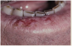
Actinic Cheilosis =
= lower lip vermillion

What are the indications for biopsy to rule out carcinoma in Actinic Cheilosis?
Crusting, ulceration, induration, thickening, leukoplakia

What does the treatment for Actinic Cheilosis depend on?
Microscopic diagnosis

What are the treatment options for high-grade dysplasia in Actinic Cheilosis?
Excision or vermilionectomy

What preventive measures should be taken for Actinic Cheilosis?
Use sunscreen and avoid sun exposure

What is the prognosis for patients with Actinic Cheilosis regarding lip squamous cell carcinoma?
~2-fold risk for lip squamous cell carcinoma (SCC)
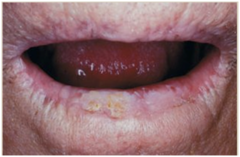
What is a precursor lesion associated with betel quid use?
Oral Submucous Fibrosis

In which demographic is Oral Submucous Fibrosis most commonly seen?
Young adults
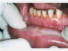
What is a common symptom of Oral Submucous Fibrosis?
Trismus (involves limited mouth opening)

What symptom of Oral Submucous Fibrosis?
Stomatopyrosis (burning sensation in the mouth)

What are two physical signs of Oral Submucous Fibrosis?
Mucosal pallor and stiffness

What are the treatment options for Oral Submucous Fibrosis?
Steroids and surgery

What is the prognosis for Oral Submucous Fibrosis after cessation of the habit?
Does not regress despite habit cessation

What percentage of patients with Oral Submucous Fibrosis may develop squamous cell carcinoma (SCC)?
~4-6%

OPMD Other Precursor Lesions: Palatal Lesions in Reverse Smoking

OPMD Other Precursor Lesions: Smokeless Tobacco
usually reversible w/ tobacco cessation
biopsy any lesion that remains despite cessation
Prognosis: low-risk (is this a true leukoplakia?)

Other sequel of smokeless tobacco use
Gingival recession
Dental caries
Extrinsic staining of teeth
SCC (conventional or verrucous) (may take several decades of exposure)

OPMD Other Precursor Lesions: "Candidal Leukoplakia"
Controversial
Hyperplastic candidiasis vs. true leukoplakia with superimposed candidiasis
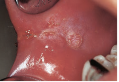
Candidal Leukoplakia - hyperplastic
white
cannot be wiped away
Candidal Leukoplakia - true leukoplakia with superimposed candidasis
colonizing fungal infection underneath lesion
OPMD: Other Conditions/Disorders - Heritable conditions
Fanconi anemia, xeroderma pigmentosum, Bloom syndrome, dyskeratosis congenita, epidermolysis bullosa
OPMD: Other Conditions/Disorders - Immune dysregulation / suppression
oral lichen planus (low risk/MTR
OPMD: Other Conditions/Disorders - Nutritional disorders
Plummer-Vinson syndrome, malnutrition, vitamin deficiencies (e.g., A,B,C)
Leukoplakia: Definitions - 1 (old)
white plaque
that cannot be wiped away
that cannot be characterized clinically or pathologically as any other disease
Leukoplakia: Definitions - 2 (new)
white plaques of questionable risk having excluded (other) known diseases or disorders that carry no increased risk for cancer (WHO 2017)
Leukoplakia: Definitions - 3
***clinical (or clinicopathologic) diagnosis / microscopic correlation often required, but NOT A MICROSCOPIC DX
"Not Leukoplakia": MORSICATIO
biting of buccal/tongue -> thickening of cells appears white
"ragged white plaque on buccal mucosa or tongue"

"Not Leukoplakia": BENIGN ALVEOLAR RIDGE KERATOSIS
very common
keratosis = thickening of keratin layer
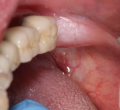
"Not Leukoplakia":WHITE SPONGE NEVUS
autosomal dominant mutations in genes for keratins 4 and 13

WHITE SPONGE NEVUS histology

"Not Leukoplakia": HEREDITARY BENIGN INTRAEPITHELIAL DYSKERATOSIS
Oral lesions similar to white sponge nevus
Ocular plaques also

"Not Leukoplakia": ORAL HAIRY LEUKOPLAKIA
Caused by EBV
Usually immunosuppressed patients
Treatment: address underlying immunosuppression
ORAL HAIRY LEUKOPLAKIA - differenetial diagnosis
hyperplastic candidiasis (burning sensation)
morsicato (biting abrasion)
"Not Leukoplakia": NICOTINE STOMATITIS
appears as inflamed cells

Why is it important to precisely define leukoplakia?
Better define those w/ risk for cancer
Impact on study results:
- Prevalence
- Malignant transformation rates
- Clinical trials
OPMD: Etiology
Tobacco
EtOH
Betel quid
Sanguinaria
UV light (lower lip)
Genetic factors
Immune factors
(Candida? Syphilis? High-risk HPV?)
Etiopathogenetic Model of nicotine
nicotine addiction -> carcinogens --(metabolic activation)-> DNA adducts -> mutations (TP53, RAS) -> CANCER
Multistep Carcinogenesis
increases genomic instability
NL -> hyperplasia -> dysplasia/CIS -> SCC
Pathogenesis: Field Cancerization
widespread carcinogen- & age-related mutations
migration of altered cells
"cancer-primed" cells without morphologic change
Epidemiology: Worldwide Prevalence Estimates
OPMD 4.5%
Oral leukoplakia 4.1%
Oral erythroplakia
Oral Leukoplakia & Erythroplakia: Key Clinical Features
M>F
Esp. middle-aged to elderly
Major risk factor = tobacco use
What are the favored sites for Oral Leukoplakia?
Lip vermilion, buccal mucosa, gingiva
Which sites are likely to harbor dysplasia or carcinoma in Oral Leukoplakia?
Ventrolateral tongue, floor of mouth, lip vermilion, buccal mucosa (betel quid users)
Is Oral Leukoplakia often symptomatic or asymptomatic?
Often asymptomatic
Oral Leukoplakia: Clinical Subtypes - HOMOGENEOUS
Thin (pictured)
Thick
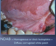
Homogeneous Thick Leukoplakia

Oral Leukoplakia: Clinical Subtypes - NON-HOMOGENEOUS
Erythroleukoplakia
Verrucous
Nodular/granular
Ulcerated
****GREATEST RISK FOR CANCER****
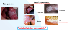
Non-homogeneous Leukoplakia (verrucous)
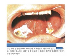
Non-homogeneous Leukoplakia (erythropleukoplakia)

Non-homogeneous Leukoplakia (nodular)
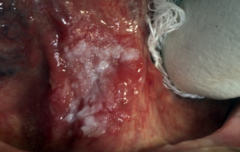
What is Proliferative Verrucous Leukoplakia (PVL)?
A variant of leukoplakia.

What is the gender prevalence for Proliferative Verrucous Leukoplakia (PVL)?
More common in females (F>M).
Is there a strong association between tobacco use and Proliferative Verrucous Leukoplakia (PVL)?
There is minimal association with tobacco use.
What are the characteristics of Proliferative Verrucous Leukoplakia (PVL) lesions?
Multiple, spreading white plaques with rough surface projections.
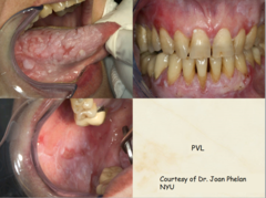
Where are Proliferative Verrucous Leukoplakia (PVL) lesions especially found?
Especially on the gingiva.
What is the risk associated with Proliferative Verrucous Leukoplakia (PVL)?
High risk for transformation into squamous cell carcinoma (SCC).
What is erythroplakia?
A red plaque or patch that cannot be attributed to any other pathophysiologic process.

What are the most common intraoral sites for erythroplakia?
Floor of mouth, buccal mucosa, and soft palate.
What is the clinical significance of erythroplakia?
High risk for high-grade dysplasia or squamous cell carcinoma (SCC) at presentation.
Correlating Clinical & Microscopic Findings
Why white?
Hyperkeratosis
Acanthosis / hyperplasia
Correlating Clinical & Microscopic Findings
Why red?
Inflammation
Increased vascularity
Epithelial atrophy
What is a typical microscopic diagnosis in OPMD? (without dysplasia)
Hyperkeratosis without dysplasia (involves thickening of the outer layer of skin)
What is a typical microscopic diagnosis in OPMD? (with dysplasia)
Hyperkeratosis with dysplasia (involves thickening of the outer layer of skin with abnormal cell changes)
What is a typical microscopic diagnosis in OPMD characterized by the presence of abnormal cells that have not invaded surrounding tissues?
Carcinoma in situ
What is a typical microscopic diagnosis in OPMD that represents invasive cancer of the skin?
Squamous cell carcinoma
What are other possible microscopic findings in OPMD besides the typical diagnoses?
Epithelial hyperplasia, acanthosis, inflammation, ulceration
Is leukoplakia considered a microscopic diagnosis in OPMD?
No
Histopathology: Hyperkeratosis without dysplasia

Histopathology: Squamous Acanthosis

Histopathology: Hyperkeratosis & Epithelial Hyperplasia

Histopathology: Hyperkeratosis with Mild Dysplasia
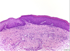
Histopathology: Hyperkeratosis with Moderate to Severe Dysplasia

Histopathology: Carcinoma in situ
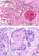
Histopathology: Squamous cell carcinoma

Oral Epithelial Dysplasia: Features
"disordered growth"
architectural & cytologic changes associated w/ increased risk for progression to SCC
~5% - 46% oral leukoplakia & nearly all oral erythroplakia
Oral Epithelial Dysplasia: Criteria - ARCHITECTURAL
Irregular epithelial stratification
Loss of basal cell polarity
Drop-shaped rete
Mitoses high in the epithelium
Premature keratinization
Keratin pearls
Loss of cohesion
Verrucous/papillary
Oral Epithelial Dysplasia: Criteria - CYTOLOGIC
Abnormal variation in cell size/shape & nuclear size/shape
Increased N/C ratio
Atypical mitotic figures
Increased # & size of nucleoli
Hyperchromasia
What is the malignant transformation rate for oral leukoplakia (OL) per year?
Approximately 1-3% per year
What is the malignant transformation rate for oral leukoplakia with mild/moderate epithelial dysplasia?
Approximately 10%
What is the malignant transformation rate for oral leukoplakia with severe dysplasia or carcinoma in situ (CIS)?
Approximately 24%
What is the risk of malignant transformation for oral leukoplakia without epithelial dysplasia?
Risk is low but not zero
What is the average time to malignant transformation for oral leukoplakia?
0.5 to 17 years
OPMD: Other Adverse Prognostic Factors
non-homogeneous
size >200mm2
subsite: ventrolateral tongue, floor of mouth
F>M
non-smoker / no known risk factors
multiple lesions
PVL (very high risk)
prior h/o head and neck squamous cell carcinoma
OPMD Evaluation: If not sure what it is?
BIOPSY or REFER FOR EVALUATION/BIOPSY
if you suspect a cause -> address cause and re-eval in 2 weeks _. persists? biopsy/refer out
OPMD Treatment - Major options:
If not cancer:
Careful observation
Surgical excision (esp. high-grade dysplasia, adverse clinical factors)
OPMD Treatment - Alternatives:
Laser ablation, cryotherapy, photodynamic therapy
OPMD Treatment
Habit cessation & long-term surveillance
What is the aim of chemoprevention?
Halt malignant transformation; may result in regression or eradication.
Name agents investigated for chemoprevention.
retinoids, beta-carotene, green tea extract, COX-2 inhibitors, EGFR inhibitors
What is an advantage of chemoprevention?
May address field cancerization; noninvasive.
What are disadvantages of chemoprevention?
Relapse and toxicity
Molecular Biomarkers
Risk asessment
Early diagnosis
Targeted therapy
Other Diagnostic Adjuncts
Autofluorescence
Chemiluminescence
In vivo microscopy
Vital staining
Brush cytology