Ch.2 Anatomy of Respiration
 Call Kai
 Learn
Learn Practice Test
 Spaced Repetition
 Match
Match Flashcards
 Knowt Play
1/89

There's no tags or description
Looks like no tags are added yet.
Name | Mastery | Learn | Test | Matching | Spaced | Call with Kai | Chat |
|---|
No analytics yet
Send a link to your students to track their progress
90 Terms
Respiration
Gas exchange between an organism and its environment
Inspiration
Inhaling oxygen
Expiration
Removing CO₂
Purposes of respiration
Biological and speech
Gas exchange occurs in ____ within the ____
alveoli, pulmonary apparatus
Inspiration is
an active, muscular process
Expiration is
passive or active depending on the context (quiet vs. forced)
Boyle’s Law
Pressure and volume are inversely related
Mechanics of inspiration
Increase thoracic volume → pressure drops → air flows in
Mechanics of expiration
Decrease volume → pressure rises → air flows out
Goal of inspiration
Brings oxygen into the lungs
Goal of expiration
Eliminates carbon dioxide from the body
Stucture of the pulmonary apparatus
lungs, trachea, bronchi, alveoli
Purpose of Pulmonary Apparatus
Structures that perform gas exchange
Purpose of Respiratory System
The functional system supporting gas exchange
If lungs are passive structures, what moves them?
Muscles
INSPIRATION – the active process
Inspiration is active: requires muscular contraction.
Primary muscle: Diaphragm.
Expands thoracic cavity, reduces internal pressure.
Air flows in to equalize pressure.
What is the primary muscle of inspiration?
Diaphragm
EXPIRATION – passive and active
Quiet expiration: passive (recoil of lungs and rib cage).
Forced expiration: active, requires abdominal and thoracic muscles.
Air pressure increases → air pushed out.
Quiet expiration
passive (recoil of lungs and rib cage)
Forced expiration
active, requires abdominal and thoracic muscles.
Formula for air pressure
Air pressure = Force / Area
AIR PRESSURE and VOLUME
↑ Volume = ↓ Pressure → air in
↓ Volume = ↑ Pressure → air out
Bony thorax
vertebrae, ribs, sternum
Visceral thorax
lungs, airways, blood vessels
Muscles
diaphragm, intercostals, accessory muscles
Structures of respiration are sorted into what subsystems?
supportive, functional, and muscular

Structures of respiration
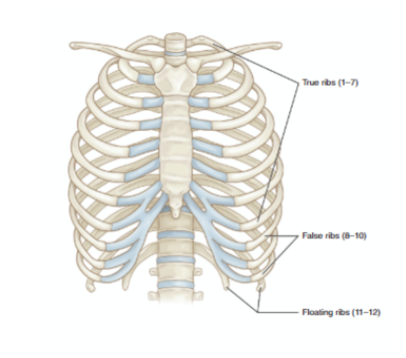
Rib Cage and Vertebrae (Bony Thorax)
BONY THORAX – Rib Cage and Vertebrae
12 pairs of ribs, connected to thoracic vertebrae
Ribs move up and out during inspiration
Ribs 1–7: true ribs, 8–10: false ribs, 11–12: floating ribs
Provides protection and mechanical leverage.
True ribs
1-7
False ribs
8-10
Floating ribs
11-12
How many pairs of ribs do we have?
12
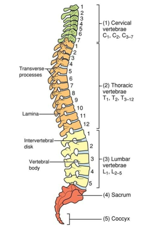
Vetebral column
5 Regions of the vetebral column
Cervical (7), Thoracic (12), Lumbar (5), Sacral (5 fused), Coccygeal (4 fused)
Vetebral Column
Supports body weight and anchors ribs.
Provides attachment points for respiratory muscles.
Cervical vertebrae
C1-C7
Thoracic vertebrae
T1-T12
Lumbar vertebrae
L1-L5
THE STERNUM aka “breastbone”
Central anterior attachment point for ribs.
Three parts: manubrium, body, xiphoid process.
Provides attachment for muscles (e.g., sternocleidomastoid).
What are the 3 parts of the sternum?
manubrium, body, xiphoid process
Which part of the sternum provides attachment for muscles?
Sternocleidomastoid
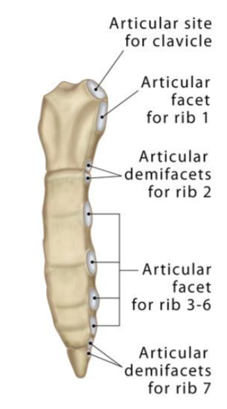
Sternum
THE RIB CAGE and COSTAL CARTILAGE
Flexible and mobile, allowing expansion and recoil.
Cartilage permits rib movement without breaking.
Protects thoracic organs while supporting breathing.
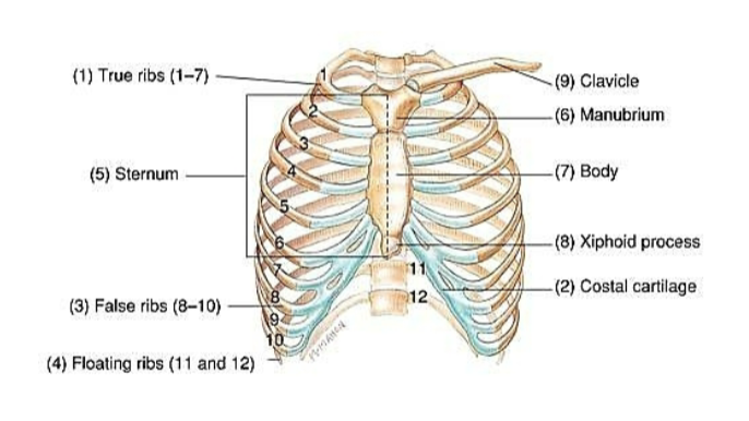
Rib cage and costal cartilage
Costal cartilage structure
Hyaline cartilage connecting ribs to sternum
Trachea and broncial tree
Flexible, cartilaginous tube ~11 cm long
Bifurcates into right and leftmainstem bronchi
Bronchi divide into secondary and tertiary branches
Lined with cilia for filtering debris
LUNGS and LUNG LOBES
Right lung: 3 lobes; left lung: 2 lobes (to accommodate heart)
Made of spongy, elastic tissue—passive in movement
Surrounded by pleural membranes to reduce friction
Contain alveoli where gas exchange occurs
Which lung only has two lobes?
Left
ALVEOLI and GAS EXCHANGE
~300 million alveoli per lung
Surrounded by capillary networks for O₂ and CO₂ exchange
Extremely thin walls to allow diffusion
Site of respiration at the cellular level
Pleural Linings
Visceral pleura, Parietal pleura, Pleural fluid
Prevents lung collapse and supports smooth movement
Visceral pleura
covers the lungs
Parietal pleura
lines the inside of the thoracic cavity
Pleural fluid
reduces friction, creates surface tension
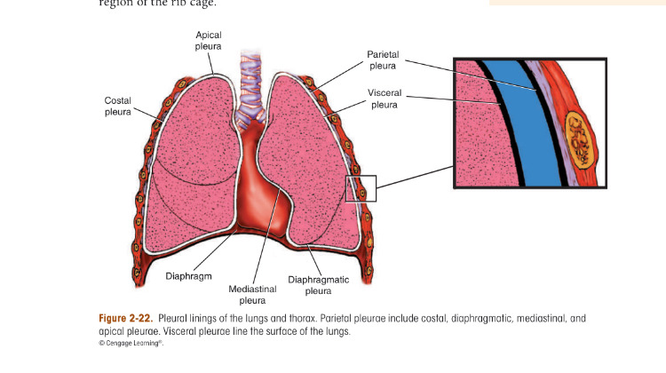
Pleural lining
Pulmonary System Main Concepts
Trachea → Bronchi → Bronchioles → Alveoli.
Lungs are passive but highly elastic.
Gas exchange = primary respiratory function.
Pleural system ensures smooth, stable expansion.
Diaphragm
Function: Expands thoracic cavity vertically; primary muscle of inspiration.
Dome-shaped, unpairedmuscle separating thorax and abdomen
Origin: xiphoid process
Course: upward and medially
Insertion: central tendon
Contraction → flattens diaphragm, increases thoracic volume
External intercostals
Located between ribs; run downward and forward
Elevate ribs during inspiration.
Increase anterior-posterior and transverse thoracic dimensions.
Active during quiet and forced inspiration.
External intercostals OCIF
Origin: Inferior surface of ribs 1–11.
Course: Downward and forward.
Insertion: Superior surface of the rib below.
Function: Elevate ribs; expand thoracic cavity in inspiration.
Accessory Muscles of Inspiration - Overview
Used during deep or forceful inspiration (e.g., speech, exercise).
Include: sternocleidomastoid, scalenes, pectoralis major/minor, serratus anterior, levator scapulae.
Elevate ribs, sternum, or stabilize the upper thorax
Sternocleidomastoid
Elevates sternum and clavicle. 2 heads, clavicular and sternal
Scalenes (anterior, middle, posterior)
elevate ribs 1 and 2.
Sternocleidomastoid and Scalenes Function
Provide vertical lift for upper rib cage
Active in speech breathing and respiratory distress
Sternocleidomastoid OCIF
Origin: Mastoid process of temporal bone
Course: Downward
Insertion: Manubrium of sternum and clavicle
Function: Elevates sternum andclavicle; expands upper thorax during inspiration
Scalenes OCIF
Origin: Transverse processes of cervical vertebrae C2–C7.
Course: Downward.
Insertion: Ribs 1 and 2.
Function: Elevate first and second ribs; stabilize upper thorax during inspiration.
What do the pectoralis major and minor do?
Assist in expanding thoracic cavity superiorly and anteriorly
Important in postural compensation during respiration
Pectoralis major
elevates sternum and rib cage (with fixed arm)
Pectoralis minor
lifts ribs 3–5
Pectoralis Major OCIF
Origin: Clavicle, sternum, and costal cartilages of ribs 1–6
Course: Laterally and upward
Insertion: Humerus
Function: Elevates sternum; expands rib cage during forced inspiration when arm is fixed
Pectoralis minor OCIF
Origin: Anterior surfaces of ribs 2–5
Course: Upward and laterally
Insertion: Coracoid process of scapula
Function: Elevates ribs 2–5 during deep inspiration when scapula is fixed
Additional acessory muscles of inspiration
Serratus anterior, Levator scapulae,Trapezius
Contribute during deep inspiration or compromised breathing.
Serratus anterior OCIF
Origin: Outer surfaces of ribs 1–9.
Course: Around the thoracic wall, posteriorly.
Insertion: Inner border of the scapula.
Function: Elevates ribs and stabilizes scapula during forced inspiration.
Levator scapulae OCIF
Origin: Transverse processes of C1–C4
Course: Downward and laterally
Insertion: Medial border of scapula
Function: Elevates scapula; stabilizes shoulder girdle during respiration
Trapezius OCIF
Origin: Spinous processes of C7–T12
Course: Fan laterally
Insertion: Clavicle and scapula
Function: Stabilizes and elevates shoulder girdle; assists indirectly in expanding thoracic cavity
Muscles of forced expiration - Overview
Expiration is passive at rest, but active during speech or exertion.
Forced expiration uses abdominal and thoracic muscles.
Compress thoracic cavity → push air out forcefully
Internal Intercostals
Located between ribs; run downward and backward.
Pull ribs down and inward.
Decrease thoracic volume → push air out.
Active in forced expiration
Internal Intercostals OCIF
Origin: Superior margin of ribs 1–11
Course: Upward and medially
Insertion: Inferior surface of the rib above
Function: Depress ribs during forced expiration
Iliac Crest
Location: Superior border of the ilium (largest bone in the pelvis).
Landmark: Easily palpable on both sides of the lower torso.
Attachments: Serves as an anchor for abdominal and back muscles (e.g., external/internal obliques, transversus abdominis, latissimus dorsi).
Function: Provides structural support and muscle leverage during posture, trunk movement, and respiration.
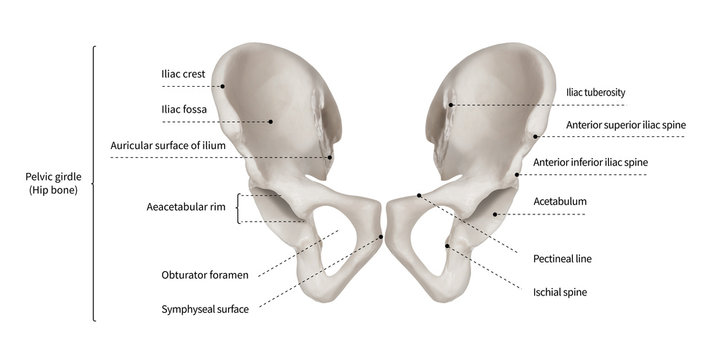
Iliac Crest
ABDOMINAL MUSCLES in EXPIRATION
Major players: rectus abdominis, transversus abdominis, internal and external obliques
Compress abdominal contents → push diaphragm upward
Provide force for controlled, sustained exhalation
Rectus Abdominis OCIF
Origin: Pubic symphysis and pubic crest
Course: Vertically upward
Insertion: Xiphoid process and costal cartilages of ribs 5–7
Function: Compresses abdominal contents; pulls sternum downward during forced expiration
External Oblique OCIF
Origin: Outer surfaces of ribs 5–12
Course: Downward and medially (“hands in pockets”)
Insertion: Iliac crest, inguinal ligament, and abdominal aponeurosis
Function: Compresses abdomen; flexes and rotates trunk; supports forced expiration
Internal Oblique OCIF
Origin: Inguinal ligament, iliac crest, and thoracolumbar fascia.
Course: Upward and medially.
Insertion: Lower ribs (10–12) and abdominal aponeurosis.
Function: Compresses abdomen; rotates and flexes trunk; assists in forced expiration
Transverse abdominis OCIF
Origin: Inguinal ligament, iliac crest, lower six ribs, thoracolumbar fascia.
Course: Horizontally across abdomen.
Insertion: Abdominal aponeurosis and linea alba.
Function: Deepest abdominal layer; compresses abdominal cavity; supports forced expiration and posture
Primary muscles of inspiration
diaphragm + external intercostals (primary), accessory muscles when needed.
Primary muscles of forced expiration
internal intercostals + abdominal muscles
Breathing for speech is not the same as breathing at rest
True
Bitch
We’re done