Retina stuff
1/64
There's no tags or description
Looks like no tags are added yet.
Name | Mastery | Learn | Test | Matching | Spaced | Call with Kai |
|---|
No analytics yet
Send a link to your students to track their progress
65 Terms
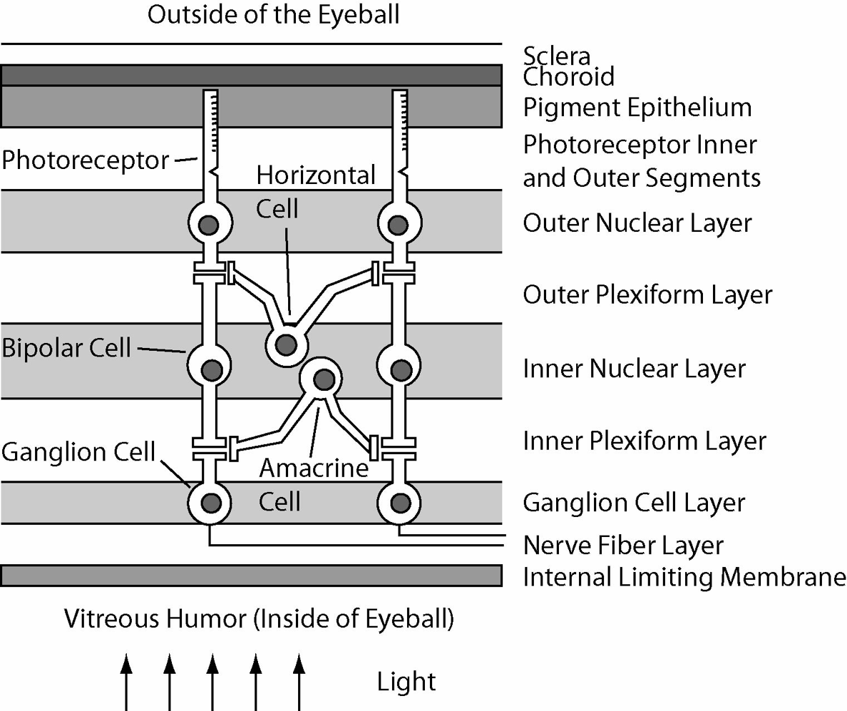
Major retinal neuron types
Photoreceptors, Bipolar Cells, Ganglion Cells, Horizontal Cells, Amacrine Cells
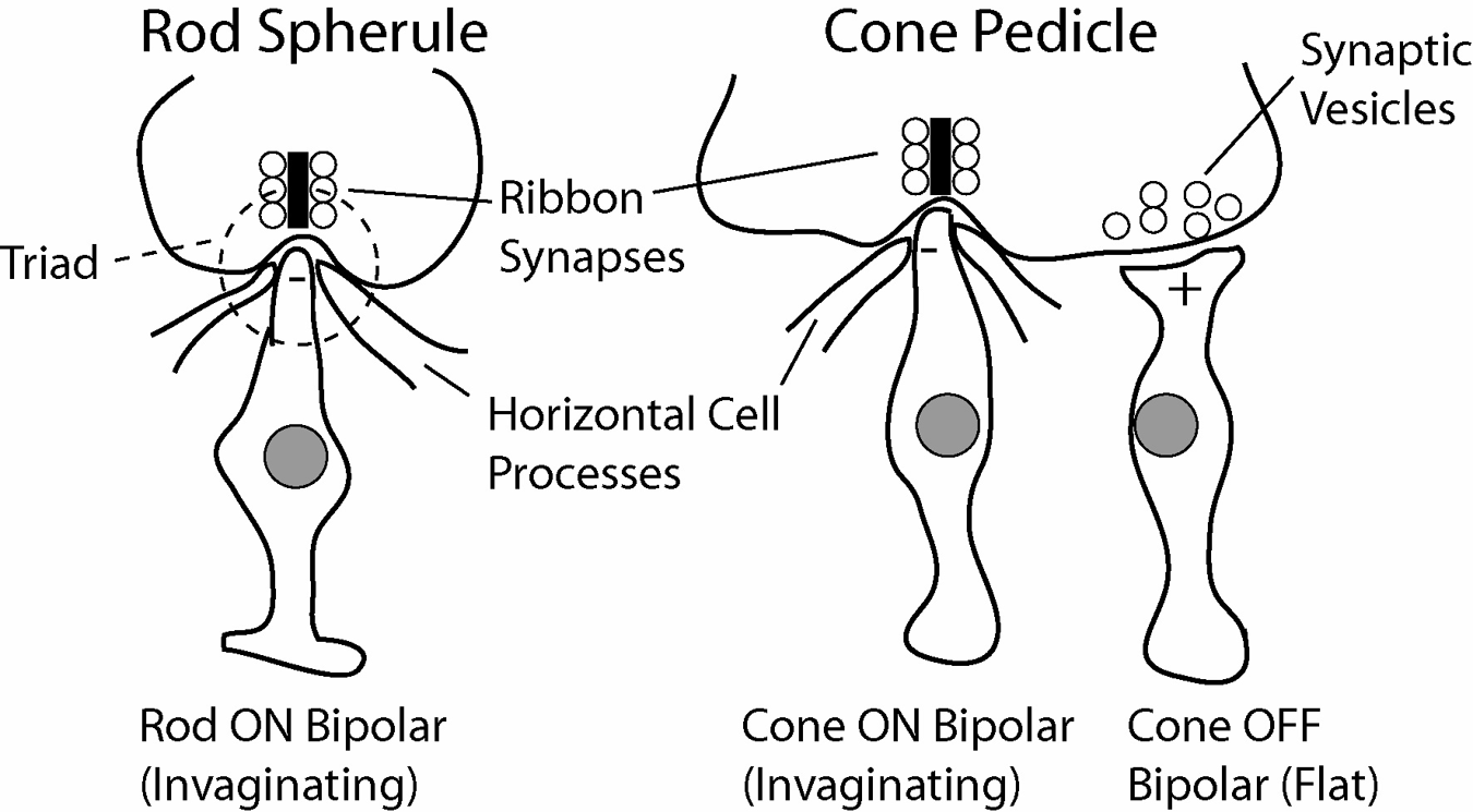
Rods/location/end/synapse type
Most numerous photoreceptor, most in peripheral retina, ends in spherule, invaginating synapses
Fovea
Contains only cones, foveola= only red and green cones
Percentage of cones that are S cones
7–10%
Why doesn’t red and green cone variability affect color vision
Color depends on relative activation of cone populations rather than absolute cone numbers
Cone terminal name
Pedicle
Triad
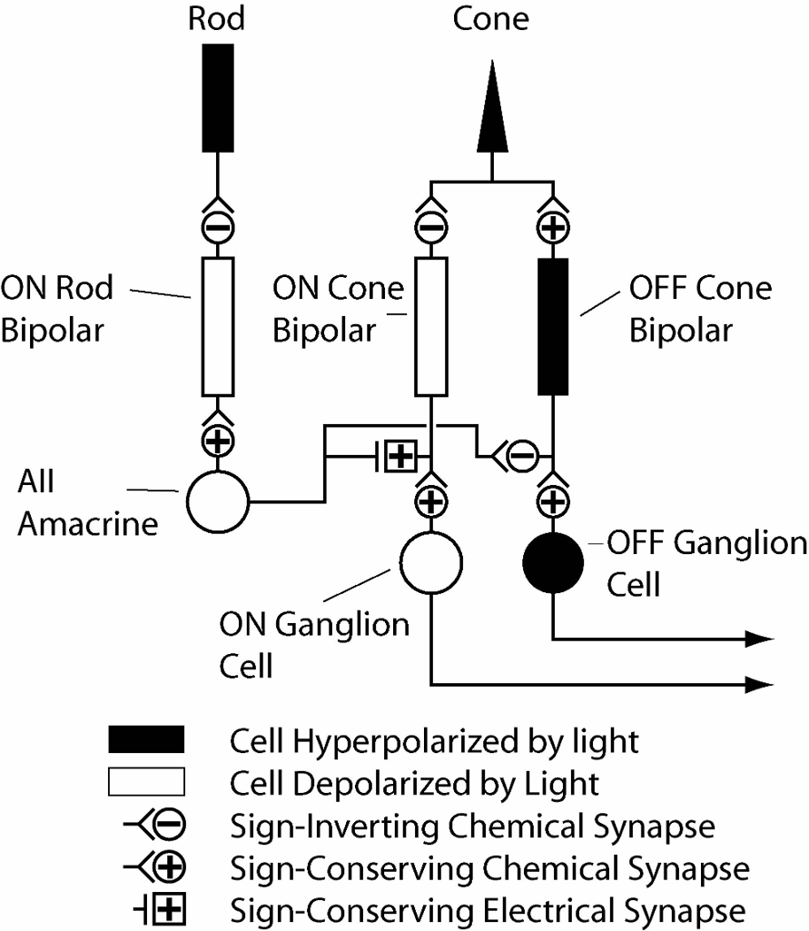
Structure formed by rod bipolar and horizontal cells in rod spherules
Cone ON bipolar synapse type
Invaginating
Cone OFF bipolar synapse type
Flat
Signal generated by bipolar cells
Graded potentials
Ribbon Synapses
Continuously release neurotransmitter vesicles, reason bipolar cells can continuously signal, present on ON BP cells
ON bipolar cell response to photoreceptor hyperpolarization
Depolarization
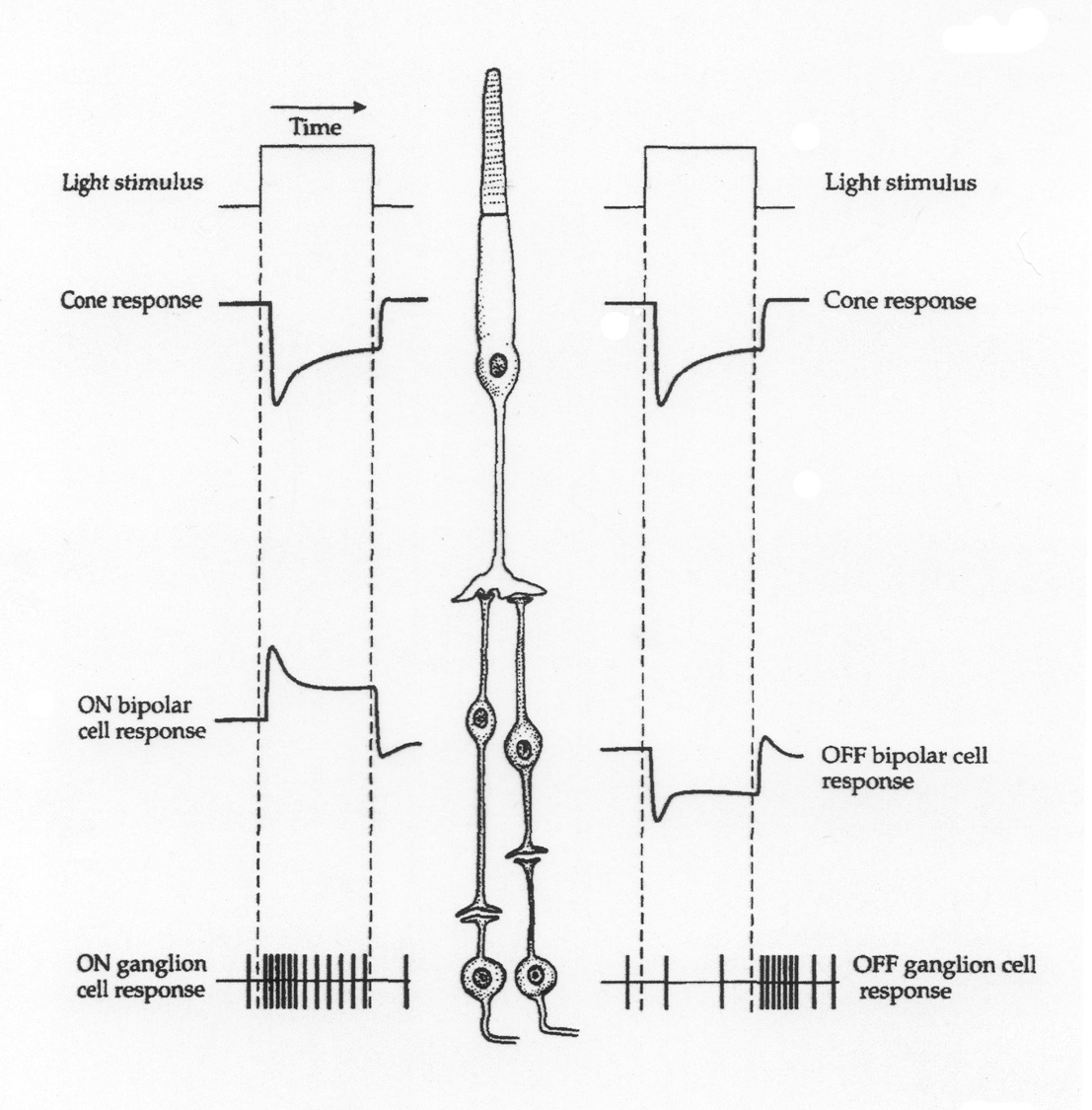
ON bipolar cell receptor type
Metabotropic glutamate receptor
OFF bipolar cell response to photoreceptor hyperpolarization
Hyperpolarization
OFF bipolar cell receptor type
Ionotropic glutamate receptor
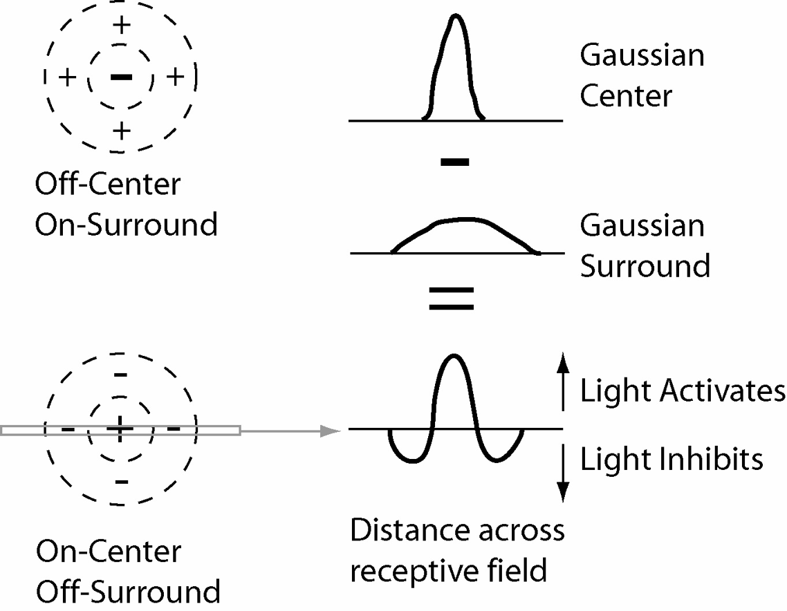
Definition of receptive field
Region of retina where stimulation affects a neuron
Function of receptive fields
Enhance edge detection and reduce responses to uniform illumination
Primary source of lateral inhibition
Horizontal cells
ON-center OFF-surround receptive field
Light in center excites light in surround inhibits

OFF-center ON-surround receptive field
Light in center inhibits light in surround excites
Primary role of amacrine cells
Inhibitory interneurons involved in retinal processing
Main neurotransmitters used by amacrine cells
GABA and glycine
Amacrine cells that use acetylcholine
Starburst amacrine cells
Amacrine cells involved in rod-cone switching
AII amacrine cells
Typical ganglion cell receptive field
Center-surround
Percentage of ganglion cells that are midgets
About 80%
Four types of midget ganglion cells
Red-ON Red-OFF Green-ON Green-OFF
Importance of midget ganglion cells
High visual acuity and color vision
Three mechanisms of light adaptation
Pupil size change, photoreceptor adaptation (slowest), network adaptation (fastest, switching from rods to cones using AII cones)
Purkinje shift
Visual sensitivity shifts toward longer wavelengths in light and shorter wavelengths in dark
Reason rods are insensitive to red light
Rod sensitivity peaks at shorter wavelengths
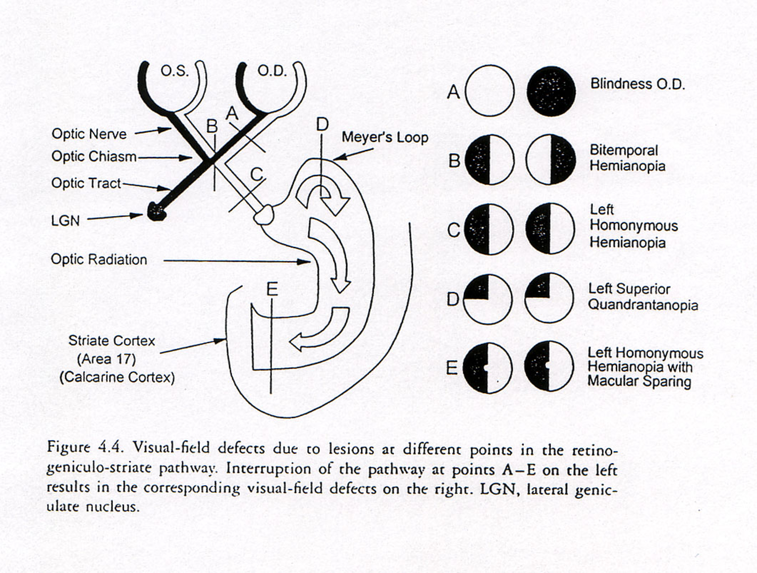
Primary visual pathway
Retina → LGN → Cortex
Percentage of ganglion cell axons terminating in LGN
About 90%
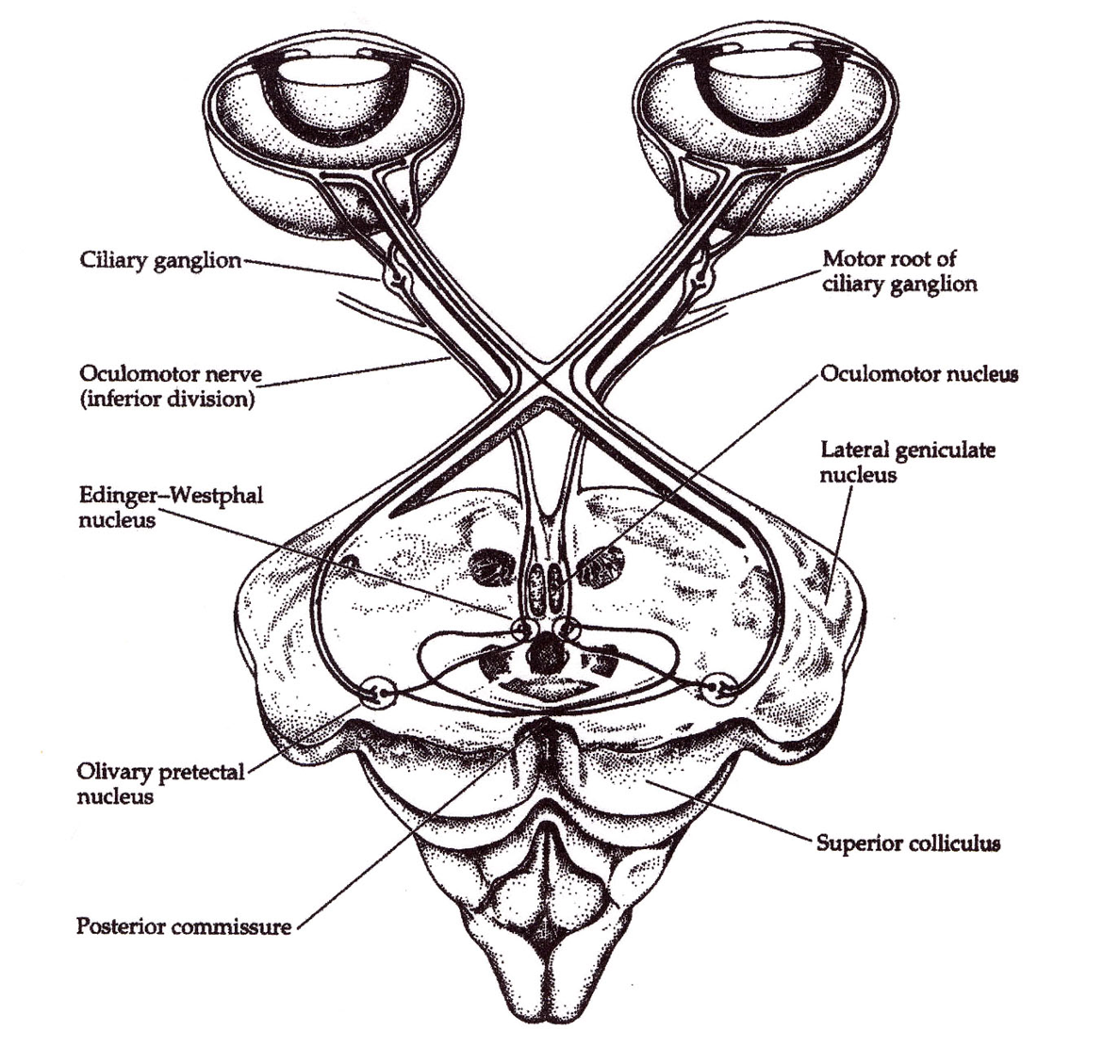
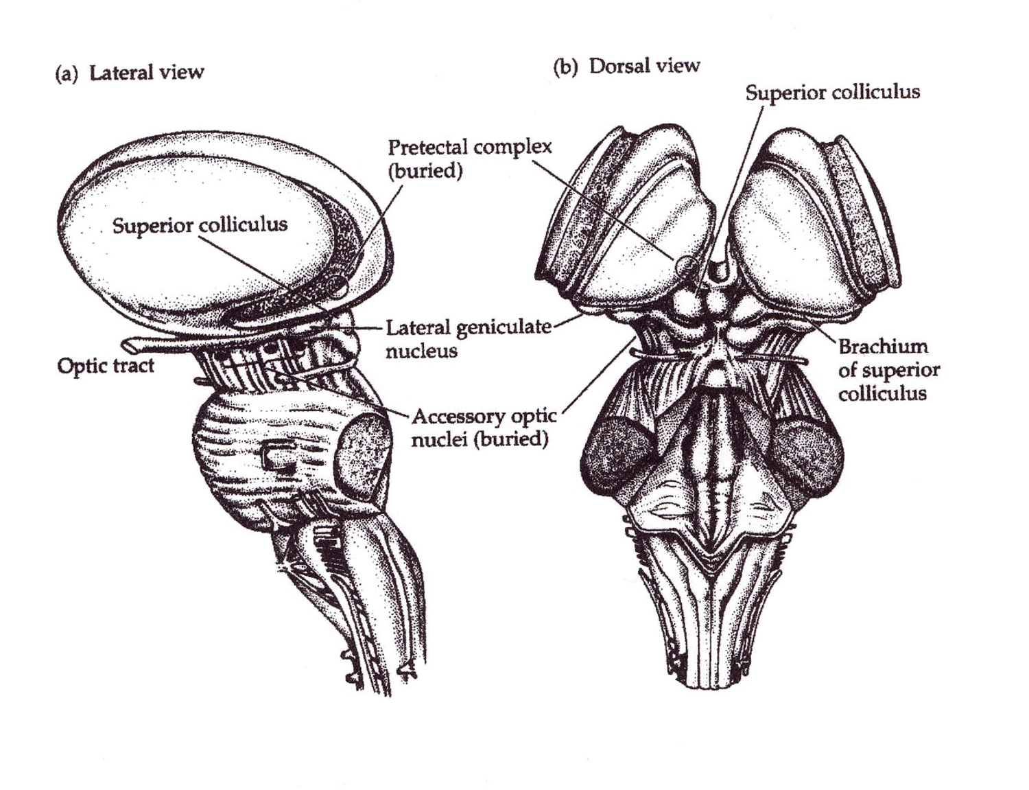
Major retinal recipient nuclei
LGN (vision)>> Superior Colliculus (saccades) > Suprachiasmatic Nucleus (circadian), Pretectum (pupillary reflex), Accessory Optic System (steadying gaze)
Blindsight
Limited visually guided behavior (reflexes) without conscious vision after damage to retina-LGN-cortex pathway
Effect of destroying geniculocortical pathway
Functional blindness with some reflexes preserved
Function of accessory optic system
Eye stabilization
Retinal slip
Motion of most of the visual scene across the retina due to unstable eye position
SCN
Hypothalamus above the optic chiasm, circadian rhythm regulation (uses light to “set” biological clock)
Pretectum
Pupillary light reflex (pupil constriction in olivary nuclei)
Major functions of superior colliculus
Saccades and orienting reflexes
Sensory maps in superior colliculus
Visual auditory, somatosensory maps, motor map
Effect of stimulating superior colliculus map location
Saccade toward corresponding location in space
Definition of LGN
Visual relay nucleus of the dorsal thalamus (part of diencephalon)
Definition of retinotopy
Neighboring retinal locations remain neighboring throughout visual pathways, allows lesions to produce predictable visual field defects
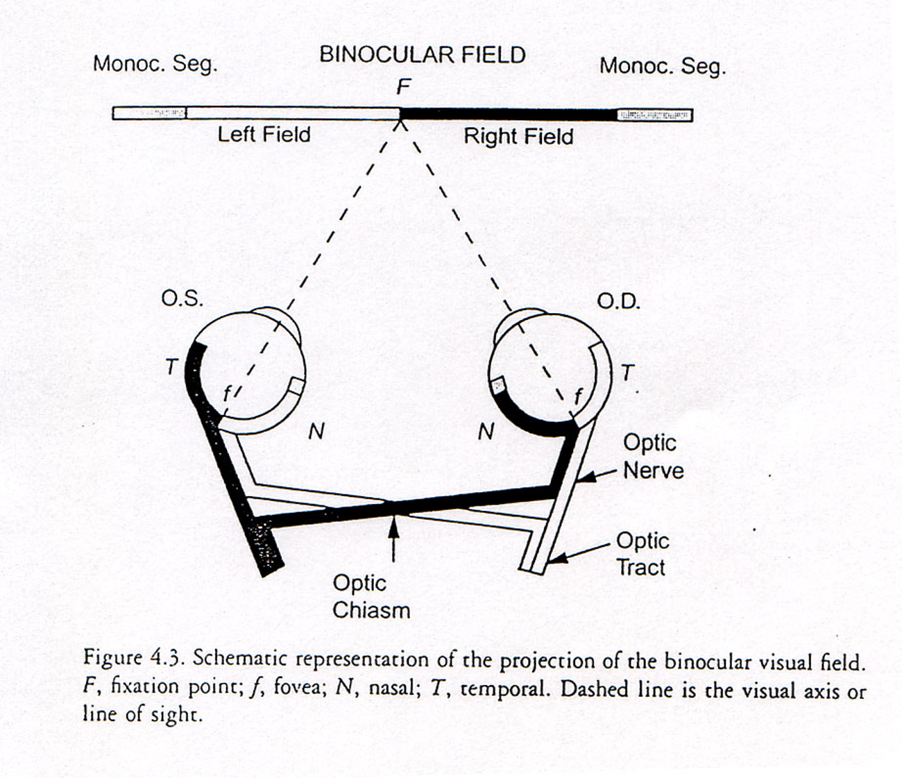
Visual field defect rule after optic chiasm
Homonymous
LGN contralateral eye input
Layers 1 4 and 6
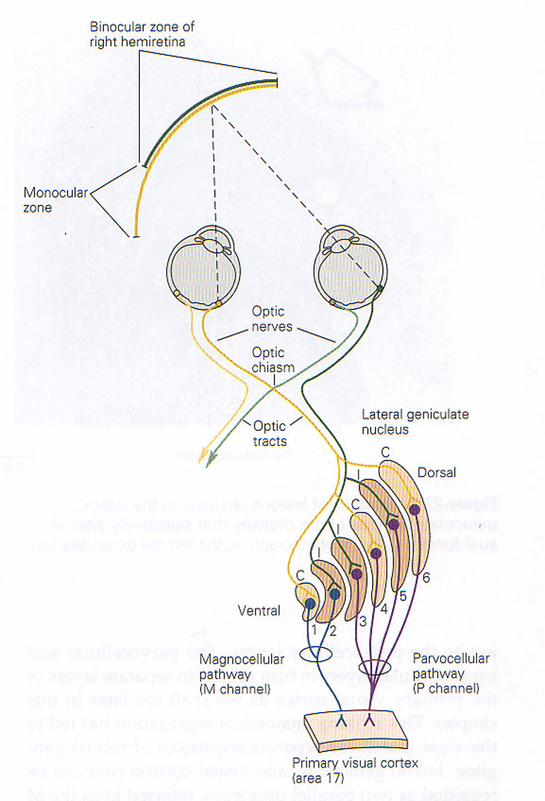
LGN ipsilateral eye input
Layers 2 3 and 5

Magnocellular LGN layers
Layers 1 and 2, parasol cells

Magno cells
Receive input from parasol ganglion cells, are “color blind”, respond well to low-contrast stimuli, low spatial acuity, very fast, sometimes called Y (homologous)

Parvocellular LGN layers
Layers 3, 4, 5, and 6, midget cells

Parvo cells
Receive input from midget ganglion cells, has color vision, fine detail, doesn’t respond to low contrast stimuli, slow, highest spatial acuity, sometimes called X (homologous)
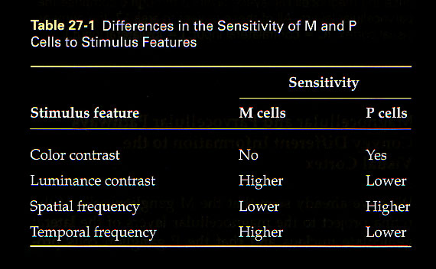
Functions of magnocellular pathway
Motion detection, low contrast sensitivity, rapid temporal responses
Functions of parvocellular pathway
Color vision and fine spatial detail
Major LGN neuron types
Relay neurons (project to cortex) and interneurons (only local connections)
Major computational strategy of retina
Information compression and feature extraction
Purpose of center-surround organization
Edge detection and contrast enhancement
Theme underlying magno and parvo pathways
Parallel processing
First location where information from both eyes converges
Cortex
LGN layers are monocular or binocular
Monocular
Superior colliculus
Recieves cortical input from frontal eye fields, part of midbrain, generates saccadic eye movements and reflexes, superficial layers= retina and cortex, deep layers= somatic and auditory inputs, head and neck orienting movements and multisensory integration
LGN lesions
*macular sparing is not obligate for visual cortex lesions
Ganglion cells
Only cells producing action potentials in the retina
First site of binocular integration
Cerebral cortex
Major theme of visual system
Specialized parallel pathways process different aspects of vision simultaneously