Screen Orientation, Direction of Flow, and Anatomy
1/27
Earn XP
Description and Tags
UT 302 - Abdomen 1
Name | Mastery | Learn | Test | Matching | Spaced | Call with Kai |
|---|
No analytics yet
Send a link to your students to track their progress
28 Terms
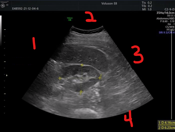
label each number and determine the imaging plane
superior
anterior
inferior
posterior
longitudinal image
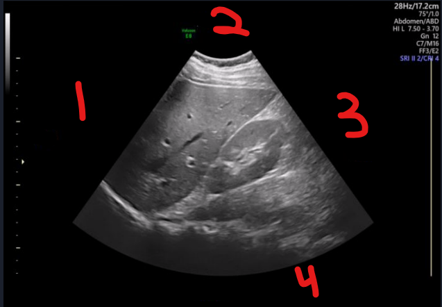
label each number and determine the imaging plane
right or medial
anterior
left or lateral
posterior
transverse image
T/F: we can only tell flow direction in long/sag vessel orientation, not transverse
true
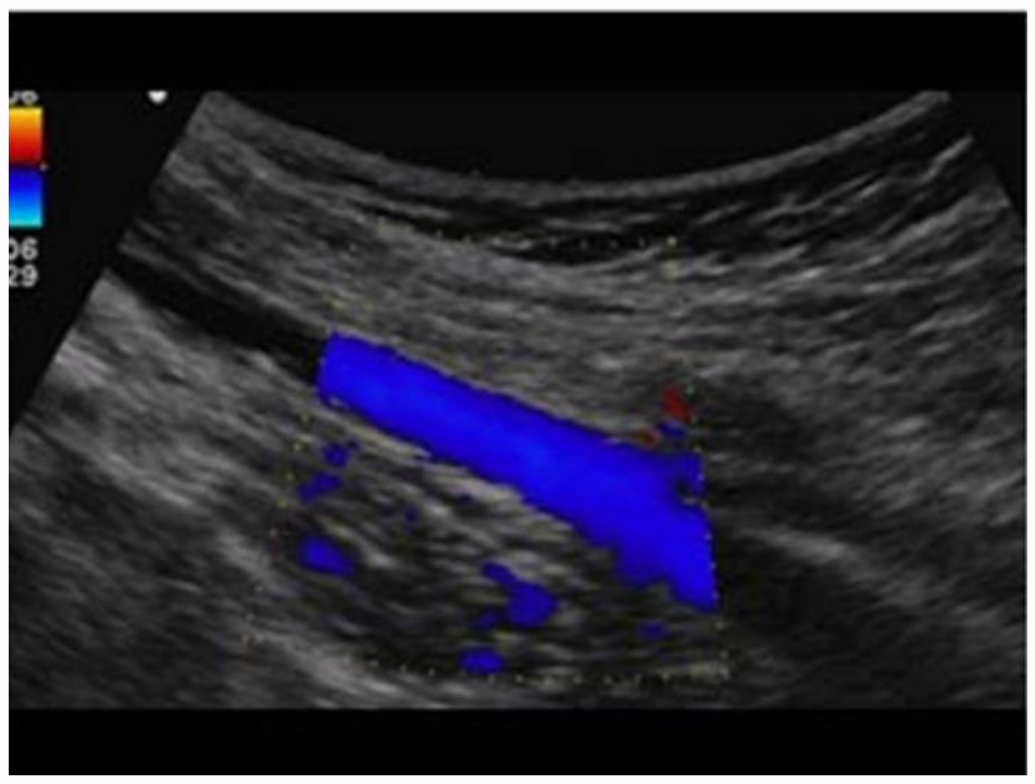
describe the blood flow occurring in this image
the anterior vessel is red, and red is on the bottom of the color bar, which means that blood is flowing away from the transducer
the posterior vessels are blue, and blue is on the top of the color bar, which means that blood is flowing toward the transducer
describe the blood flow occurring in this image
when imaging the carotid artery, we are above the heart, so
blood flowing from inferior to superior is going away from the transducer/heart
blood flowing from superior to inferior is going toward the transducer/heart
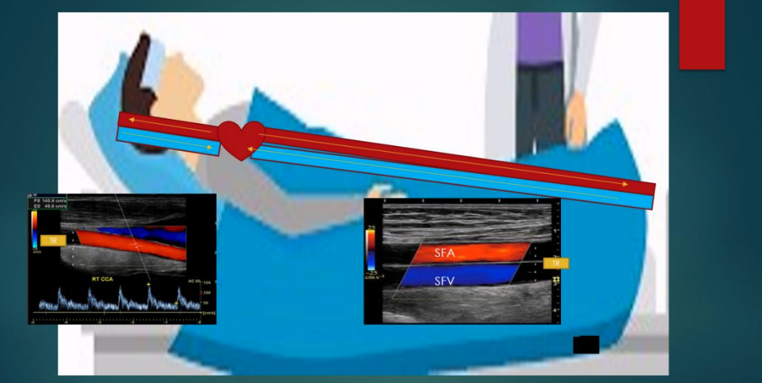
when imaging the SFA/SFV, we are above the heart, so
blood flowing from inferior to superior is going toward the transducer/heart
blood flowing from superior to inferior is going away from the transducer/heart
where is the color detector/transducer in a blunted sector abdominal image?
it is at the highest point in the vessel
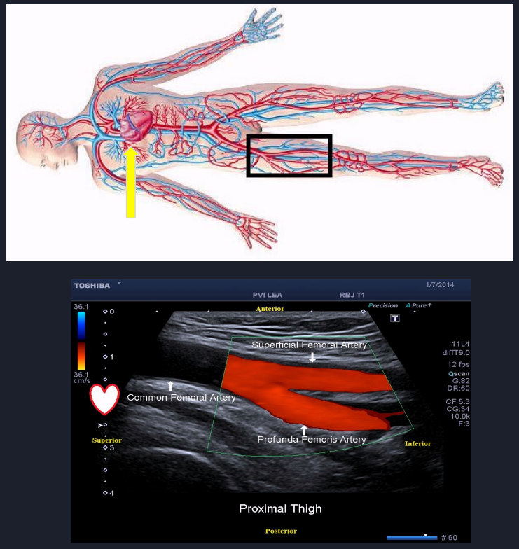
describe the blood flow occurring in this image
the heart is superior to the transducer
the color detector/transducer is at the highest point in the vessel (top left of vessel in image)
red is on the bottom of the color bar, which means that blood is flowing away from the transducer
blood is flowing from superior to inferior
this is normal blood flow for the femoral artery
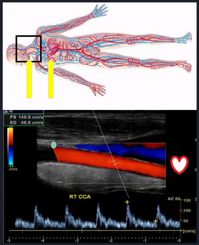
describe the blood flow occurring in this image
the heart is inferior to the transducer
the color detector/transducer is at the top left edge of the color box
red is on the top of the color bar, which means that blood is flowing toward the transducer
blood is flowing from inferior to superior
this is normal blood flow for the carotid artery
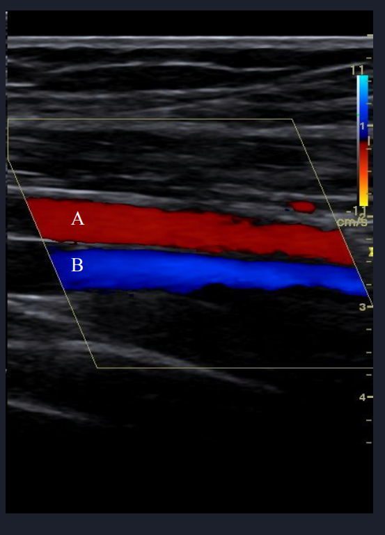
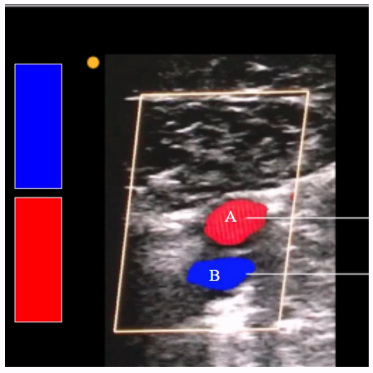
which is the CFV and which is the IJV and why?
A is the IJV because blue is on top of the color bar, which means that color is flowing toward the transducer (indicator is at the right top edge of the color box)
blood is flowing from superior (head) to inferior (heart)
B is the CFV because blue is on the bottom of the color bar, which means that color is flowing away from the the transducer (indicator is at the right top edge of the color box)
blood is flowing from inferior (legs) to superior (heart)
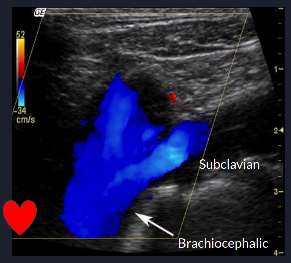
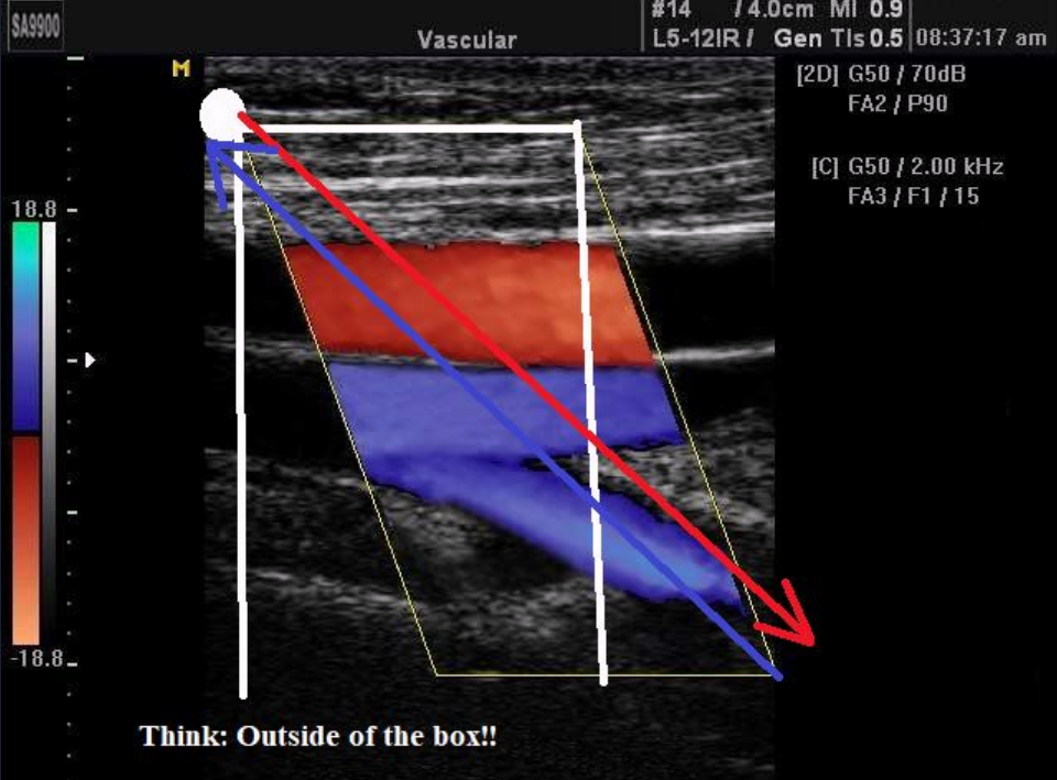
describe the blood flow in this image and what is unique about vascular evaluation of the brachiocephalic artery and vein? is this the brachiocephalic artery or vein?
it is the only vascular study that is done in trans
the color indicator is at the top right of the color box, and the heart is at the bottom left
blue is on the bottom of the color bar, which means blood is flowing away from the transducer
this is the brachiocephalic vein
is this the femoral artery or vein?
this is the femoral vein
blue is on the top of the color bar, which means that it is going toward the transducer
blood is flowing from inferior to superior
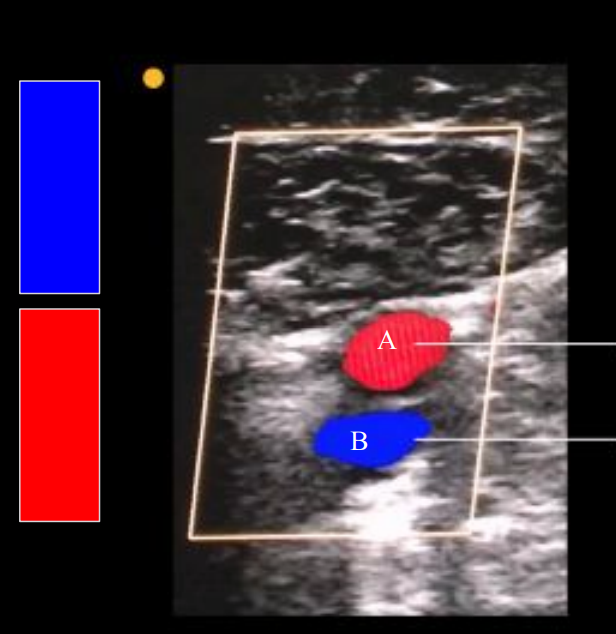
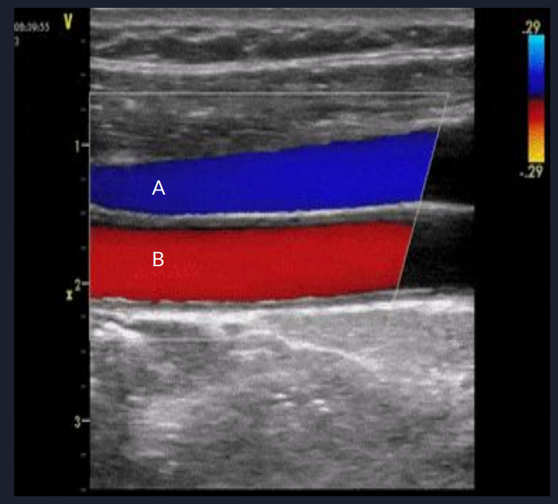
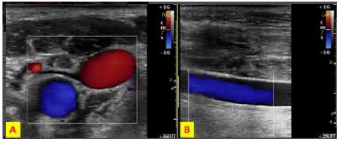
which vessel is the femoral artery and which is the femoral vein?
A is the femoral artery
red = bottom = away
B is the femoral vein
blue = top = toward
is this normal flow?
yes, this is normal antegrade flow
red = top = toward
blood is flowing from inferior (heart) to superior (brain)
is this normal flow?
yes, this is normal antegrade flow
blue = top = toward
blood is flowing from superior (head) to inferior (heart)
using direction of flow, explain which vessel is the CCA
blue = top = toward = blood is flowing from superior to inferior
red = bottom = away = blood is flowing from inferior to superior
the red (posterior) vessel is the CCA
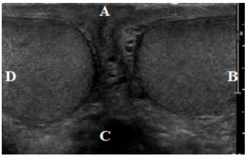
what directions do the labels represent?
D: superior
A: anterior
B: inferior
C: posterior
what directions do the labels represent?
D: right
A: anterior
B: left
C: posterior
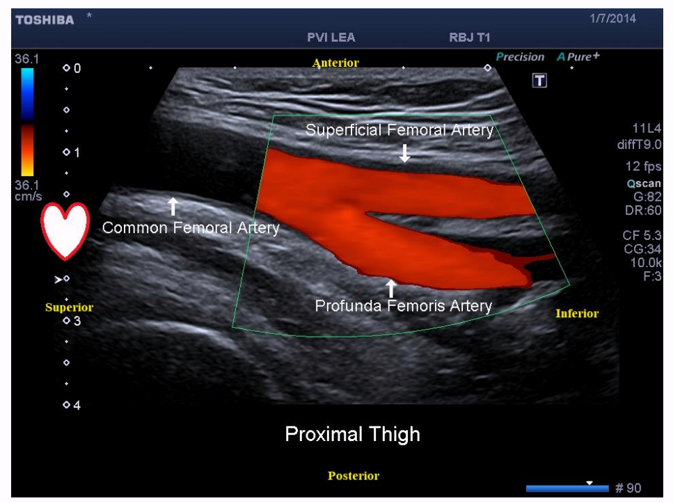
the flow in this femoral artery goes from
superior to inferior
the direction of flow for this IJV in this image is
superior to inferior
the direction of flow for this carotid artery in this image is
inferior to superior
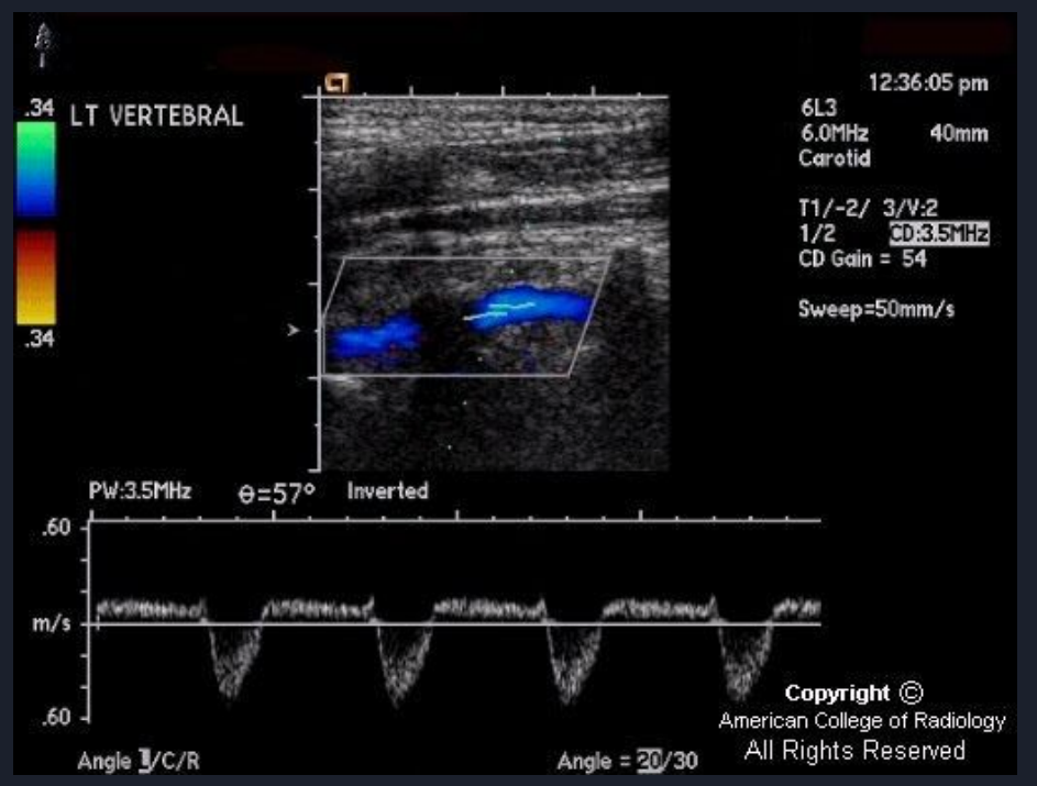
the direction of flow for this internal carotid artery in this image is
inferior to superior
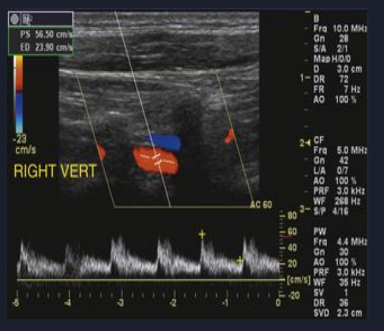
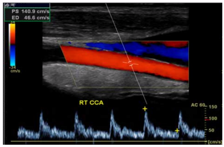
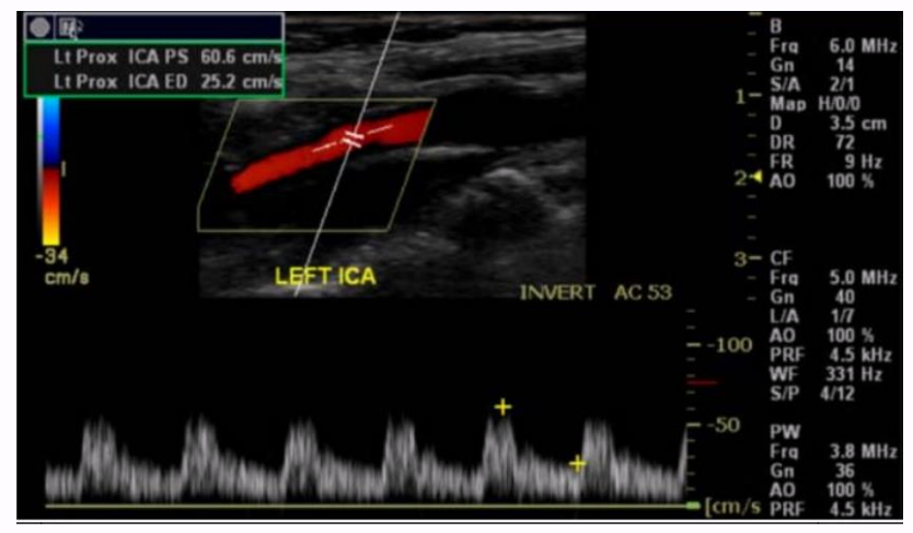
the direction of this common carotid artery blood flow is
inferior to superior
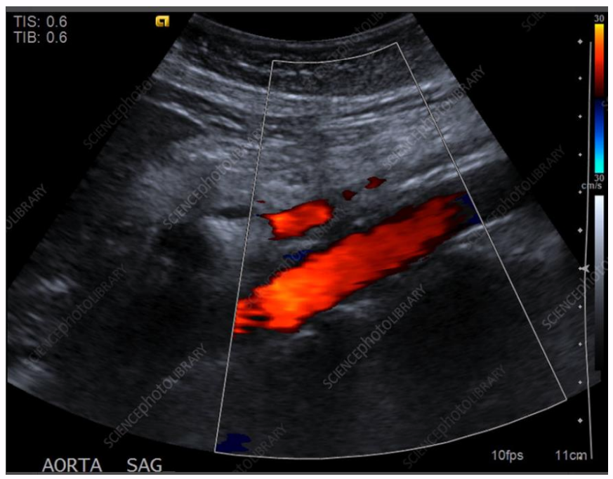
the direction of flow in this aorta is
superior to inferior
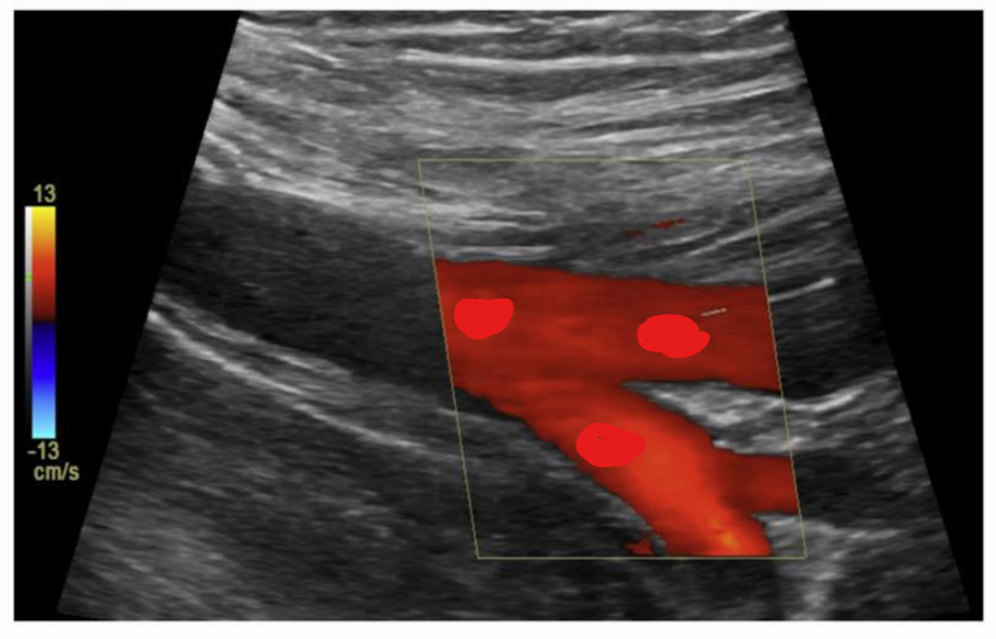
the direction of flow in these vessels is
inferior to superior

what vessels are these by looking at the color flow?
veins
the direction of flow in this femoral vein is? and is it antegrade or retrograde flow?
superior to inferior
flow for the femoral vein is supposed to go from inferior (legs) to superior (heart)
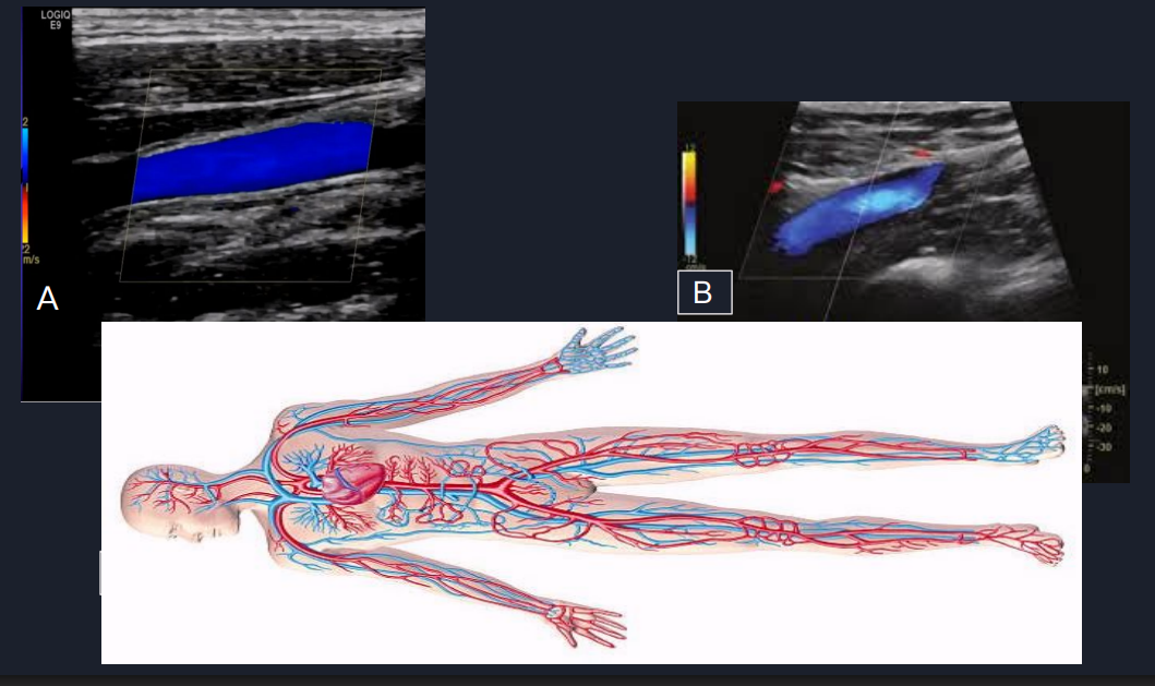
what is the direction of flow in vessel A?
cannot determine direction of flow
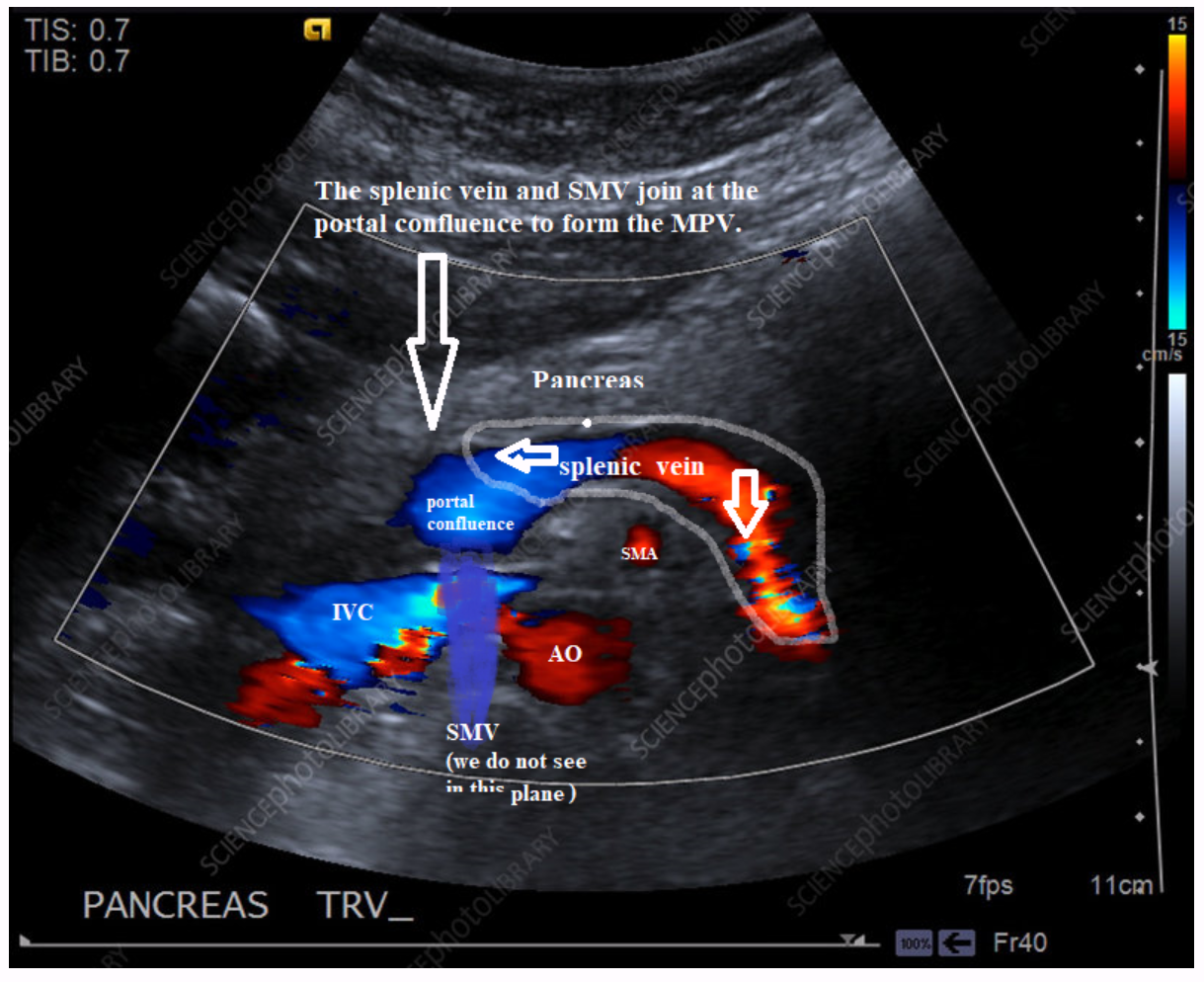
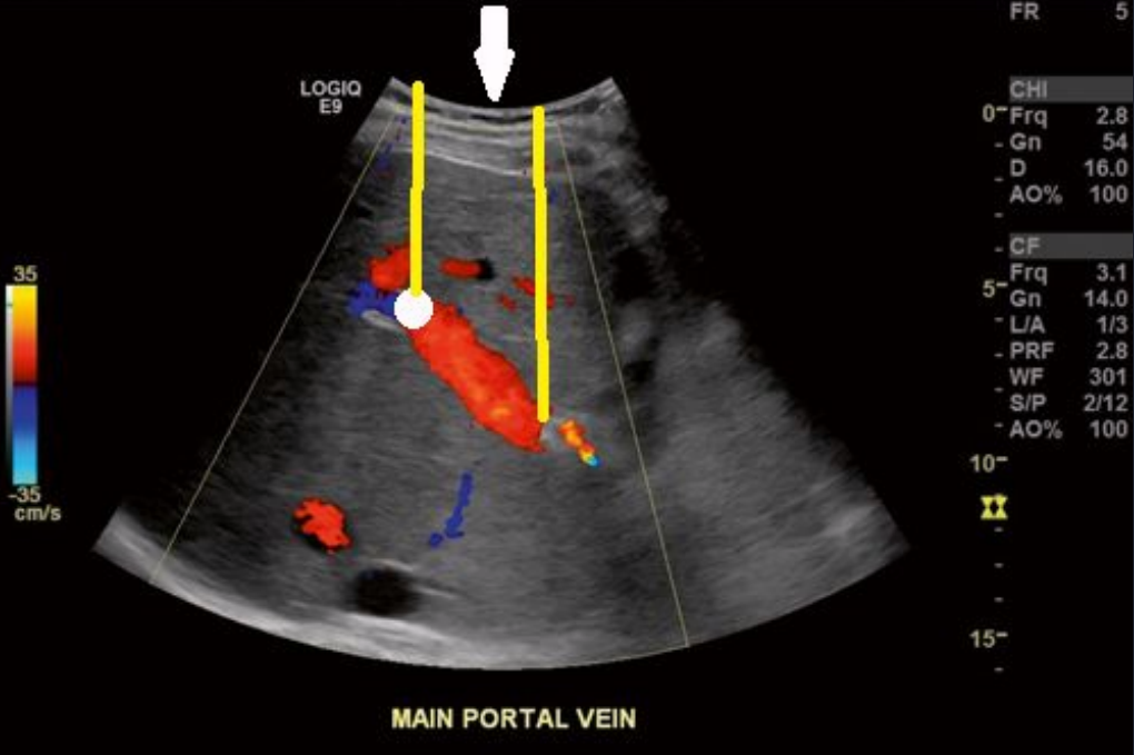
Explain the direction of flow in this image. Where is the blood coming from and where is it going? Is this image normal?
the vessel in red is the splenic vein, and the red part is going toward the transducer
the vessel in blue is the splenic vein, and the blue part is going away from the transducer
it joins with the SMV (not pictured but drawn in) at the portal confluence to create the MPV
this is normal flow