Biology A Level - Biological Molecules summary questions
 Call Kai
 Learn
Learn Practice Test
 Spaced Repetition
 Match
Match Flashcards
 Knowt Play
1/16

There's no tags or description
Looks like no tags are added yet.
Name | Mastery | Learn | Test | Matching | Spaced | Call with Kai | Chat |
|---|
No analytics yet
Send a link to your students to track their progress
17 Terms
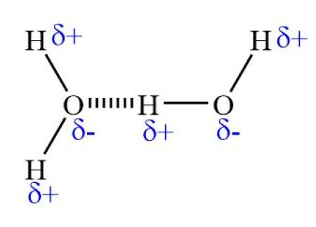
How does hydrogen bonding occur between water molecules? Include diagram.
Water molecule are polar (meaning charge is unevenly distributed), hence contain d+ hydrogen atoms and d- oxygen atoms. Between water molecules, there is a force of attraction between the d+ H and d- O, so a weak hydrogen bond forms.
Name the 6 properties of water that are important for life processes. Relate each one to living organisms.
Cohesion/ surface tension - supports small insects eg. pond skaters.
Adhesion - Capillary action: movement of H2O through blood vessels and xylem.
Latent heat of evaporation - Acts as a coolant eg. sweat
High specific heat capacity - Maintains constant temperatures in cellular environments.
Density of ice - Insulating layer above lake water so fish can survive in winter.
Universal solvent - Medium for chemical reactions and transport eg. blood.
Explain hydrolysis and condensation reactions and provide examples in ALL biological molecules
Condensation: joining of two monomers - releases water
Hydrolysis: breaking down of a polymer - requires water
Lipids - H on glycerol and OH on fatty acid react x3 to form 3 ester bonds and 3 H2O
Carbs - OH groups on C1 and C4 react to form a 1,4 glycosidic bond and H2O
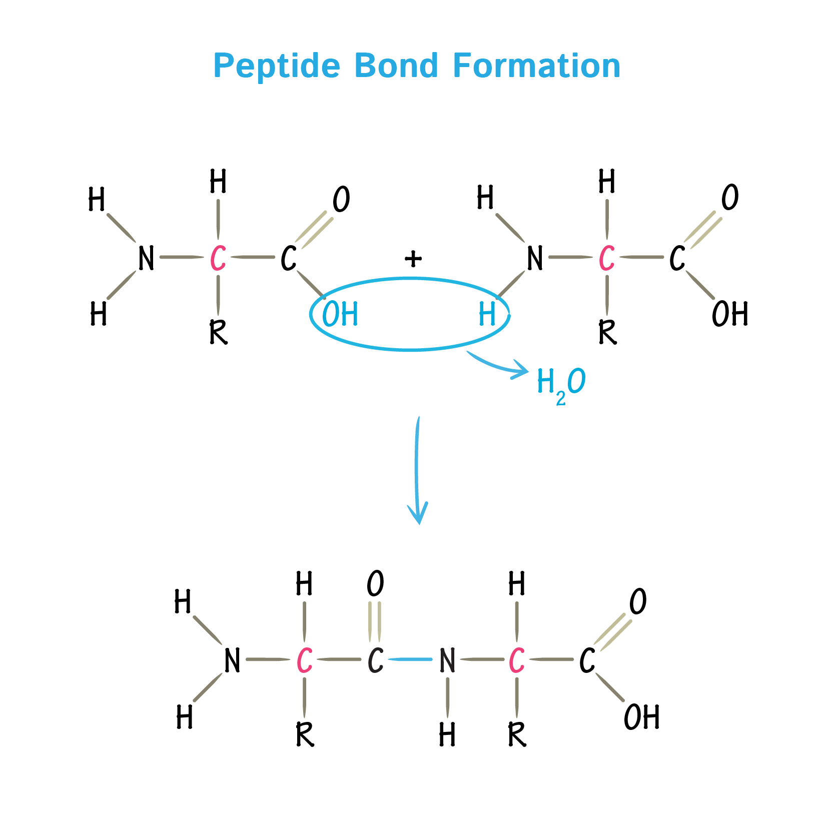
Protein - OH group (in carboxyl group) and H (on amine group) react to form peptide bond and H2O
What are the chemical elements that make up each biological molecule?
Protein - C,H,O,N,S
Lipids - C,H,O
Carbs - C,H,O
Nucleic acid - C,H,O,N,P
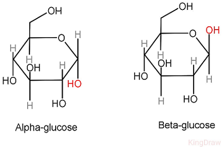
Draw an alpha and beta glucose molecule. Describe the differences and similarities between them.
Similarities:
Hexose ring structure
Same molecular formula - C6H12O6
Same structure on C2,3,4,5 + 6
Difference:
Alpha - C1 has same position of OH and H as C4 (OH below)
Beta - C1 has opposite position of OH and H as C4 (OH above)
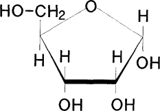
Draw a ribose molecule. Describe the difference between glucose and ribose.
Ribose only has 5 carbons - pentose molecule
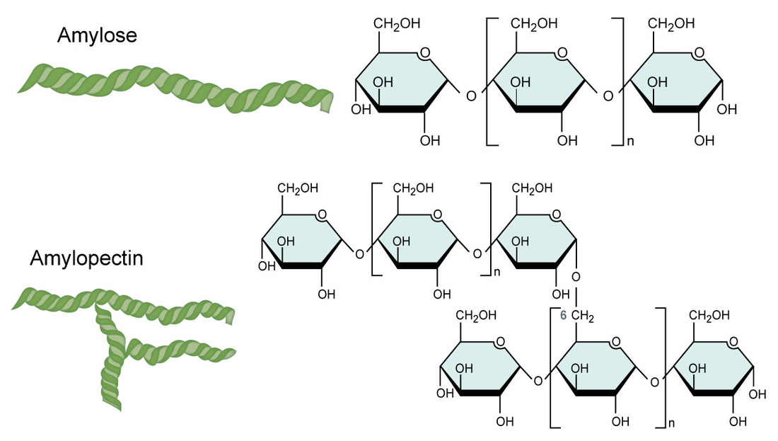
Describe how the structure of starch (amylose/amylopectin) relates to its function.
Amylose: long chain of a-glucose molecules joined together by 1-4 glycosidic bonds to form an alpha helix (strengthened by H bonds
Function: Storage molecule in plants - compact, water insoluble
Amylopectin: Branched chain of a-glucose molecules with 1-4 bonds in chain and 1-6 bonds at branching points
Function: Storage molecule in plants - Compact, insoluble, branched so many free ends where glucose can be broken off for reactions (hydrolysis)
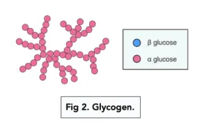
Describe how the structure of glycogen relates to its function.
Structure: Branched chain of a-glucose molecules with 1-4 bonds in chain and 1-6 bonds at branching points. Branches more frequently than amylopectin
Function: Storage molecule in animals and fungi - compact, many free ends to release glucose (hydrolysis)
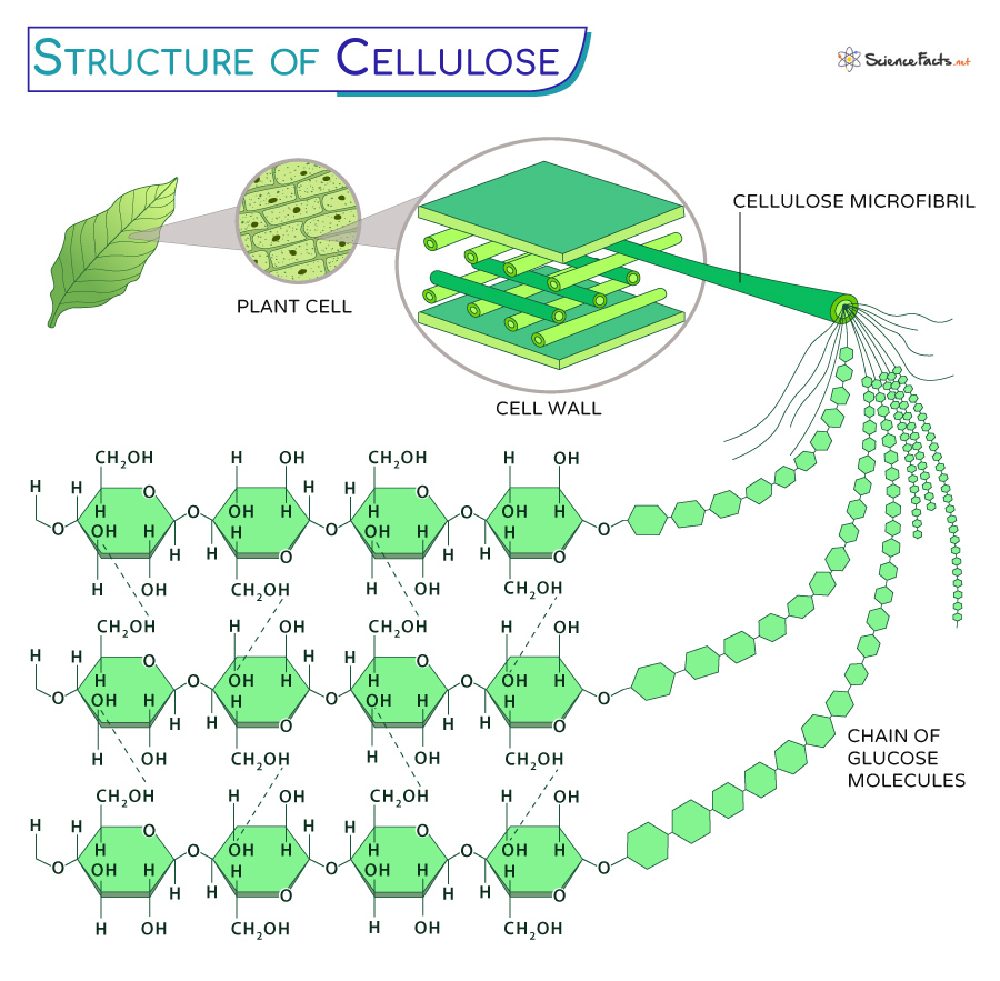
Describe how the structure of cellulose relates to its function.
Structure: chains of b-glucose where every other molecule is rotated 180º. Many chains joined together by H bonds to form large sheet of cellulose
Function: Cell wall of plants - strong (many H bonds) and water insoluble.
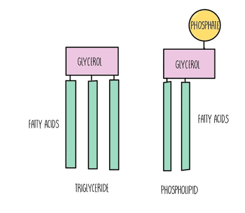
Compare and contrast the structure of triglycerides to phospholipids.
Triglycerides: glycerol and 3 fatty acids joined by ester bonds, can be saturated or unsaturated (contains C=C), non-polar, stores energy.
Phospholipids: glycerol, 2 fatty acid and a phosphate group, hydrophilic head and hydrophobic tail, found in plasma membrane.
Explain the differences between saturated and unsaturated fatty acids
Saturated - all C-C, no C=C, higher melting point as they are more compact, so bonds harder to break
Unsaturated - contains C=C, lower melting point as they are less compact, so bonds easier to break
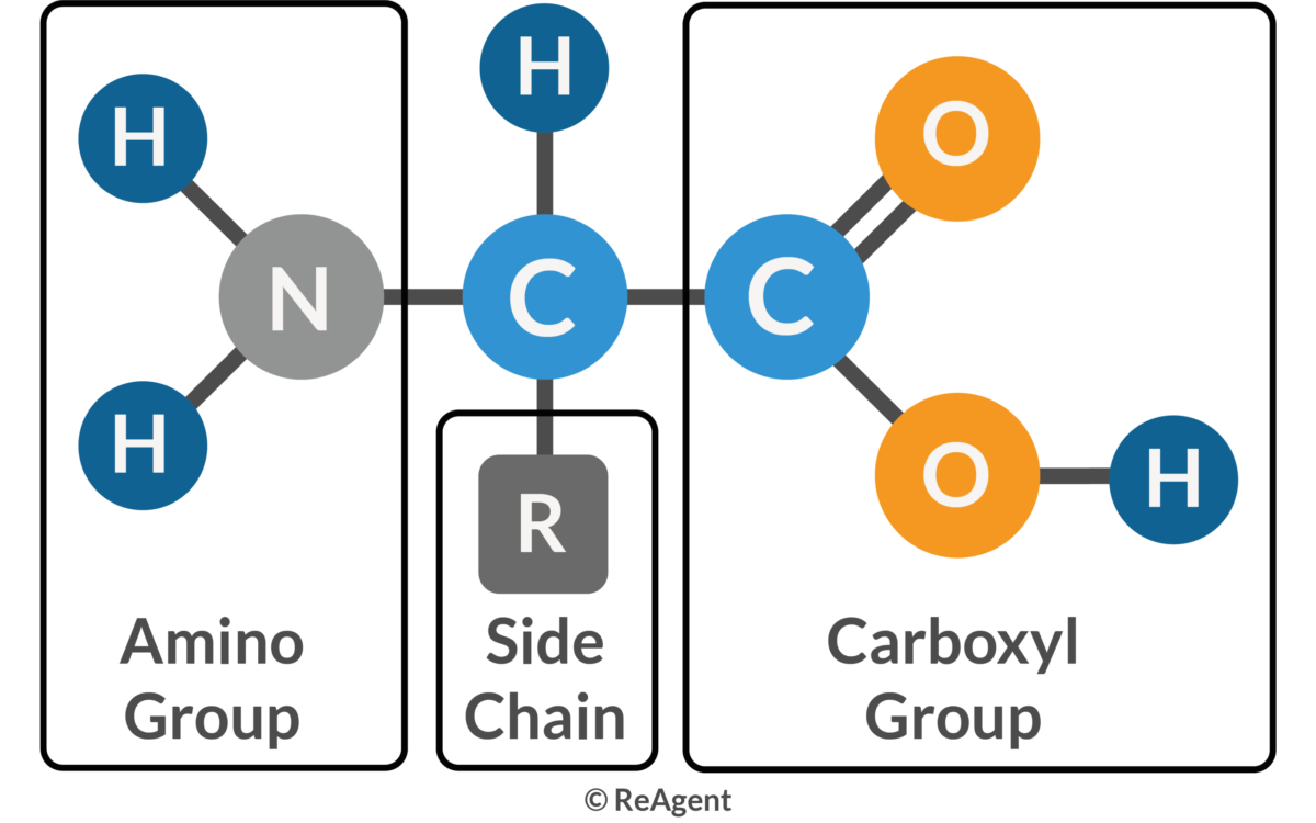
Describe the general structure of an amino acid.
Amine group - NH2
Central Carbon - CH
R group - a range of chemical groups different in each amino acids. 20 different amino acids naturally occuring in the human body.
Carboxyl group - COOH
Explain how a peptide bond forms.
Condensation reaction: bond forms between the C in carboxyl group and N in amine group, releasing H2O and forming peptide bond
Broken via hydrolysis reaction.
Outline the four different levels of protein structure.
Primary - the sequence of amino acids in a polypeptide chain. Determines all other levels of structure
Secondary - Polypeptide chain fold into a helix or b pleated sheet due to hydrogen bonding within the peptide chain.
Tertiary structure - 3D shape held in place by bond between the R groups in the polypeptide chains: covalent disulphide, ionic bonds, hydrophobic and hydrophilic interactions. Vital for function.
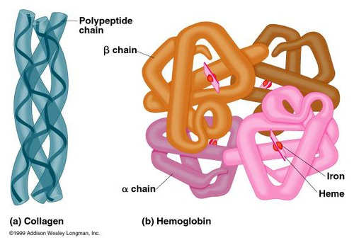
Quaternary - NOT ALL HAVE: Composed of 2+ polypeptide chains/subunits interacting and the interactions between peptide chains and prosthetic groups
Compare conjugated, globular and fibrous protein structures providing examples of each.
Conjugated - contains a prosthetic group ie. non-protein component. Example: Haemoglobin - contain Fe2+ in haem group
Globular proteins - compact, water soluble, spherical shape. Hydrophilic R groups on outside, hydrophobic inside. Example: Insulin - transported in bloodstream
Fibrous proteins - Long and insoluble. contain many hydrophobic R groups. Repetitive and organise structure with simpler 3D structure. Examples: Keratin, Elastin and Collagen - all have strength and flexibilty function so must be strong
Explain how to chemically test for reducing and non reducing sugars
Benedict’s test for reducing sugars:
Add equal parts Benedict’s solution to sample and warm in water bath (~80ºC)
Benedict’s test for non - reducing sugars:
Add 2cm of ~1.5M HCl and heat in water bath for 3 mins (to hydrolyse glycosidic bonds)
Cool and neutralise with NaHCO3, checking with pH paper
Carry out Benedict’s test as above
Results:
Red - Large, Orange - moderate, Green - traces, Blue - none
Explain how to chemically test for proteins, lipids and starch
Proteins - Biuret test for proteins
Add Biuret A and B. Lilac colour indicates protein present
Lipids - Emulsion test
Add ethanol, shake and add distilled water. Milky white emulsion indicates presence of lipid.
Starch - Iodine test
Add iodine solution. Blue black colour indicates presence of starch.