SECTION 01: INTRODUCTION TO RESPIRATORY PHYSIOLOGY
1/46
There's no tags or description
Looks like no tags are added yet.
Name | Mastery | Learn | Test | Matching | Spaced |
|---|
No study sessions yet.
47 Terms
By the end of Section 01, you should be able to:
Describe the purpose of the respiratory system, and the four steps involved in external respiration.
Describe the role of the respiratory muscles in both inspiration and expiration.
Describe the purpose of the pleural space
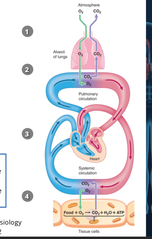
🫁 External Respiration – Overview
What it is: The process that brings oxygen into the body and removes carbon dioxide.
Goal: Deliver O₂ to tissues and remove CO₂ from tissues.
Main system involved: Respiratory system (lungs, airways) + circulatory system (blood transport).
✅ External Respiration: 4 Main Steps 1. Ventilation (Breathing)
What happens? Air goes in and out of lungs.
Why? To move fresh air (with O₂) into the alveoli and remove stale air (with CO₂).
✅ External Respiration: 4 Main Steps 2. Gas Exchange between Air and Blood
Where? In the alveoli (air sacs in lungs) and pulmonary capillaries.
What happens?
O₂ diffuses from alveoli → blood.
CO₂ diffuses from blood → alveoli.
✅ External Respiration: 4 Main Steps 3. Transport of Gases in the Blood
O₂: Carried from lungs → body tissues.
CO₂: Carried from tissues → lungs (to be exhaled).
How? Via red blood cells in circulation.
✅ External Respiration: 4 Main Steps - 4. Gas Exchange between Blood and Tissues
Where? In body capillaries near tissues.
What happens?
O₂ diffuses from blood → tissues.
CO₂ diffuses from tissues → blood.
🎯 Key Concept: Internal vs. External Respiration
Internal respiration = Cell-level usage of O₂ + production of ATP + release of CO₂.
External respiration = Body-level process of getting O₂ in and CO₂ out.
🫁 Other Functions of the Respiratory System 🗣 1. Speech Production
How? Air passes over vocal cords in the larynx (voice box).
Why important? This airflow lets us talk, sing, shout, etc.
🫁 Other Functions of the Respiratory System 🛡 2. Defense Against Foreign Particles
What does it do? Filters out dust, bacteria, and allergens from air.
How?
Mucus traps particles.
Cilia (tiny hairs) push them out of the airways.
🫁 Other Functions of the Respiratory System 💪 3. Helps in Pushing Movements (Parturition & Defecation)
Parturition = childbirth
Defecation = bowel movement
How? The respiratory muscles (like the diaphragm) contract and help increase pressure in the abdomen, which pushes things out (baby or stool).
🫁 Other Functions of the Respiratory System 🩸 4. Lung as a Blood Reservoir
What? Lungs can hold extra blood.
Why? Helps balance blood flow between the right and left sides of the heart quickly when needed.
🫁 Other Functions of the Respiratory System ⚖ 5. Acid-Base Balance
What does it do? Helps control blood pH.
How? By controlling how much CO₂ is breathed out (less CO₂ = less acid).
Will be explained more in Module 05.
🫁 Anatomy of the Respiratory System: Overview
Main Parts:
Lungs
Chest Wall
Pleural Space (space between lungs & chest wall)
Focus here: Lungs → Upper & Lower Tract
🛤 Airway vs. Alveoli
Airways = Passages that move air in & out.
Alveoli = Tiny air sacs where O₂ and CO₂ exchange happens.
🔼 Upper Respiratory Tract (Airway Part) - 3 main part —> 👃 1. Nose & Nasal Cavities
Entry point for air
Warms, moistens, and filters the air
🧠 2. Pharynx (Throat)
Shared tube for both air and food
Sends air to lungs and food to esophagus
🗣 3. Larynx (Voice Box)
Located just below the pharynx
Contains vocal cords (for speech)
Directs air into the lower airways
💡 Summary:
Air flows:
Nose → Nasal Cavity → Pharynx → Larynx → Lower Tract
🔽 Lower Respiratory Tract (Airway Part) - 5 parts to this —> 🧵 1. Trachea (Windpipe)
A tube that carries air down from the larynx.
Divides into two bronchi—one for each lung.
🌳 2. Bronchi
Right & Left main branches from the trachea.
Each bronchus supplies one lung.
Further split into smaller tubes inside each lung.
🌿 3. Bronchioles
Tiny branches of the bronchi.
Get narrower the deeper you go.
No cartilage, just smooth muscle.
🫧 4. Respiratory Bronchioles
Smallest bronchioles with very thin walls.
Start to allow some gas exchange.
🫁 5. Alveoli
Tiny air sacs at the ends of bronchioles.
Main site of gas exchange (O₂ in, CO₂ out).
Surrounded by capillaries.
📍 Air Path Summary:
Trachea → Bronchi → Bronchioles → Respiratory Bronchioles → Alveoli
🩸 What Are Capillaries?
Capillaries are the smallest blood vessels in your body.
They connect arteries (which carry blood away from the heart) to veins (which return blood to the heart).
Their walls are super thin—just one cell thick—so that gas exchange (O₂ and CO₂) can happen easily.
🔄 What Do Capillaries Do in the Lungs?
In the lungs, capillaries surround the alveoli.
O₂ diffuses from the alveoli into the blood.
CO₂ diffuses from the blood into the alveoli to be exhaled.
❓ Are Capillaries Part of the Lower Respiratory Tract?
Not exactly.
They are part of the circulatory system, not the airway.
But they work very closely with the lower respiratory tract (especially the alveoli) to exchange gases.
🌳 The Respiratory (Tracheobronchial) Tree
🫁 Airway Branching Pathway
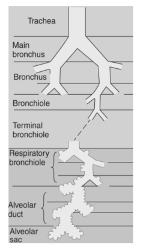
Trachea
Main Bronchus
Bronchus
Bronchiole
Terminal Bronchiole
Respiratory Bronchiole
Alveolar Duct
Alveolar Sac
💨 Types of Air Flow in the Respiratory Tree 🔄 Convective Flow (Needs energy)
Happens from Trachea → Terminal Bronchioles
Air is pushed by muscle contractions (like the diaphragm)
Similar to how you blow air through a straw
🌬 Diffusive Flow (Passive)
Starts at Respiratory Bronchioles
Air moves by diffusion (no energy needed)
Gas spreads naturally into alveoli where gas exchange happens
📏 Why Does Branching Matter?
Even though the tubes get smaller, the total surface area increases
This large area slows airflow, making it ideal for diffusion in the alveoli
🧍♂ Chest Wall: What Is It?
Includes everything that helps with breathing:
Thorax (chest) + AbdomenHouses: Lungs & heart
Protected by: Rib cage
Key Muscles: Found between ribs and at the base of the chest (diaphragm)
💪 Muscles of the Chest Wall 🩻 Intercostal Muscles (Between Ribs)
External intercostals = outer layer
Internal intercostals = deeper layer
Help move the ribs to change chest size
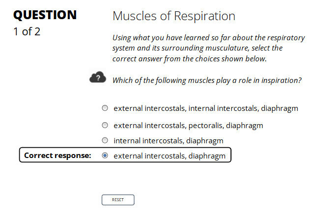
🧘♀ Muscles Used in Inspiration (Breathing In)
Diaphragm
Moves down to make room for lungs
Main muscle for inhaling
External Intercostal Muscles
Lift the ribs upward and outward
Makes chest cavity bigger
💨 Muscles Used in Expiration (Breathing Out)
Internal Intercostal Muscles
Pull ribs down and in
Decrease chest size
Abdominal Muscles
Push up against diaphragm
Help force air out during coughing, sneezing, exercise, etc.
🧠 Note: In normal relaxed breathing, expiration is passive—these muscles are only used when needed!
🫁 What Is the Pleural Space?
A thin fluid-filled space between the lungs and the chest wall.
Only about 2 mm wide.
Helps lungs move smoothly when you breathe.
pleural space - 🧼 Why Is It Important?
Filled with pleural fluid.
Reduces friction as the lungs expand and contract.
Acts like lubricant between the lung and the chest wall.
🩻 Layers of the Pleura (Membranes)
1. Visceral Pleura
Sticks directly to the surface of the lungs.
2. Parietal Pleura
Lines the inside of the chest wall (thoracic wall).
🟦 Pleural Space = The tiny gap between the visceral and parietal pleura.
📍Visual Reference from the Image:
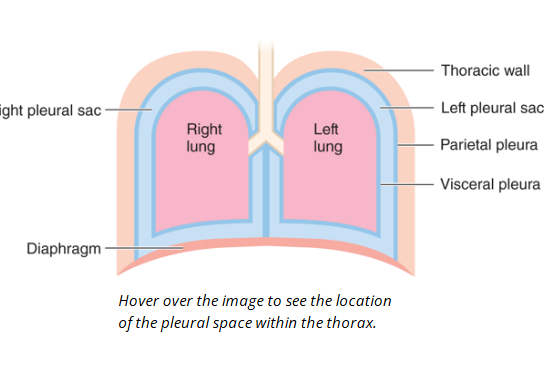
Right/Left Pleural Sacs: Each lung is inside its own sac.
Pleural Space: Located between the two blue linings (parietal & visceral pleura).
Diaphragm: Sits underneath both lungs, helps pull lungs down when inhaling.
🫁 External Respiration: Two Main Processes
1. Pressure-Driven Airflow
Muscles (like the diaphragm) create a pressure gradient.
Air flows from high pressure → low pressure.
Must overcome airway resistance to get into lungs.
2nd processs - 2. Gas Diffusion Across Alveoli
Oxygen (O₂) moves from alveoli → blood.
Carbon dioxide (CO₂) moves from blood → alveoli.
Happens through thin alveolar-capillary barrier by diffusion.
🧮 Key Equation:
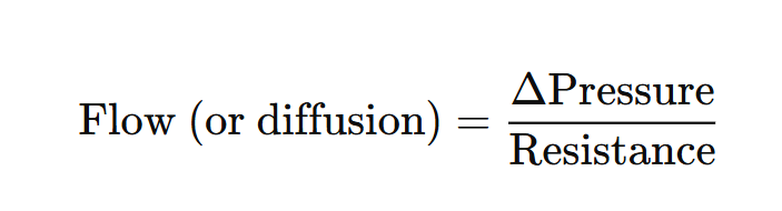
equation - 🧠 What This Means:
Δ Pressure = Difference in pressure between two points (e.g., inside vs. outside lungs)
Resistance = How hard it is for air (or gas) to move
More pressure or less resistance = more flow
🎯 Real-Life Analogy:
Think of it like blowing through a straw:
Harder you blow (more pressure) = more air flows out.
Thicker the straw (more resistance) = harder to get air through.
🫁 Path of Air Through the Respiratory System
📍 Step-by-Step Airflow:
Nares → Nostrils (openings to the nose)
Nasal Cavity
Cilia = tiny hairs that filter dust
Mucous membranes = add moisture
Blood vessels = warm the air
Pharynx → Throat (shared by food & air)
Larynx → Voice box (contains vocal cords)
Epiglottis closes during swallowing to block food
Trachea → Windpipe (has cartilage rings to keep it open)
Bronchus (pl. bronchi) → Left & right branches into lungs
Bronchioles → Smaller branches inside the lungs
Alveolar Sacs → End of bronchioles
Alveoli → Tiny air sacs where gas exchange happens
🌬 Gas Exchange: How It Works
Happens in the alveoli, which are:
Lined with one cell layer (very thin!)
Surrounded by blood capillaries
↔ Diffusion Process:
Oxygen (O₂) moves:
Alveoli → Capillaries → BloodCarbon Dioxide (CO₂) moves:
Capillaries → Alveoli → Exhaled
🔁 Respiration Summary
Inhalation path:
Nose → Pharynx → Larynx → Trachea → Bronchus → AlveoliExhalation path:
CO₂ follows the reverse path out
Activity
🫁 Inspiration = Inhalation = Breathing In
What happens?
The diaphragm contracts and moves downward.
External intercostal muscles lift the rib cage.
This increases the space in your chest (thoracic cavity).
Air flows into your lungs because the pressure inside is now lower than the outside air.
Activity pt 2
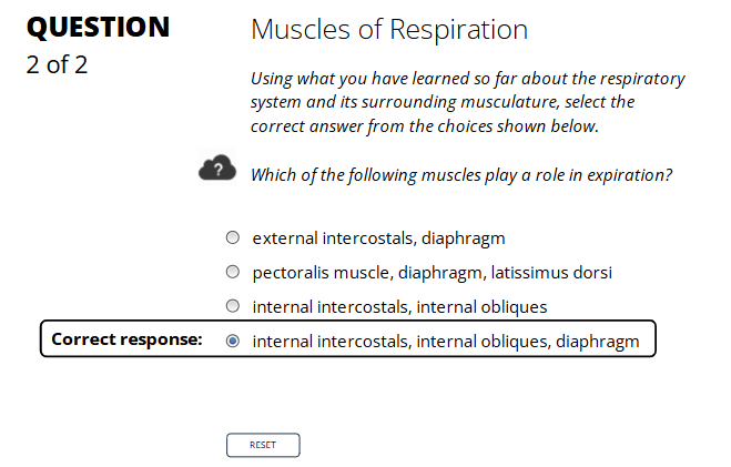
🏋♂️ 2. Forced (Active) Expiration
(Occurs during exercise, coughing, sneezing, singing, shouting)
🔽 Muscles That Contract:
Muscle | Action in Expiration |
|---|---|
Internal Intercostals | Pull ribs down and inward to shrink chest cavity |
Diaphragm (already relaxed) | Passive part — stays relaxed & dome-shaped |
Internal Oblique (abdominal muscle) | Compresses abdomen → pushes diaphragm up |
External Oblique (also abdominal) | Same as internal oblique — helps push air out |
Rectus Abdominis | Pushes diaphragm up by squeezing abdominal contents |
Transversus Abdominis | Deepest core muscle — tightens abdominal wall to force air out |
(dont need to know the words above tbh) in passive thoMuscles used:
❌ No active muscle contraction
✅ Diaphragm relaxes (moves up)
✅ External intercostals relax