BCH4024 - Exam 1
 Call Kai
 Learn
Learn Practice Test
 Spaced Repetition
 Match
Match Flashcards
 Knowt Play
1/106

There's no tags or description
Looks like no tags are added yet.
Name | Mastery | Learn | Test | Matching | Spaced | Call with Kai | Chat |
|---|
No analytics yet
Send a link to your students to track their progress
107 Terms
Foundations in Biochem
cellular
chemical
physical
genetic
evolution
Cellular Foundations
cells are fundamental units of life
at each level of organization novel properties emerge; life emerges at cellular level
Common characteristics of living things
organization
expression and transmission of genetic info
transfer and transformation of energy and matter
interact w/ their system and enviro
evolution - conservation of function and diversity in structure
Phylogeny of Life
Bacteria
Archaea
Eukarya
Prok and Euks contain
DNA
ribosomes
cytosol
plasma membrane
euks contain
DNA in membrane bound nucleus and membrane bound organelles
Cells are limited by diffusion
upper limit of cell size is set by rate of diffusion
as size increase, surface to volume ration decreases
many animal cells have highly convoluted/folded surfaces
Cell Cytoskeleton
dynamic and made of protein filaments
Actin filaments (smallest) - 6nm wide, made from G-actin
polymerizes to F-actin through noncovalent bonds
Intermediate filaments (medium) - 10 nm wide, made from a-keratin subunits
microtubules (largest) - 23nm wide, made from tubulin
Each filament composed of protein monomers that are bound NONcovalently
provide shape, organization, and help things move

Nucleic Acids
DNA and RNA are polymers of nucleotides
store and transmit genetic info
some RNAs have structural and catalytic roles
Genome = entire seq of cell’s DNA or RNA
Genomics = characterization of structure, function, evolution, and mapping of genomes
Proteins
Long polymers of AAs
function as enzymes, structural elements, signal receptors, transporters
Proteome = sum of all proteins functioning in a cell
Proteomics = systematic characterization of this prot complement under a specific set of cond
Polysaccharides
Polymers of simple sugars
Energy rich fuel stores, rigid structural components, extracellular recognition elements
Glycome = entire complement of carbohydrate-containing molecules
Lipids
Water-insoluble hydrocarbon derivatives
structural comps, energy-rich fuel stores, pigments, intracell signals
Lipidome = lipid containing molecules in a cell
Chemical Foundations
Less than 30 of naturally occurring elements essential to life
H, O, N, C make up 99% of organisms
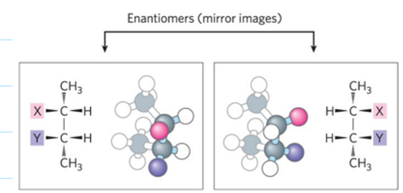
Enantiomers
non-superimposable mirror images
Physical Foundations
molecules are consistently synthesized and broken down
Living cells are in a dynamic steady state distant from eq with its surroundings
maitaining requires constant investment of energy
Organisms either make energy or extract it (photoautotrophs vs chemotrophs)
Laws of Thermodynamics
total amount of energy in universe remains constant
randomness (S, entropy), in universe is constantly increasing
creating and maintaining order requires work and energy
Free Energy
Want deltaG to be less than zero for a rxn to happen spontaneously
DeltaG = deltaH - TdeltaS
Keq > 1
prods > reactants
G < 0
rxn is spont
Keq < 1
prods < reactants
G > 0
rxn is NOT spont
Keq = 1
prods = reac
G = 0
rxn @ eq
standard free energy
G0 = -RTlnK
Actual free energy change
G = G0 + RTlnK
K = Q
rxn is at eq
G = 0
K < Q
rxn is past eq
more prods/reacts now then at eq
G > 0
rxn is not spont
K > Q
rxn has not reached eq
less prods/reacts now then at eq
G < 0
rxn is spont
H bonds are fleeting
1-20 ps
H bonds are ___ and ____ than covalent bonds
longer and weaker
23 kj/mol vs 470 kj/mol
liquid water has _ h bonds vs ice has _
3.4 vs 4
Weak noncovalent interactions compared to covalent
Hydrophobic and aromatic ring stacking and van der waals (weak but many)
0.4 - 4 kj/mol (weakest)
H bonds and electrostatic
4-40 kj/mol
Salt bridge (H bond + electrostatic)
40-400 kj/mol
carboxylate AA side chain (Asp, Glu) to basic AA (Arg, Lys)
Hydrophobic Effect
Water molecules forming cage around nonpolar substances and releases ordered water as it clusters together
increases entropy
amphipathic molecules form micelles
similar interactions w/ enzymes and substrates
Kw of water
1.0 × 10⁻¹⁴ M2
henderson-hasselbalch
pH = pKa + log [A-]/[HA]
Keq of water at 25C
1.8 × 10⁻¹⁶ → can normally assume concentration is 55.5M
optimal pH for enzymes
have max catalytic activity
pepsin - pH of 1.5 (digestive enzyme)
Trypsin - neutral pH in lumen (digestive enzyme in SI)
Alkaline phosphatase - pH of 8-9 (hydrolytic enzyme of bone tissue)
Diabetes results in
acidosis
accumulation of high conc of beta-hydroxybutyric acid and acetoacetic acid which lowers blood pH to less than 7.35
AA structure
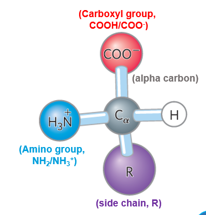
Proteins consist almost entirely of what stereoisomer
L-amino acids
rare D-amino acids but converted after prot syn
optically active due to chirality
Zwitterion
have both positive and negative charge at certain pH ranges
****MAJOR FORM
AAs also called ampholytes due to having + and - charge (acidic and basic groups)
Isoelectric point of AAs
where net charge EQUALS ZERO
pI = (pka1 + pka2) / 2
Glycine
Nonpolar, achiral, hydrophobic
G, Gly

Alanine
nonpolar,hydrophobic
A, Ala

Proline
nonpolar, hydrophobic
P, Pro
can be in cis or trans conformation

Valine
hydrophobic, nonpolar
V, Val
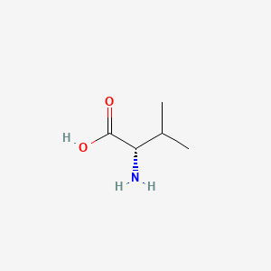
Leucine
Hydrophobic, nonpolar
L, Leu
Isoleucine
hydrophobic, nonpolar
I, Ile

Methionine
hydrophobic, nonpolar
M, Met

Pheylalanine
aromatic, rel hydrophobic
F, Phe

Tyrosine
aromatic, rel hydrophobic
Y, Tyr
can H bond, plays important functional role in some enzymes

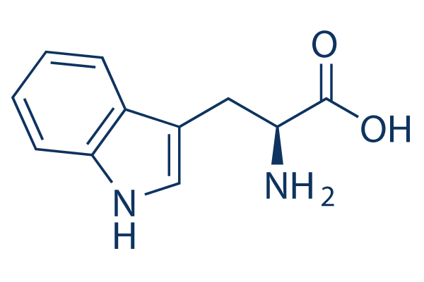
Tryptophan
aromatic, rel hydrophobic
W, Trp
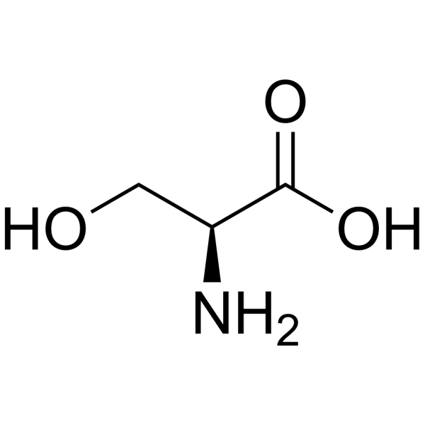
Serine
polar, uncharged
S, Ser
Threonine
polar, uncharged
T, Thr

Cysteine
polar, uncharged
can form cystine through disfulfide bonds → not under physiological pH (pka = 8.3)
C, Cys

Asparagine
polar, uncharged
N, Asn

Glutamine
polar, uncharged
Q, Gln

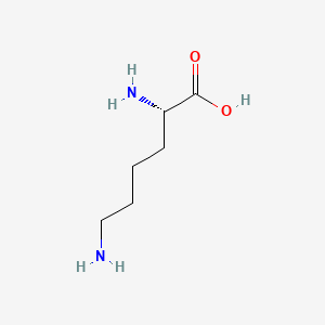
Lysine
positively charged, basic
K, Lys
Arginine
positively charged, basic
R, Arg

Histidine
positively charged, basic
H, His
only AA w/ pka near neutrality
can be H+ donor or acceptor

Aspartate / Aspartic Acid
negatively charged, acidic
Asp, D

Glutamate / Glutamic Acid
negatively charged, acidic
Glu, E

Characterize biomolecules by light absorption
Absorbance of wavelengths
aromatic AAs have distinct wavelengths
C labeling in AAs

Peptide Bond
molecule of water eliminated for each peptide bond formed
covalent bond
amide linkage
requires energy to form
to break raise temp or use enzyme that lowers activation energy
Peptide Bond Structure
Linus Pauling and Robert Corey Discovered
C-N bond is 10% shorter than usual amide bonds due to double bond character (40%) from resonance
makes it stronger and shorter (1.32 A)
planar
Peptide bond angles
phi = Ca-N
psi = Ca-C=O
omega = peptide bond

Peptide Nomenclature
N-terminus on the left and C-terminus on the right
AAs are added onto C-terminus
Protein Size
avg MW of nucleotide is 110 Da
to estimate # residues in a protein take proteins MW and divide by AA MW
Number of Possible Prot Seqs
20a
a = peptide length
orgs typically rely on 30,000 - 50,000 seqs so necessary function will determine which sequence is constructed
Folding is directed by
AA side chains
Determine peptide sequence by
Mass Spec
Proteases
Conjugated proteins
permanently associated chemical components (prosthetic groups)
lipoproteins
glycoproteins
metalloproteins
proteins are stabilized by
noncovalent interactions and forces
primary structure
backbone of protein, sequence of AA residues
3 covalent bonds separate alpha C of adjacent AA residues
dihedral angles (phi and psi) define secondary strucs
Secondary Structures
spatial arrangement of main chain atoms
alpha helix, beta conformation, b turn, random coils
phi and psi remain same throughout a segment to give uniform structures
stabilized by weak noncovalent interactions
r groups ALWAYS protrude out from backbone
alpha helix
simplest arrange, max number of H bonds
every 4 AAs: H of n and O of n+4 (of main chain backbone)
each turn is 5.4A and 3.6 residues
polar residues on one side and hydrophobic on the other
Pro and Gly destabilize the helix
Pro introduces a kink and Gly allows too much flexibility
Generally right-handed, left-handed is less stable
small electric dipoles align through H bonds in each peptide bond (N → C)
310 = smallest, 3 residues
alpha = medium, 3.6 residues
pi = largest, 4.4 residues
Beta conformation
Backbone extends into zigzag (7 A), organizes polypep into sheets, R groups are trans
antiparallel (most common due to linear H bonding between strands)
Parallel (less common, distorted H-bonding)
forms between backbone atoms of adjacent strands
Beta turns
connect ends of two adjacent segments of an antiparallel B sheet
180 degree turn
Type 1 → 4 residues, proline is the 2nd residue
Type 2 → 4 residues, glycine is the 3rd residue
Gamma turns → 3 residues, proline is the 2nd residue
H bond forms btwn 1st and last residue
Ramachandran Plots
Visualizes phi and psi angles and tests quality of 3D protein structures

Circular Dichroism
assesses common 2nd structures
measures differences in molar absorption of left vs right-handed circularly polarized light
AAs found in alpha helix
Kristin Has Marvelous LACE Q tips
AAs found in Beta Sheet
IVY For the Win
AAs found in reverse turns
SPeeDiNG
What AA is found in all secondary stucture
Arginine
Tertiary Structure
overall 3D structure
arrangement of all atoms in a prot
weak interactions and covalent bonds hold interacting segments in position
Quarternary Structure
arrangement of TWO+ polypeptide chains in 3D complexes
Types of Proteins
Fibrous
Globular
Membrane
Intrinsically Disordered
Fibrous Proteins
give strength/flexibility to structures
simple repeating element of 2nd struc
water insoluble due to high conc of hydrophobic residues
alpha helix cross-linked with disulfide bonds
beta sheets
collagen triple helix
alpha-keratin
two right-handed alpha helices wrapped around each other to form a super-twisted coil (overall left-handed_
Common AAs: Angry Vikings Make Fantastic Iced Lattes
cross-links stabilized by disulfide bonds
Collagen
found in connective tissue
3 left-handed alpha-helices twisted around each other right-handed
rich in Gly-X-Y
Y = hydroxyproline
X = proline
Cross-linked by covalent bonds involving Lys, hydroxylysine, histidine
super strong
Scurvy
general degradation of connective tissue
lack of vitamin C
Vit C required for hydroxylation of proline and lysine in collage
Globular Proteins
fold back on each other
more compact
distinct structure, unique for biological function
Myoglobin
Intrinsically Disordered Proteins
lack defineable structure
lack hydrophobic core
high densities of charged residues
Every Kid Reeks of Poop
facilitates a protein to interact w/ multiple binding partners
disordered segments can assume diff structures
Example: p53
Proteostasis
Protein homeostasis: folding, unfolding, and degradation of proteins
Native proteins
synthesized from intermediates or chaperone assisted folding
Thermodynamics of Prot Folding
Unfavorable entropy change (negative S)
folding decreases protein’s internal S
Favorable enthalpy (negative H)
folding creates stable internal bonds → release heat
Hydrophobic effect
increases S of water, MAIN driving force for folding
Denaturation
by heat, pH, organic solv, solutes, detergents, etc.
lead to protein precipitation
loss of function
determine stability of prot by playing with one of these factors
Anfinsen Experiment
Showed that the primary struc (AA seq) contains all info needed to fold a protein chain into its tertiary struc
renaturation
Denatured RNase A w/ urea and mercaptoethanol (to break disulfide bonds)
Removed solvents and RNase A became catalytically active again
Folding is a rapid stepwise process
local secondary strucs fold first
ionic interaction and H bonds play important role
longer range interactions follow
hydrophobic effect
process continues until entire polypep folds
Levinthal’s Paradox
mathematically impossible for protein folding to occur randomly by trying every conformation until the lowest energy one is found
Free-Energy Funnel
multiple stable intermediates leading to final fold
high degree of conformational entropy
high free energy
Chaperone Proteins
facilitate correct folding pathways or ideal microenvironments