Looks like no one added any tags here yet for you.
cell theory
all living organisms are made of cells, sharing
Cell surface membrane
Cytoplasm
DNA
Ribosomes
organelle
a component in a cell that carries out a specific task
cells
basic functional and structural units in a living organism
tissues
group of similar cells of similar structure working together to perform a particular function
organ
made from a group of different tissues working together to perform a particular function
organ system
made from a group of organs w related functions, working together to perform body functions within organism
Eukaryotic cells diameter
10-100 μm
Prokaryotic cells diameter
0.1-5 μm
differences between eukaryotic and prokaryotic cells
Eukaryotic cells have a more complex ultrastructure
Eukaryotic cells are larger
eukaryotic cells is divided up into membrane-bound compartments
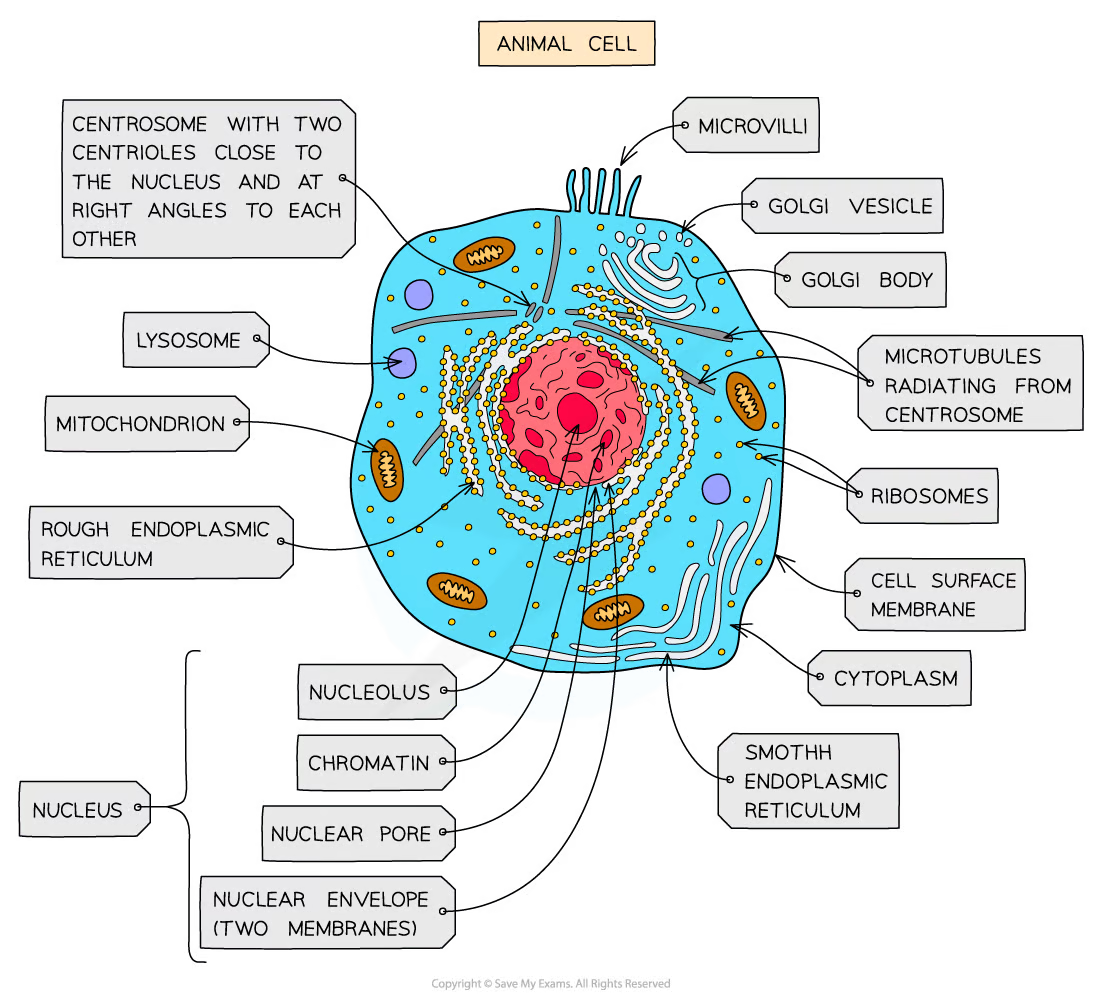
Animal and plant cells are both types of eukaryotic cells that share key structures such as
Membrane-bound organelles, including a nucleus
Larger ribosomes known as 80S ribosomes
Key differences between animal and plant cells include
Animal cells have centrioles and some have microvilli
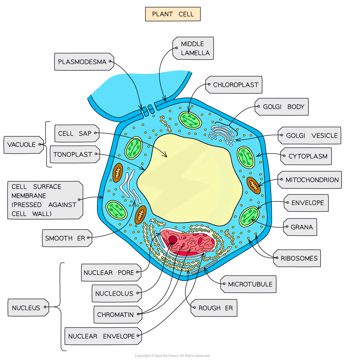
Plant cells have a cellulose cell wall, large permanent vacuoles, and chloroplasts
labelled animal cell
labelled plant cell
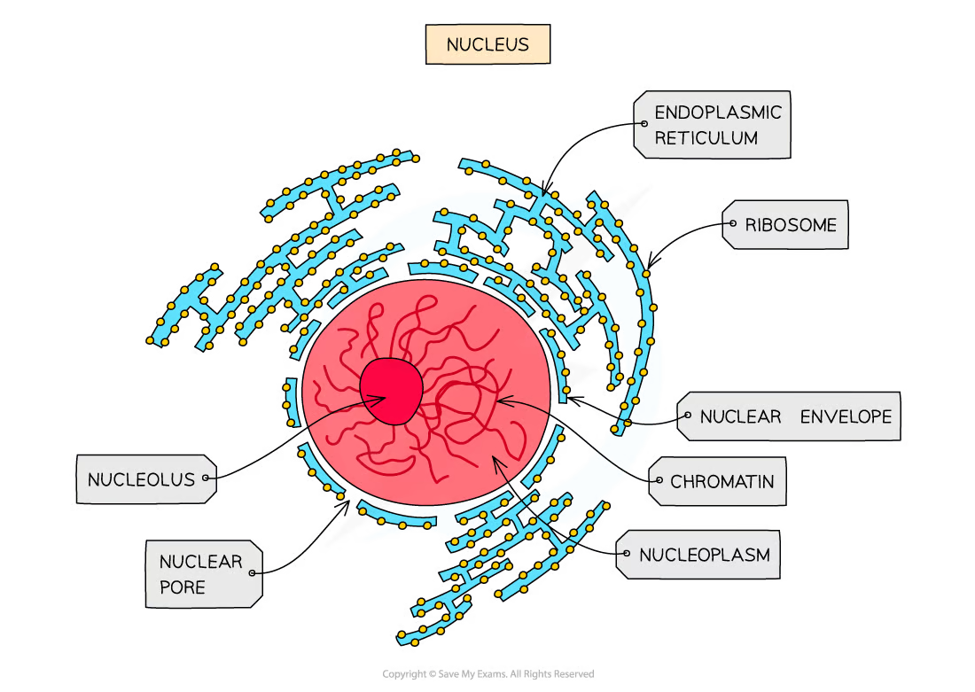
nucleus
relatively large
separated from the cytoplasm by a double membrane called the nuclear envelope
contains nuclear pores - important channels for allowing mRNA/ribosomes/enzymes to travel out of the nucleus
chromatin - the material from which chromosomes are made (Chromosomes are made of sections of linear DNA)
nucleolus - makes ribosomes
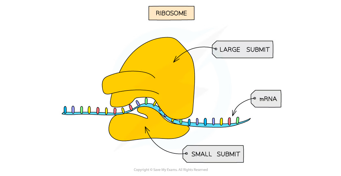
ribosomes
They aren’t surrounded by a membrane
It’s made up of ribosomal RNA (rRNA) and proteins
80s ribosomes are found in eukaryotic cells
70s ribosomes are found in prokaryotes, mitochondria, and chloroplasts
the site of translation
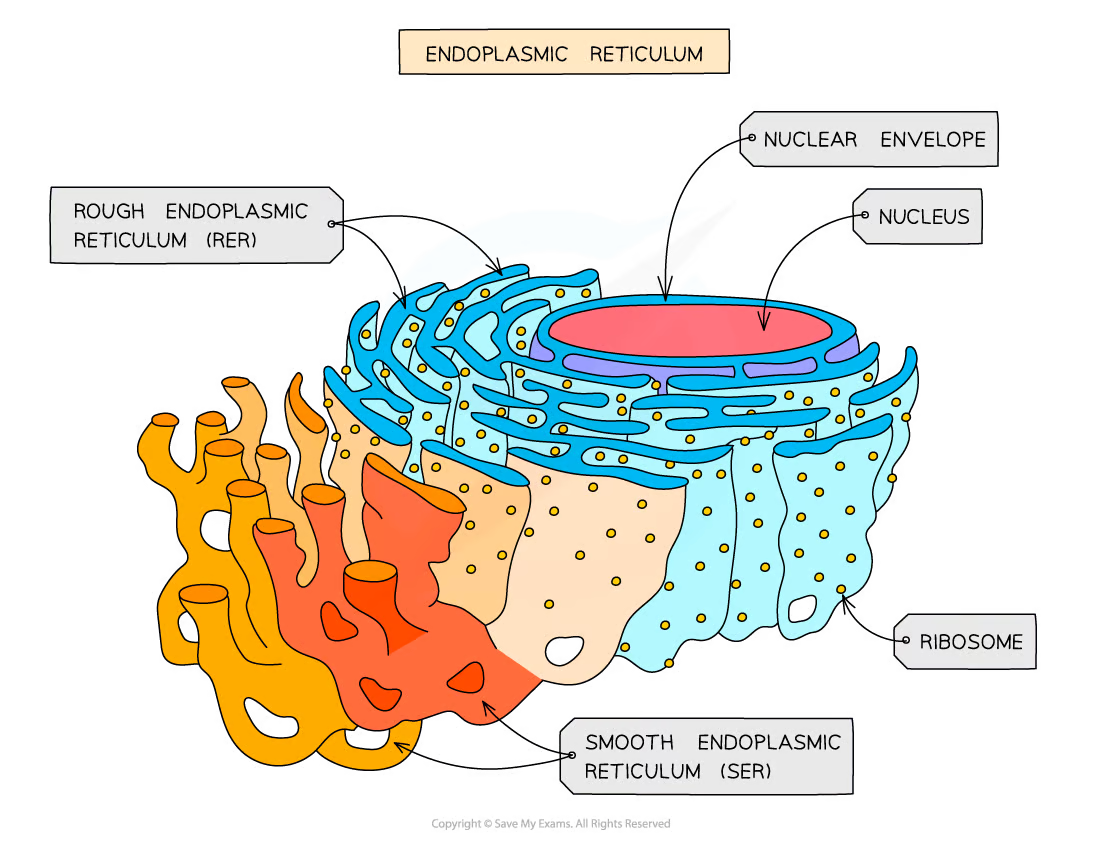
Rough Endoplasmic Reticulum (RER)
RER is formed from folds of membrane continuous with the nuclear envelope
surface is covered in ribosomes
to process proteins made on the ribosomes
Smooth Endoplasmic Reticulum (SER)
also formed from folds of membrane
involved in the production, processing and storage of lipids, carbohydrates and steroids
doesn’t have ribosomes on its surface
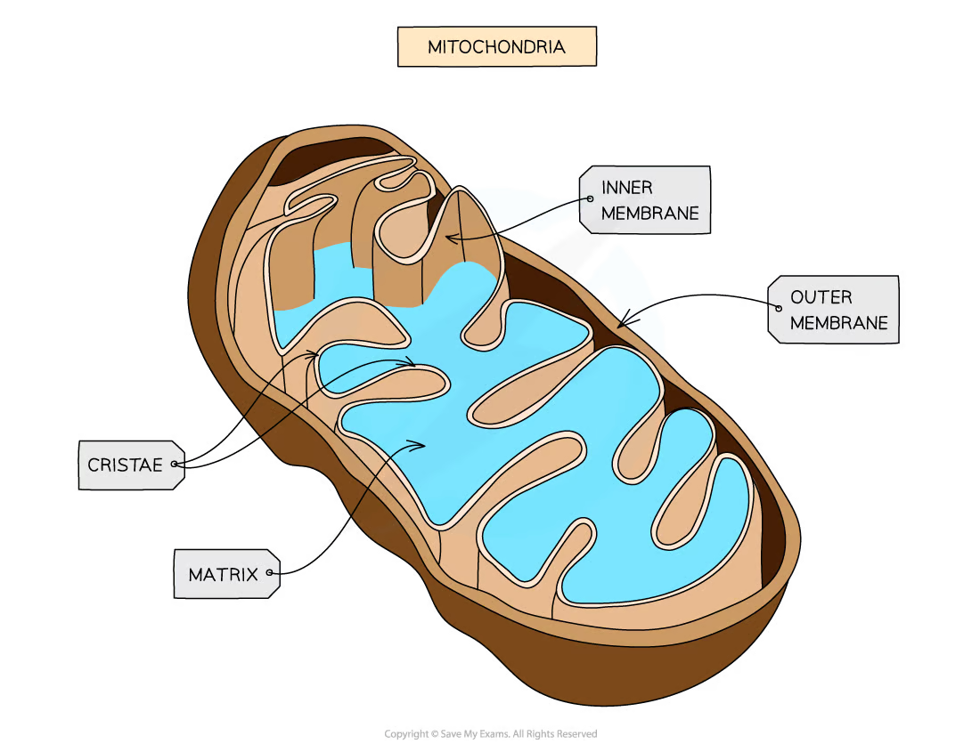
The site of aerobic respiration within eukaryotic cells - visible with a light microscope
surrounded by a double-membrane with the inner membrane folded to form structures called cristae
matrix contains enzymes needed for aerobic respiration producing ATP - also contain DNA and ribosomes
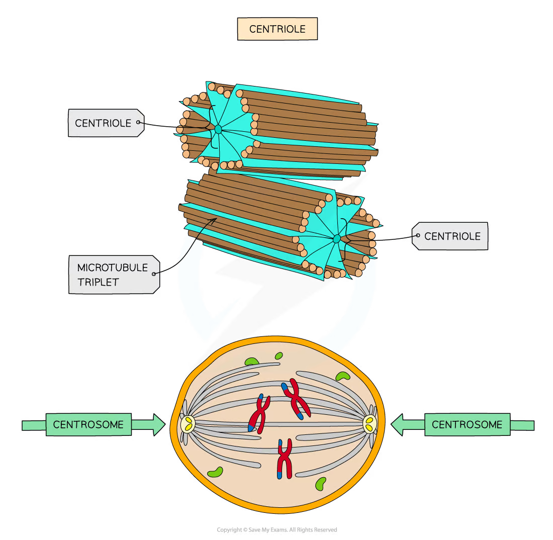
made of hollow fibres - microtubules
used to move substances around inside a cell and to support the shape of a cell
arranged at right angles
involved in the separation of chromosomes during cell division
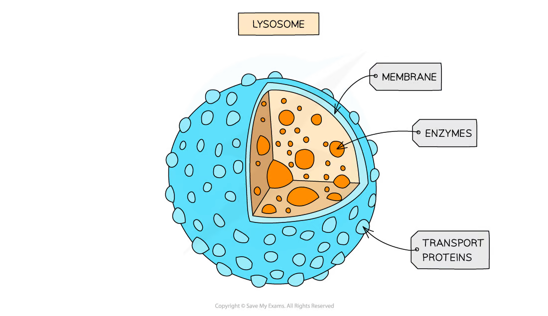
forms of vesicle which contain hydrolytic enzymes
break down waste materials such as worn-out organelles
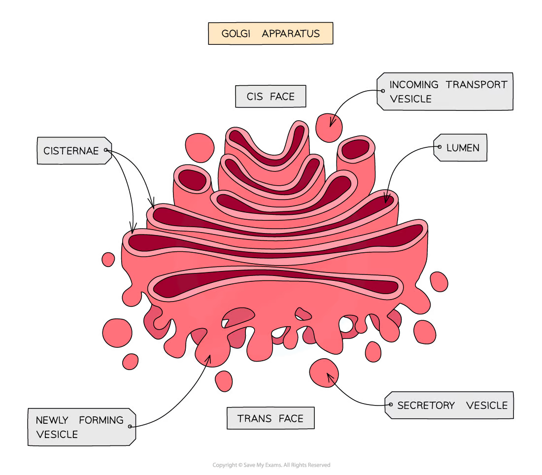
consists of flattened sacs of membrane
modifies proteins and lipids before packaging them into Golgi vesicles
The vesicles then transport the proteins and lipids to their required destination
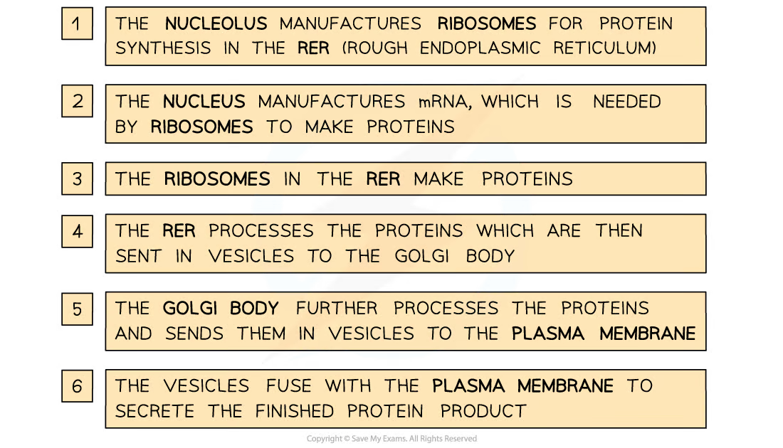
organelles involved in protein production and transport
proteins first go to the Golgi apparatus from the RER
the Golgi apparatus modifies the protein
then packages it into a secretory vesicle
the secretory vesicle then goes to the cell membrane
which then fuses w the membrane
releases the proteins through exotyocisis
free ribosomes
found within the cytoplasm make proteins that stay within the cytoplasm
Proteins that go through the Golgi apparatus are usually
Exported, e.g. extracellular enzymes
Put into lysosomes, e.g. hydrolytic enzymes
Delivered to other membrane-bound organelles
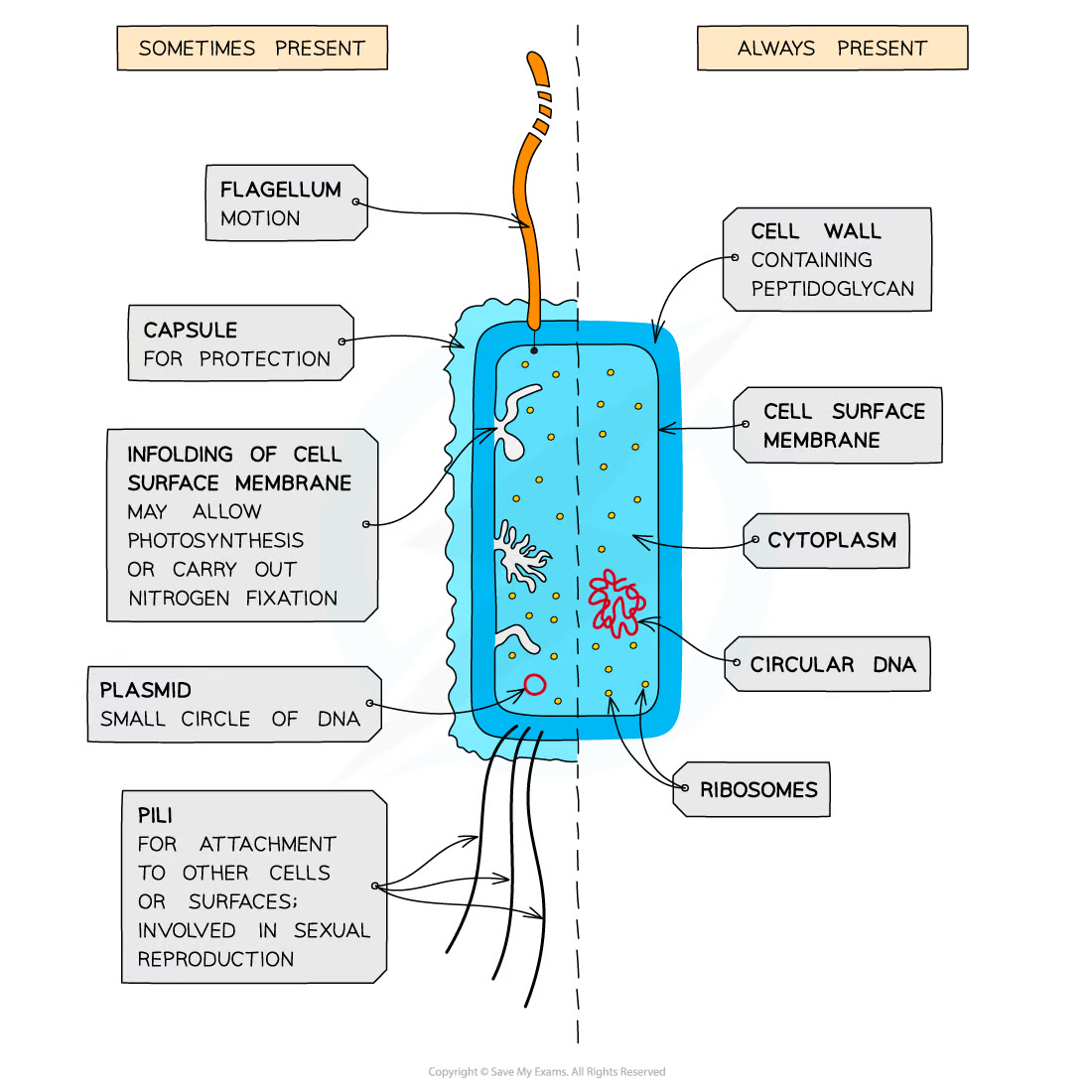
differences b/w prokaryotic and eukaryotic
A cytoplasm that lacks membrane-bound organelles
Ribosomes that are smaller (70 S)
No nucleus, instead having a single circular bacterial chromosome that is free in the cytoplasm and is not associated with proteins
A cell wall that contains the glycoprotein murein
Loops of DNA known as plasmids
Capsules
Flagella
Pili
A cell membrane that contains folds known as mesosomes
plasmid
small loops of DNA
contain genes that can be passed b/w prokaryotes
not present in all prokaryotes
capsule
surrounds prokaryote
helps to protect from bacteria from drying out and from attack by cells of the immune system of the host organism
not present in all
flagellum
long hair like structure that rotates
allows mobility
sometimes theres more than one
not present in all
pilli
thread like structures on the surface of some bacteria
enables bacteria to attack to other cells or surfaces
mesosomes
infoldinfgs of the inner membrane which contains enzymes required for respiration
ciruclar DNA
the genetic material of prokaryotic cells consists of a single circular strand of DNA
the area where this molecule is found is known as nucleoid
ribosomes
70S ribosomes which are smaller
the site of protein synthesis
Transmission Electron Microscopes
use electromagnets to focus a beam of electrons
which is transmitted through a thin specimen
Denser parts of the specimen absorb more electrons; which appear darker on the image
The internal structures can be seen as a 2D image
very high resolution
Scanning Electron Microscopes
scan a beam of electrons across a specimen
This beam bounces off the surface of the specimen and the electrons are detected, forming an image
can produce 3D images that show the surface of specimens
specimen viewed does not have to be thin
lower resolution than TEMs
magnification
how many times bigger the image of a specimen observed is in comparison to the actual size of the specimen
how to calculate the total magnification in light microscope
total magnification = eyepiece lens magnification (10) x objective lens magnification
Resolution
The ability to distinguish between two separate points
Relationship bw magnification and resolution
The resolution of a microscope limits the magnification that it is capable of
why do electron microscopes have a better resolution than light microscopes
electrons have a much smaller wavelength than visible light
The resolution of a light microscope is limited by the wavelength of light; the wavelength of light is too long to allow for high resolution
comparing light and electron microscopes
vacuum is needed for EM while its not needed for LM
EM has a magnification over x500000 and LM has up to x2000
EM has a resolution of 0.5nm while LM has 200nm
EM observes dead specimens while LM observes dead or living
why are specimens stained in microscopy
as the cytoplasm and other cell structures may be transparent or difficult to distinguish
light microscope stains
most of the colours seen in images taken using a light microscope are due to added stains. Except for chloroplasts which show up as their natural colour (green)