Chapter 14
1/158
There's no tags or description
Looks like no tags are added yet.
Name | Mastery | Learn | Test | Matching | Spaced | Call with Kai |
|---|
No analytics yet
Send a link to your students to track their progress
159 Terms
Brain
the center for registering sensations,
correlating them with one another and with stored information, making decisions, and taking actionIt is also the center for intellect, emotions, behavior, and memory, it also directs our behavior towards others
2% of body weight
utilizes about 20% of the body’s oxygen supply
one of the most metabolically active organs of the body, and the amount of oxygen it uses varies with the degree of mental activity
neural tube
has three regions called primary brain vesicles: prosencephalon (forebrain), mesencephalon (midbrain), rhombencephalon (hindbrain)
major parts of the brain
brain stem, diencephalon, cerebrum, and cerebellum
parts of the diencephalon
thalamus, hypothalamus
parts of the brain stem
midbrain, pons, medulla oblongata

A
thalamus (diagram)

B
hypothalamus (diagram)
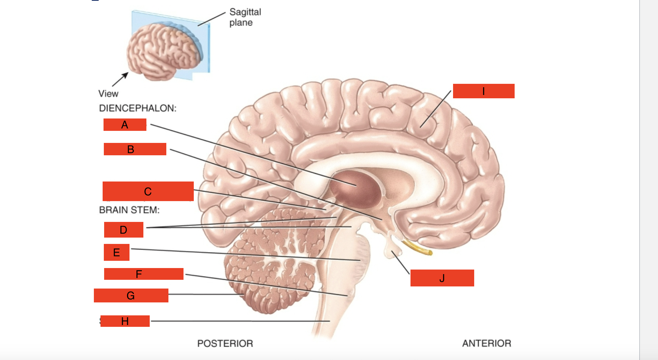
C
pineal gland (diagram)

D
midbrain (diagram)

E
pons (diagram)

F
medulla oblongata (diagram)

G
cerebellum (diagram)

H
spinal cord (diagram)

I
cerebrum (diagram)

J
pituitary gland (diagram)
Cranial bones, Cranial meninges, cerebrospinal fluid
Protects the brain
Cranial meninges
Pia mater (inner), arachnoidea (middle), and dura mater(outer)
Cranial dura mater
composed of 2 layers: periosteal layer (external) and meningeal layer (internal)
Extensions of the cranial dura mater
separate parts of the brain:
1. The falx cerebri: separates the two hemispheres of the cerebrum
2. The falx cerebelli: separates the two hemispheres of the cerebellum
3. Tentorium cerebelli: separates the cerebrum from the cerebellum
falx cerebri
separates the two hemispheres of the cerebrum
The falx cerebelli
separates the two hemispheres of the cerebellum
Tentorium cerebelli
separates the cerebrum from the cerebellum
cerebrospinal fluid
a clear, colorless liquid composed primarily of water that protects the brain and spinal cord against chemical and physical injuries
Flows over and around the brain and cord in the subarachnoid space (SAS). In essence, the brain "floats" in it
It carries oxygen, glucose, and other important substances from the blood to nervous tissue cells
Total volume is 80-150mL, composed of glucose, proteins, lactic acid, ions (Na+, K+, Ca2+, Mg2+, Cl-, HCO3-) some white blood cells
There are four cavities within the brain
called ventricles that are filled with thiscontributes to hemostasis by providing
mechanical protection(shock absorption), chemical protection (pH affects pulmonary ventilation), and circulation
•The majority comes from ependymal cells
in the choroid plexuses networks of blood
capillaries in the walls of the ventriclesMost is absorbed by the arachnoid villi of the superior sagittal blood sinus. This absorption normally occurs at the same rate at which it is produced in the
choroid plexuses, thereby maintaining a relatively constant volume and pressure
vertebral (posteriorly) and carotid arteries
blood flows to the brain via
jugular veins
blood flows back to the heart from the brain via
interruption of oxygen to the brain
results in weakening, permanent damage, or death of brain cells
Interruption of the mother’s blood supply to a child during childbirth before it can breathe
may result in paralysis, mental retardation, epilepsy, or death
Glucose deficiency
produce mental confusion, dizziness, convulsions, and unconsciousness
because carbohydrate storage in the brain is limited, the supply of this to the brain must be continuous
Blood-Brain Barrier
consist of tight junctions that seal together the endothelial cells of blood capillaries and a thick basement membrane against which astrocytes press
protects brain cells from harmful substances and pathogens by serving as a selective barrier to prevent passage
of many substances from the blood into the braincan prevent the entry of therapeutic drugs
Glucose and lipid soluble substances
can cross
lateral ventricles
are separated by a thin membrane –septum pellucidum
The 3rd ventricle
lies superior to the hypothalamus and between the right and left halves of the thalamus
The 4th ventricle
lies between the brain stem and cerebellum
bidirectional
The secretory capacity of ependymal
cells is…
ependymal cells
From blood capillaries substances are filtered and secreted by _______ _______ to produce CSF and metabolites from nervous tissue are transported back to blood through ependymal cells
Because of tight junctions between __________ _______ material cannot leak from choroid capillaries, they must pass through the ________ ________
blood-cerebrospinal fluid barrier(BCFB)
formed by tight junctions of ependymal cells
Flow of cerebrospinal fluid
Lateral ventricles → interventricular foramina → 3rd ventricle → cerebral aqueduct (aqueduct of Sylvius)→ 4th ventricle → median aperture (of Majendie) and the lateral apertures (of Luschka) → subarachnoid space (SAS) and central canal
arachnoid villi (glandulae Pacchioni)
fingerlike extensions of the arachnoid mater that project into the dural venous sinuses especially the superior sagittal sinus
How the CSF maintains volume and pressure
Absorption normally occurs at the same rate at which CSF is produced in the choroid plexuses
The medulla oblongata
continuous with the superior aspect of the spinal cord
It forms the inferior part of the brain stem
It is part of the hindbrain (rhombencephalon)
Contains portions of both motor and sensory tracts
has two pyramids formed by large corticospinal (motor) tracts controlling voluntary movements in the body
contain nuclei which control vital body functions
pyramids
Some white matter forms bulges or _______ on the
anterior aspect of the medulla
decussation of pyramids
Axons from the left pyramid cross over to the right and axons on the right cross over to the left so that the left hemisphere of the brain controls the right side muscles, while the right hemisphere controls the left side
nuclei
collections of neuronal cell bodies
Nuclei of the medulla oblongata
cardiovascular center, respiratory center, vomiting center, deglutition center (swallowing), gustatory, cochlear (auditory), vestibular(equilibrium)
Olive
lateral to each pyramid
oval-shaped swelling
in the medulla oblongata
inferior olivary nucleus
within the olive
receives input from the cerebral cortex and regulate the activity of cerebellar neurons
gracile nucleus
Nuclei associated with sensation of touch, pressure,
vibration and conscious proprioception
cuneate nucleus
are continuations of the ascending sensory (start out as) gracile fasciculus and cuneate fasciculus
Posterior column-medial pathway
Nuclei associated with sensation of touch, pressure, vibration and conscious proprioception are the gracile nucleus and cuneate nucleus are continuations of the ascending sensory(start out as) gracile fasciculus and cuneate fasciculus
•They continue(white matter-axons) to thalamus as medial lemniscus and the entire pathway is known as
nuclei in the medulla associated with cranial nerves
Vestibulocochlear (VIII) nerve: sensory and motor impulses related to hearing
Glossopharyngeal (IX) nerve: motor impulses related to swallowing and salivation
Vagus (X) nerve: motor impulses related to thoracic, abdominal viscera
Accessory (XI) nerves: impulses related to swallowing
Hypoglossal (XII) nerves: impulses controlling tongue movement and swallowing
Pons
known as the bridge or ______ Varolii
located superior to the medulla oblongata and anterior to the cerebellum
part of the hindbrain
links parts of the brain with one another by way of tracts
It consist of both nuclei and tracts
It relays nerve impulses related to voluntary skeletal movements from the cerebral cortex to the cerebellum
major structural components of the pons
ventral and dorsal regions
ventral region of the pons
forms a large synaptic relay station with pontine nuclei
dorsal region
contains ascending and descending tracts
pontine respiratory group
within the pons
helps control breathing, the pneumotaxic and apneustic areas
nuclei of cranial nerves associated with the pons
Trigeminal (V) nerve: sensory impulses from head and face, motor impulses governing chewing
Abducens (VI) nerve: motor impulses for eyeball movement
Facial (VII) nerve: sensory impulses for taste, secretion of saliva, tears and facial muscles
Vestibulocochlear (VIII) nerve: sensory and motor impulses for balance and equilibrium
midbrain
also called the mesencephalon
the superior portion of the brain stem and extends from the pons to the diencephalon
conveys motor impulses from the cerebrum to the cerebellum and spinal cord, sends sensory impulses from the spinal cord to the thalamus, and regulates auditory and visual reflexes
Structures within the midbrain include the cerebral aqueduct, the cerebral peduncles (pedunculus =little feet)
cerebral aqueduct
connects the 3rd ventricle above and 4th ventricle below
cerebral peduncles (pedunculus =little feet)
consist of the corticospinal, corticobulbar, and corticopontine tracts
anterior part of the midbrain
corticopontine tracts
part of the cerebral peduncles
convey nerve impulses from motor areas in the cortex to spinal cord, medulla and pons
Tectum
posterior part of the midbrain
contains 4 rounded elevations
Superior colliculi (little hills)
serves as reflex centers for visual activities: scanning, tracking images
Inferior colliculi
part of the auditory pathway
Substantia nigra
left and right;
pathway to basal ganglia have neurons with
dopaminergic tracts - control subconscious muscle movements
Red nuclei
control voluntary muscular movement
nuclei of cranial nerves in the midbrain
Oculomotor (III) nerve: motor impulses for eyeball
Trochlear(IV) nerve: motor impulses for eyeball
Reticular formation
is a netlike arrangement consisting of small areas of gray matter interspersed among fibers of white matter and has both sensory and motor functions
extends from the superior part of the spinal
cord throughout the brain stem and into the
inferior part of the diencephalonhelps regulate muscle tone, alerts the cortex to incoming sensory signals (reticular activating system, or RAS), and is responsible for maintaining consciousness and awakening from sleep
cerebellum
“little brain”
attached to the brain stem by three pairs of
cerebellar pedunclesthe second largest part of the brain, has a highly folded surface for neuron accommodation and their connections
part of the hindbrain
Nearly half of all the neurons are located in here
functions in the coordination of skeletal muscle contractions and in the maintenance of normal muscle tone, posture, and balance
occupies the inferior and posterior aspects of the cranial cavity and consists of two
hemispheres and a central vermisseparated from the cerebrum by the transverse fissure and tentorium cerebelli
has a butterfly shape with central vermis and cerebellar hemispheres or wings or lobes
anterior and posterior lobe govern the subconscious aspects of muscle movements
The flocculonodular lobe
part of the cerebellum
contributes to equilibrium and balance
cerebellar cortex
The superficial layer of the cerebellum
consist of the gray matter in a series of folds
slender and parallel called folia
arbor vitae
in the cerebellum
Deep are the tracts of white matter
cerebellar nuclei
Deeper in arbor vitae of the cerebellum
cerebellar peduncles
The superior cerebellar peduncle, the middle cerebellar peduncle, the inferior cerebellar peduncle
The superior cerebellar peduncle
axons from cerebellum to red nuclei and thalamus
The middle cerebellar peduncle
axons carrying impulses for voluntary movements from pontine nuclei into the cerebellum
The inferior cerebellar peduncle
Spinocerebellar sensory tracts from trunk and limbs into cerebellum
Axons from vestibular apparatus and vestibular nuclei of medulla and pons into cerebellum
Axons from inferior olivary nucleus of the medulla into cerebellum
Axons from cerebellum to vestibular nuclei of medulla and pons
Axons from cerebellum to reticular formation
The diencephalon
inner brain
forms the central core of the brain just superior to the midbrain
extends from the brain stem to the cerebrum and surrounds the 3rd ventricle
composed of the:
Thalamus
Hypothalamus
Epithalamus
The thalamus
inner chamber
3cm in length and makes up 80% the diencephalon
is located superior to the midbrain and contains nuclei that serve as relay stations for all sensory impulses (except smell) to the cerebral cortex
the interthalamic adhesion
A bridge of gray matter
connects the two halves of thalamus
internal medullary lamina
The Y shaped white matter in the thalamus
internal capsule
Axons that connect the thalamus and cerebral cortex pass through this
a thick white band lateral to the thalamus
seven major groups of thalamic nuclei
The anterior nucleus, the medial nucleus, nuclei in the lateral group, five ventral nuclei, intralaminar nuclei, the midline nucleus, the reticular nucleus
The anterior nucleus
Input from hypothalamus, output to limbic system
Function in emotions and memory
The medial nucleus
input from limbic system and basal ganglia(nuclei), output to cerebral cortex
function in emotions, learning, memory, cognition
Nuclei in the lateral group
Input from limbic system, superior colliculi and cerebral cortex, output to cerebral cortex.
types: the lateral dorsal nucleus(emotions), the lateral posterior nucleus and pulvinar nucleus
Five ventral nuclei
Ventral anterior nucleus: input from basal ganglia output to cerebral cortex
Ventral lateral nucleus: input from cerebellum and basal ganglia, output to cerebral cortex
Ventral posterior nucleus relays impulses from touch, pressure, vibration, itch, tickle, temperature, pain, proprioception to cerebral cortex
Lateral geniculate nucleus relays visual impulses to the primary visual area of the cerebral cortex
Medial geniculate nucleus relays auditory impulses from ear to primary auditory area of the cerebral cortex
Ventral anterior nucleus
input from basal ganglia output to cerebral cortex
Ventral lateral nucleus
input from cerebellum and basal ganglia, output to cerebral cortex
Ventral posterior nucleus
relays impulses from touch, pressure, vibration, itch, tickle, temperature, pain, proprioception to cerebral cortex
Lateral geniculate nucleus
relays visual impulses to the primary visual area of the cerebral cortex
Medial geniculate nucleus
relays auditory impulses from ear to primary auditory area of the cerebral cortex
Hypothalamus
is found inferior to the thalamus, composed of ~ dozen nuclei in four major regions (mammillary, tuberal, supraoptic, and preoptic)
controls many body activities, and is one of the major regulators of homeostasis
functions of the hypothalamus
Control of the ANS
Production of hormones
It functions in regulation of emotional and behavioral patterns
It regulates eating and drinking through the feeding center, satiety center, and thirst center
It aids in controlling body temperature
It regulates circadian rhythms and states of consciousness
The mammillary region of hypothalamus
the most posterior part of the hypothalamus
Includes the mammillary bodies and posterior hypothalamic nuclei
The mammillary bodies are two rounded projections: relay station for sense of smell
The tuberal region of the hypothalamus
widest part of the hypothalamus
Includes the dorsomedial nucleus, ventromedial nucleus and arcuate nucleus and the infundibulum which connects the pituitary gland to the hypothalamus
The supraoptic region of the hypothalamus
lies superior to the optic chiasm
Includses the paraventricular nucleus, supraoptic nucleus, anterior hypothalamic nucleus and suprachiasmatic nucleus
The paraventricular and supraoptic nuclei are part of the hypothalamohypophyseal tract
The pre-optic region of the hypothalamus
contains the medial and lateral pre-optic nuclei
The epithalamus
lies superior and posterior to the thalamus and contains the pineal gland which secretes melatonin and habenular nuclei which are involved in olfaction
pineal gland
secretes melatonin
habenular nuclei
involved in olfaction
Circumventricular Organs (CVOs) of the Diencephalon
They lie in the wall of the 3rd ventricle
Parts of the diencephalon
monitor chemical changes in the blood
because they lack a blood-brain barrierinclude the hypothalamus (a portion of
it), pineal gland, and the pituitary glandcoordinate homeostatic activities of the endocrine and nervous systems