Histology lecture 1 & 2
 Call Kai
 Learn
Learn Practice Test
 Spaced Repetition
 Match
Match Flashcards
 Knowt Play
1/96

Earn XP
Description and Tags
8/10/26
Name | Mastery | Learn | Test | Matching | Spaced | Call with Kai | Chat |
|---|
No analytics yet
Send a link to your students to track their progress
97 Terms
Function of the nucleus
Store genetic material, controls cellular activity
function of the Plasma membrane
regulates what enters/exits cell, phospholipid bilayer, hydrophilic heads, hydrophobic tails
function of the Rough ER
synthesizes proteins that are exported out of the cell (sent to golgi) and contains attached ribosomes.
Function of the smooth ER
makes lipids, stores calcium and assists in detox
Function of mitochondria
energy production (ATP)
Golgi apparatus
packages and processes proteins for transport into vesicles
function of centrioles
paired microtubules inside the centrosome
FUNCTION of the centrosomes
organizes centrioles and mitotic spindle for division
Major components of the plasma membrane
phospholipid bilayer, cholesterol, transmembrane proteins, glycocalyx
Glycocalyx
make up of polysaccharides
help with cell signaling, adhesion and immune recognition
Types of nucleoprotein
Histone protein, non-histone protein
Histone protein
package DNA around histones (stuctural)
Non-histone proteins
enzymes that synthesize DNA and RNA, gene replication, regulate proteins
Heterochromatin
inactive/stored DNA, tightly coiled, looks dark and dense
Euchromatin
actively transcribing DNA, uncoiled, looks light
Major steps of protein synthesis
DNA→ mRNA→ proteins
transcription (nucleus) → translation (cytoplasm)
which organelle is responsible for mRNA translation to proteins
ribosomes
Free floating ribosome make
proteins that stay in the cell
rER ribosomes make
proteins for export and for plasma membrane
Exocytosis
when rER proteins are made and sent outside the cell
hormone secretion, digestion enzymes
Endocytosis
when vesicles bring proteins into the cell, 2 types
phagocytosis, receptor mediated
Phagocytosis
Cell eating, not picky
Receptor mediated
Cell eating after molecule binds to receptor
types of cellular transport
passive, facilitated, active, bulk transport
Passive diffusion
small, non polar molecules
facilitated diffusion
with concentration gradient, larger hydrophilic molecules, requires binding to carrier proteins to go through channels
Active transport
uses energy to cross membrane
Bulk transport
vacuoles containing large molecules M
Mitosis
cell division, results in 2 diploid daughter cells, identical
Meiosis
results in 4 haploid daughter gametes, sperm and eggs
components of cytockeleton
microfilaments, intermediate filaments, microtubules
Microfilaments
actin filaments for cell shape, movement and support
intermediate filaments
Purely structural and for support
Microtubules
active, form cilia and flagella, important for cell division, allow cell shape to change and be flexible
Chiasmata
chromosomal crossing over, only in meisosis
Cilia
hair-like projections that move things along the cell surface
cell types- ciliated epithelial cells
locations- respiratory system, uterus, ependymal cells lining brain ventricles
microvilli
finger like projections designed to increase surface area and absorb
cell types- absorptive epithelial cells
locations- small intestine, kidney tubules
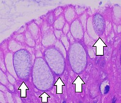
goblet cell function
secrete mucus
Goblet cells
connective tissue components
cells and extracellular matrix
extracellular matrix
3 types of fibers, ground substance
3 types of extracellular fibers
collagen, elastin, reticular
Collagen
most abundant protein in the body
found in tendons, ligaments, skin and cartilage
gives tissue strength\
Elastin
found in skin, heart and lungs
allows tissue to stretch
ground substance
gel-like part of extracellular matrix that fills space
includes water, glycosaminoglycans, and hyaluronic acid (type of glycosaminoglycan)
basement membrane
thin supporting layer between epithelium and underlying connective tissue, act as a limiting membrane and are made of collagen 4
type 1 collagen
most abundant, provides strength
tendons, ligaments, bones, skin
type 3 collagen
forms reticular fibers to support cells and organs
type 4 collagen
forms basement membrane, supports cells and filters
type 6 collagen
anchors cells to extracellular matrix (velcro)
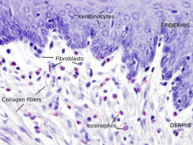
fibroblast
main connective tissue cell, make tropocollagen, repair tissues
fibroblast
Types of collagenous tissue
loose, dense irregular, dense regular, granulation
Loose collagenous tissue
found in areolar
Dense irregular collagenous tissue
found in dermis, sheaths of nerves, organ capsules
Dense regular collagenous tissue
found in ligaments and tendons
Granulation tissue collagenious tissue
wound healing and repair
Adipose tissue types
storage and fat processing, found in loose connective tissue, have receptors for growth hormones, insulin
White adipose, brown adipose
white adipose vs brown adipose
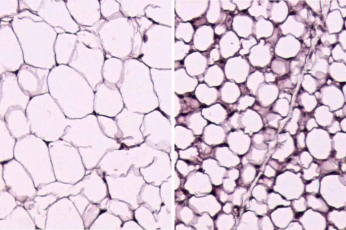
white adipose
energy storage
brown adipose
mostly in baby animals and hibernating animals, thermal insulation
cartilage structures
chondroblast
Chrondocytes
Perichondrium
Matrix
cartilage is avascular, so little blood supply
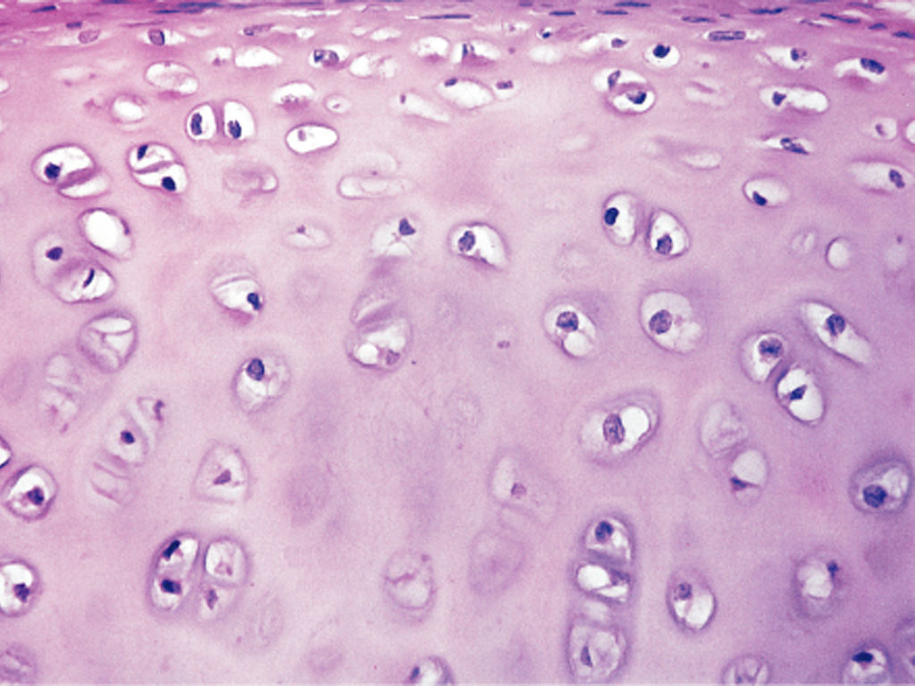
Hyaline cartilage
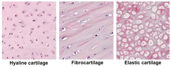
most common, in bone plates, joints, contains type 2 collagen
Hyaline cartilage (histology)
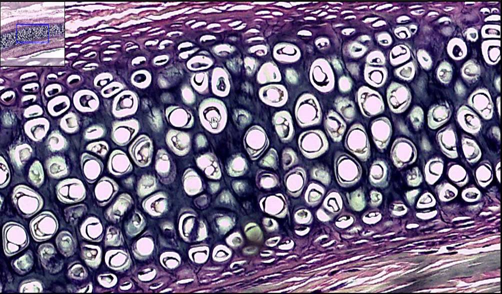
Elastic cartilage
contains elastin and elastic fibers, found in ear pinna, epiglottis and larynx
Elastic cartilage (histology)
Fibrocartilage
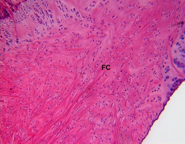
an intermediate between cartilage and dense connective tissue, tough, found in intervertebral discs
Fibrocartilage (histology)
Cartilage comoparison
Bone
composed of mineralized collagenous cells, type 1 collagen, hydroxyapatite
support system
bone cells
osteoblast, osetoclast, osteocyte
Osteoblast
bone formation, on the end of bone trabeculae
Osteocyte
resting stage of the osteoblast trapped in the matrix
Osteoclast
help in bone turnover, create channels in the bone, digest bone
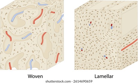
Woven bone
immature bone, also seen in repairs, can become lamella, lots of collagen, unorganized
Lamellar bone
mature healthy bone, organized
2 types
Compact (cortical) bone
outter wall of bone
Cancellous (medullary) bone
interior, surrounds bone marrow
Woven vs lamenar bone (histology)
Endochondral ossification
Occurs in long bones, cartilage is growing from growthplate and replaced by bone
Intramembranous ossification
occurs in skull bones, grows from the mesenchymal tissue
dominant function of muscle tissue? and what causes it?
contraction and movement
caused by the interaction of actin and mycin
3 types of muscle tissue and where are they found
skeletal- voluntary muscles, make up the appendicular skeleton
smooth- involuntary/gastric muscles, also found in vessel walls
cardiac- found in the heart
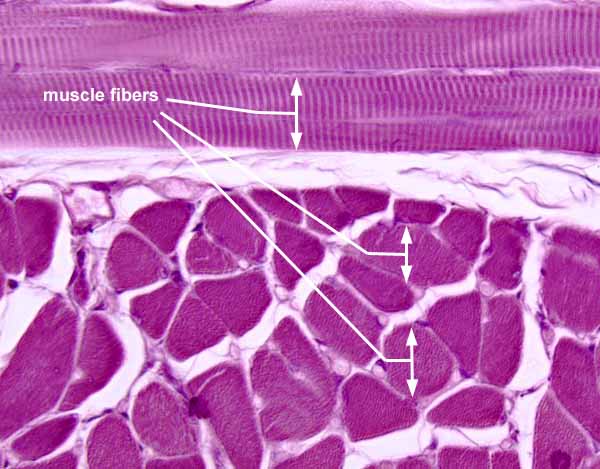
skeletal muscle histology
striated, nucleus is off to the side, white endomysium
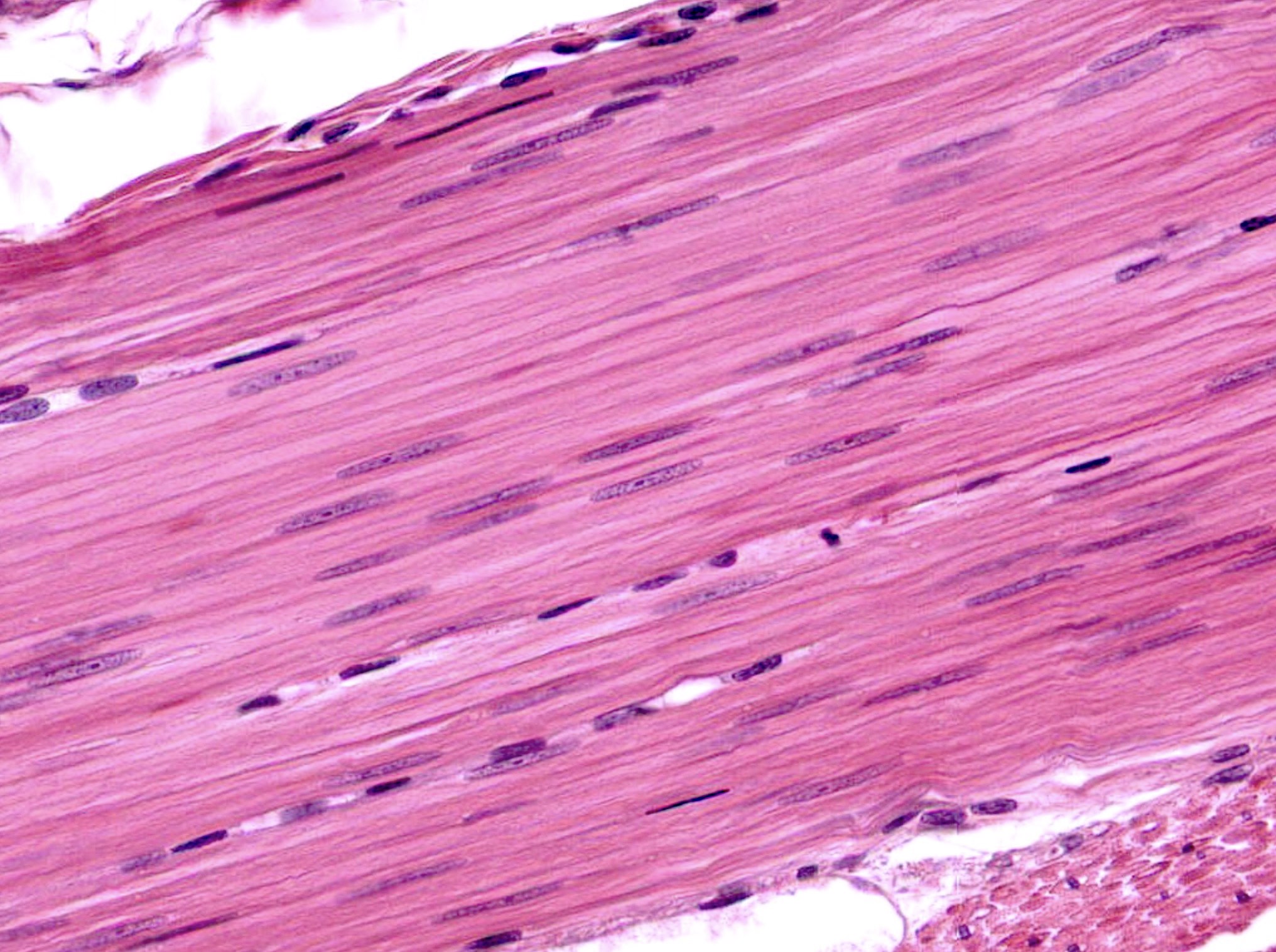
smooth muscle histology
long cell with tapered edge, central nucleus, no cross striations, has cartilage between cells
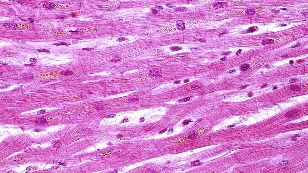
Cardiac muscle histology
striated, branching fibers that end in intercalating discs, central nucleus
what neurotransmitter is found in the neuromuscular junctions
acetylcholine
this also releases calcium ions
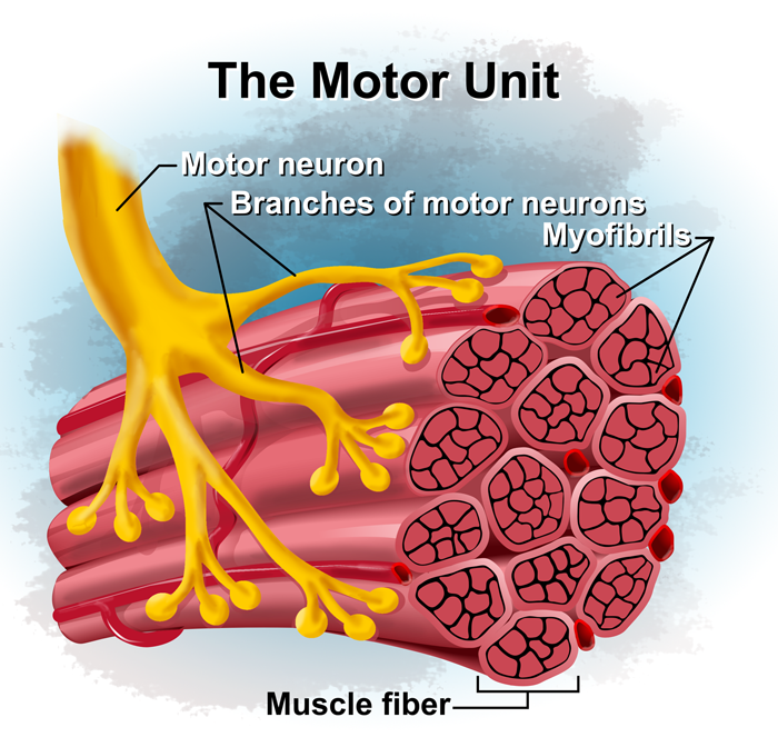
What is a motor unit
one motor neuron and all the muscle fibers it integrates
what is the organization of striated muscle
skeletal and cardiac have the same orginization
myofibers make up myofibrils which make up sarcomeres
what is a sarcomere
the contractile element of the myofiber
sarcomeres contract z-line to z-line
what happens when a myofiber depolarizes
it releases calcium ions from the sarcoplasmic reticulum, this shortens and contracts the muscle
what are the thick filaments of the sarcomere made up of
myosin
what are the thin filaments of the sarcomere made up of
actin
what is the function of smooth muscles in vessels and bronchioles
blood vessel- constricts, vasoconstriction
broncioles- bronchoconstriction
which layer of the blood vessel contains the smooth muscle
tunica media
what is the function of intercalating discs
transmits contractile forces between cells
contain gap junctions which allow rapid spread of excitatory stimuli with low electrical resistance.