Lecture 2-5
 Call Kai
 Learn
Learn Practice Test
 Spaced Repetition
 Match
Match Flashcards
 Knowt Play
1/374

There's no tags or description
Looks like no tags are added yet.
Name | Mastery | Learn | Test | Matching | Spaced | Call with Kai | Chat |
|---|
No analytics yet
Send a link to your students to track their progress
375 Terms
What do postmortem changes result from?
Autolysis
Are postmortem changes considered lesions?
No
What are decompositions?
Dead organic substances that are broken down
What are the two mechanisms of decompostion?
Putrefaction
Autolysis
What is putrefaction?
State of decomposition where microorganisms cause dissolution of tissues into gases, liquids and simpler molecules
What is autolysis?
Destruction of cells/tissues by their own enzymes (self-digestion)
What tissues die faster than others? How long do they take?
Neurons: 3-4 minutes
What is the difference between autolysis and necrosis?
Autolysis occurs in dead tissue, involves the entire body and there is no inflammation
Necrosis occurs in live tissue in local/regional areas and inflammation is present
Autolysis begins in ______ of the last heartbeat and involves physical and chemical changes that are ______ and ______.
minutes, irreversible, unavoidable
What is the general process of autolysis beginning from the last hearbeat?
Last heartbeat → Hypoxia → CO2 accumulation → Drop in tissue pH → Rupture of cytosolic lysosomes → Lysosomal enzymes enter cytosol and cause autolysis
Postmortem autolysis can mask what?
Antemortem lesions
What does autolysis look histologically?
Lack of cellular architecture and detail. Saprophytic bacteria.

What is rigor mortis?
Muscle stiffening that occurs 2-36 hours postmortem
What position does rigor mortis leave muscles?
Hyperextension
How does rigor mortis occur?
Muscles are flaccid at death. Sarcoplasmic reticulum degrades and releases calcium, which floods the sarcomere and binds actin and myosin. No ATP to disengage, so body can’t relax.
What is livor mortis?
Purple tissue discoloration postmortem due to intravascular blood pooling due to gravitational pull
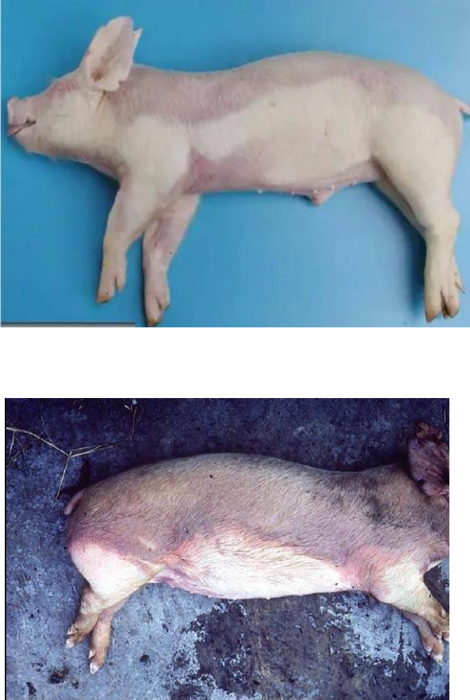
What is another word for intravascular pooling?
Hypostatic congestion
How does livor mortis occur?
Hypoxic capillaries dilate, causing blood to accumulate in skin and organs
How long after death does livor mortis develop?
30 minutes to 2 hours
How long after death does livor mortis become fixed?
8-12 hours
What is algor mortis?
Body cooling postmortem to match environment
Which is faster: external cooling or internal cooling?
External cooling
What characteristics allow a body to autolyze more rapidly?
Animals with thick fur or that are hyperthermic
What is hemoglobin imbibition?
Staining of tissues by free hemoglobin, causing a red discoloration to tissues
What is bile imbibition?
Staining of tissues near the gallbladder/ducts by bile, causing yellow/green discoloration
What is pseudomelanosis?
Putrefactive bacteria rapidly diffuse through tissues and react with iron in hemoglobin to create iron sulfide, causing black discoloration to tissues

Where does pseudomelanosis mostly occur?
GIT
What makes the black discoloration associated with pseudomelanosis?
Reaction of the iron in hemoglobin with putrefactive bacteria that grows on the organ post-mortem to create iron sulfide
Does pseudomelanosis an antemortem or postmortem reaction?
Postmortem
What is melanosis?
Normal melanin pigment that causes black discoloration to tissues in dark colored animals. Typically occurs in focal or multifocal spots.

How do postmortem clots appear?
Blood is stagnant and separates. RBCs stick towards the ground while plasma and platelets remain on top. AKA “chicken fat clot”
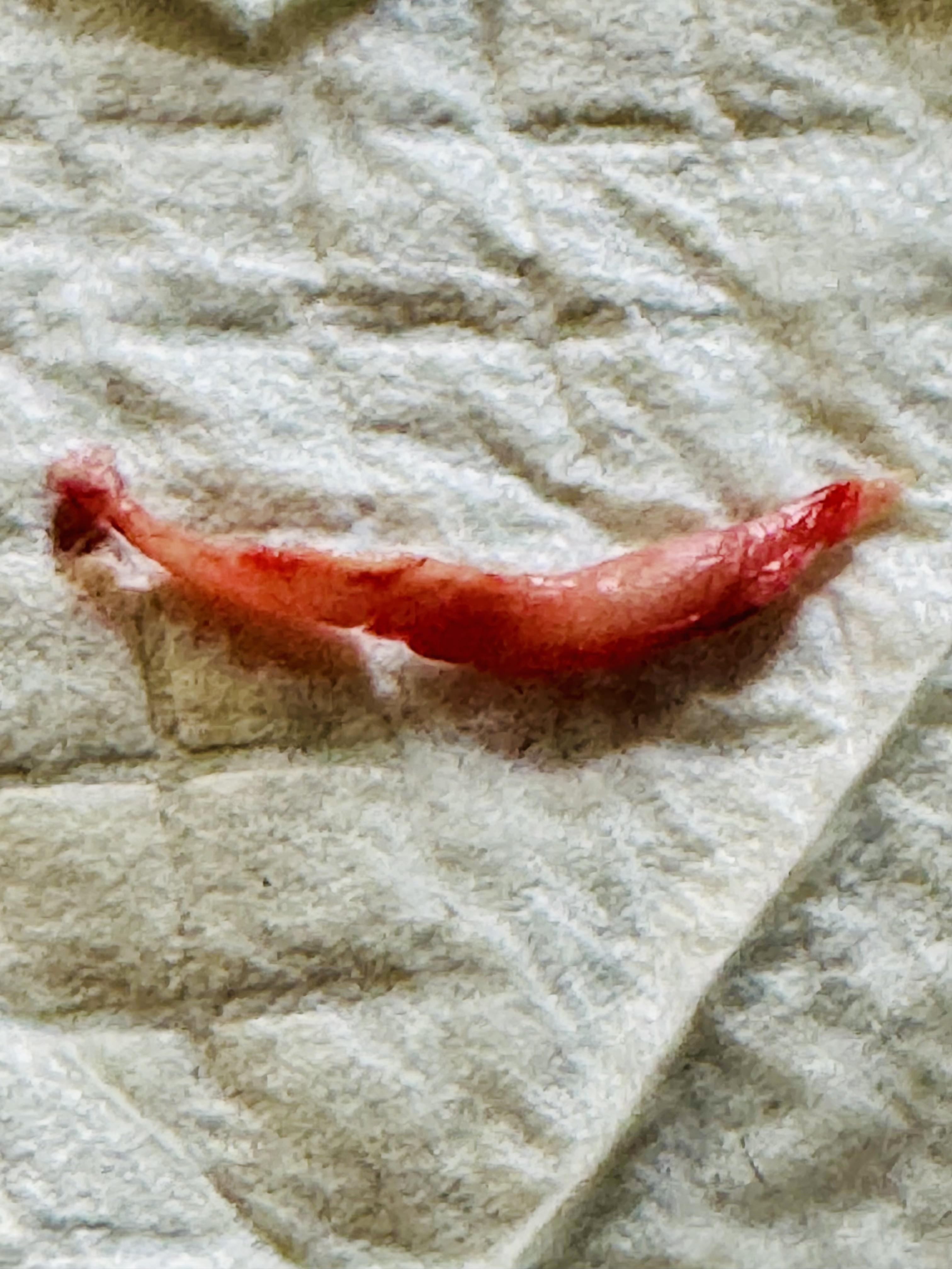
How long after dead do postmortem clots resolve?
2 hours
What causes postmortem clots to dissolve?
Fibrinolysin is released from endothelium and prevents clots from forming
What causes an antemortem clot?
Any element of Virchow’s trid (ex. hypercoagulability, endothelial damage, turbulent blood flow)
What are the characteristics of an antemortem clot?
Rough
Difficult to remove (friable, falls apart, loosely adhered to endothelial wall)
Red and uniform in color

What are the characteristics of a postmortem clot?
Smooth and shiny
Simple to remove
“Chicken Fat” appearance and feel

You’re doing a necropsy on an animal and notice pink coloration to the teeth. Is this a postmortem autolysis or pathology?
Postmortem autolysis → Hemoglobin imbibition

Is this considered an antemortem or postmortem clot? Why?
Antemortem → rough, uniform red color, no separation of RBCs and plasma

Pathology or non-lesion? Why?
Non-lesion. Pseudomelanosis, which occurs in GIT and is a postmortem condition (Putrefactive bacteria = putrefaction!)

Pathology or non-lesion? Why?
Non-lesion. Hemoglobin imbibition, results from free hemoglobin, but a pathological condition

Pathology or non-lesion? Why?
Non-lesion. Livor mortis, occurs post-mortem not due to pathology.

Pathology or non-lesion? Why?
Non-lesion. Bile imbibition due to GB/duct leakage, not pathological.
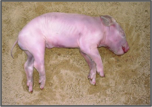
Livor mortis
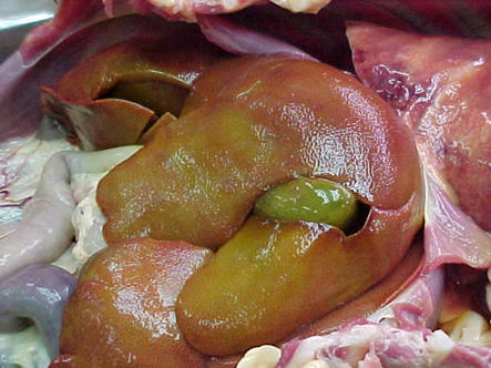
Bile imbibition
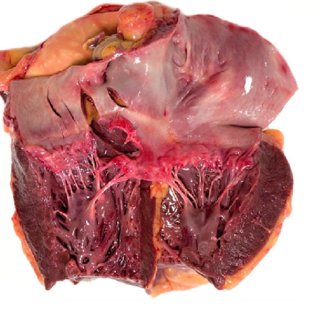
Hemoglobin imbibition
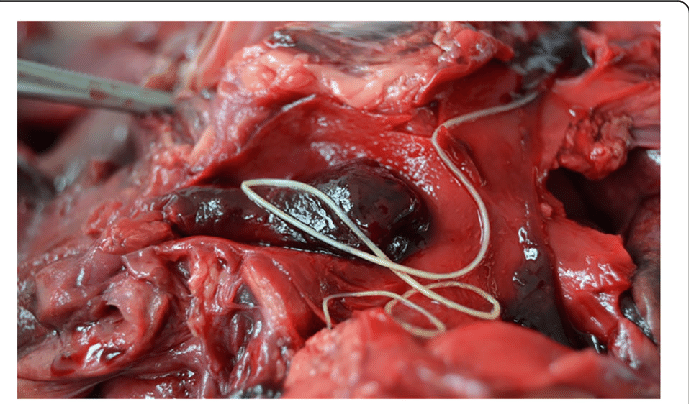
Antemortem clot with heartworm
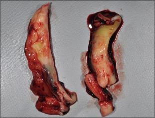
Postmortem clots
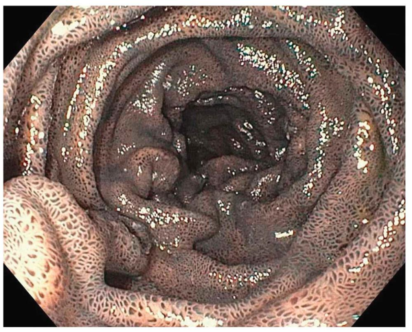
Pseudomelanosis
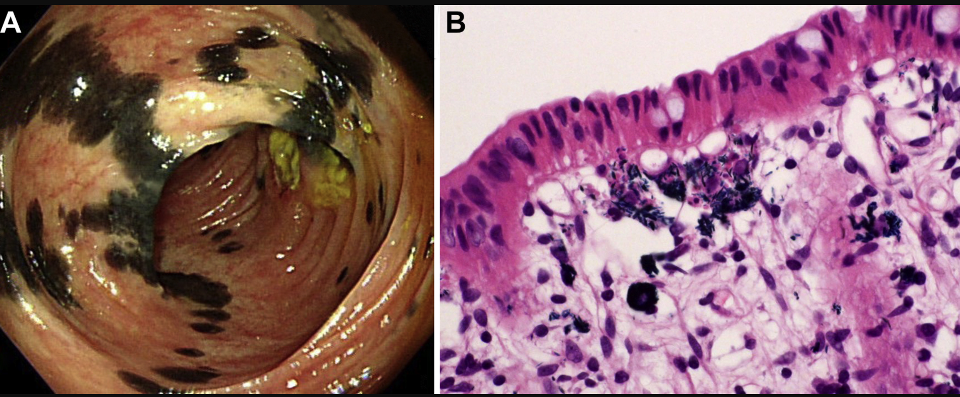
Pseudomelanosis
What is cell stress?
changes that are nonlethal and disrupts homeostasis
What is cellular injury?
alteration in cell structure or biochemical functioning that overwhelms the cell’s ability to compensate and adapt
When does injury occur?
Cell can’t adapt to the changes and cannot maintain normal, critical functions
How does the cell respond to stressors/injurious stimuli?
Adapts or dies
What does cell response vary based on?
cell type, nature and intensity of injury, metabolic state at injury
What’s the difference between reversible vs. irreversible cell injury?
Reversible the cell can reestablish function
Irreversible the cell cannot reestablish function
What can lead to irreversible cellular injury?
longer duration or higher intensity injuries
Which occurs faster in the cell: morphological/histological changes or functional changes?
Functional
Microscopic lesions develop ___ hours after an ischemic event
4-12
In what type of tissue/cell is acute cell swelling is worse?
Neurons
What is the most common reversible cellular injury?
Acute Cellular Swelling
What causes cell to swell?
Na/K pump failure causing influx of Na and H2O
How does hypoxia cause acute cell swelling?
Lack of ATP production → Na/K pump failure
What can cause cell membrane injury?
Free radicals, lytic pores, bacterial cytolysins, NK Cells
How does acute cellular swelling show macroscopically?
Rounded margins, bulges at the surface, pale, heavy, wet
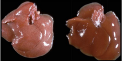
What is being shown
hydropic degeneration
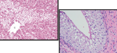
What is being shown? Parenchymal organ.
Hydropic degeneration
What characterizes irreversible cell injury?
Inability to repair cell damage
Inability to restore cell or mitochondria function
What cells are most susceptible to hypoxia causing irreversible cell injury?
Neurons, cardiomyocytes, renal tubular epithelium
What is the most crucial in the transition from reversible to irreversible damage?
membrane damage and calcium homeostasis
What things cause cytoplasmic changes in irreversible cell damage?
Mitochondrial swelling and increased membrane permeability
What are characteristics of mitochondrial swelling?
loss of dense granules, formation of amorphous densities, calcium deposits
What are signs of increased membrane permeability?
myelin figure formation, leakage of enzymes, inflammation
What are the irreversible nuclear changes?
pyknosis, karyorrhexis, karolysis
What is pyknosis?
chromatin condensation, nuclear shrinking
What is karyorrhexis?
nuclear fragmentation
What is karyolysis?
nuclear dissolution
What are the 4 mechanisms of cellular injury
Hypoxia
Free radicals
Chemical/traumatic
Infectious
What is hypoxia due to?
ischemia usually
Where do free radicals do most damage?
cell membrane
What are the 2 pathways of ATP production?
oxidative phosphorylation and glycolytic pathway
How is ATP made in oxidative phosphorylation?
Oxygen is reduced to transition ADP to ATP
Oxidative phosphorylation is a major pathway in what species?
mammals
How does oxidative phosphorylation cause reduction of oxygen?
by electron transport chain in mitochondria
How is ATP made in glycolytic pathway?
Hydrolysis of glucose in absence of oxygen
What is produced as a by product in glycolytic pathway?
lactate
ATP depletion as little as _____ has widespread effects and is the fundamental cause of ____.
5-10%, necrotic cell death
What does a loss of ATP do?
Na/K and Ca pumps malfunction
No oxidative phosphorylation
Structure disrupted
Accumulation of misfolded proteins
What are the major causes of ATP depletion?
reduced supply of oxygen and nutrients
mitochondrial damage
toxins
How can the mitochondria be damaged?
increased cytosolic Ca, free radicals, oxygen deprivation
What are the consequences of mitochondrial damage?
Membrane proteins, cytochrome C and apoptosis-inducing enzymes (Capases) are released into the cytosol and causes apoptosis.
What does mitochondrial permeability transition pore cause?
loss of membrane potential, stopping oxidative phosphorylation, loss of ATP then necrosis
What does cytochrome C in cytosol do?
causes apoptosis
Cytosolic Ca is maintained at ___ concentrations
very low
Where is most intracellular Ca sequestered?
in mitochondria and ER
How does increased intracellular Ca cause cell injury?
Opening of mitochondrial transmission pore
Activation of phospholipases, proteases, endonucleases and ATPase
Induction of apoptosis by caspases
What is a free radical?
any molecule with an unpaired electron
What do free radicals do?
react with substances to damage cells or create abnormal cells
What are reactive oxygen species (ROS)?
an oxygen free radical
What are the most important reactive oxygen species?
hydrogen peroxide, hydroxyl, nitric oxide
Where are hydrogen peroxide, hydroxyl, nitric oxide produced?
in mitochondrial respiration in cell