MERRILL'S POSITIONING HIP AND PELVIS
1/49
There's no tags or description
Looks like no tags are added yet.
Name | Mastery | Learn | Test | Matching | Spaced |
|---|
No study sessions yet.
50 Terms
HIP LATERAL PROJECTION (LAUENSTEIN AND HICKEY METHODS) MEDIOLATERAL: PURPOSE
to show the hip joint and the relationship of the femoral head to the acetabulum
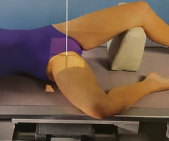
HIP LATERAL PROJECTION (LAUENSTEIN AND HICKEY METHODS) MEDIOLATERAL: POSITION OF PT
from the supine position, rotate the pt slightly toward the affected side to an oblique position
HIP LATERAL PROJECTION (LAUENSTEIN AND HICKEY METHODS) MEDIOLATERAL: POSITION OF PART
pt flex affected knee and draw the thigh up to a position at nearly a right angle to the hip bone
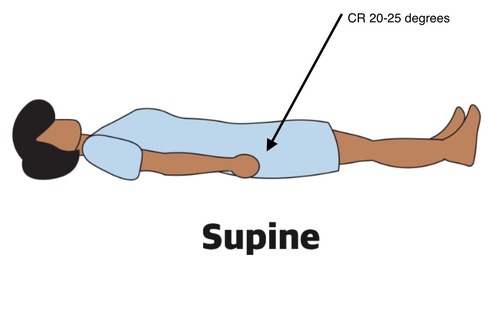
HIP LATERAL PROJECTION (LAUENSTEIN METHOD) MEDIOLATERAL: CENTRAL RAY
perpendicular through the hip joint, which is located midway between the ASIS and the pubic symphysis
HIP LATERAL PROJECTION (HICKEY METHOD) MEDIOLATERAL: CENTRAL RAY
perpendicular through the hip joint and at a cephalic angle of 20 to 25 degrees and an additional 1 inch more inferior
HIP LATERAL PROJECTION (LAUENSTEIN AND HICKEY METHODS) MEDIOLATERAL: STRUCTURES SHOWN
lateral projection of the hip, including acetabulum, the proximal end of the femur, and the relationship of the femoral head to the acetabulum
HIP AXIOLATERAL PROJECTION (DANELIUS-MILLER METHOD): NOTE
projection is often called the cross-table or surgical-lateral projection
HIP AXIOLATERAL PROJECTION (DANELIUS-MILLER METHOD): POSITION OF PART
for thin pt’s or lying on a soft bed, elevate the pelvis on a firm pillow or folded sheets sufficiently to center the most prominent point of the greater trochanter to the midline of the IR
HIP AXIOLATERAL PROJECTION (DANELIUS-MILLER METHOD): POSITION OF PART
when the pelvis is elevated, support the affected limb at hip level on sandbags or firm pillows
HIP AXIOLATERAL PROJECTION (DANELIUS-MILLER METHOD): POSITION OF PART
flex the knee and hip of the unaffected side to elevate the thigh in a vertical position
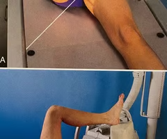
HIP AXIOLATERAL PROJECTION (DANELIUS-MILLER METHOD): POSITION OF PART
unless contraindicated, grasp the heel and medially rotate the foot and lower limb of the affected side approximately 15 to 20 degrees
HIP AXIOLATERAL PROJECTION (DANELIUS-MILLER METHOD): POSITION OF IR
place the IR in the vertical position with its upper border in the soft tissue crease above the iliac crest
HIP AXIOLATERAL PROJECTION (DANELIUS-MILLER METHOD): POSITION OF IR
angle the IR away from the body until it is exactly parallel with the long axis of the femoral neck
HIP AXIOLATERAL PROJECTION (DANELIUS-MILLER METHOD): CENTRAL RAY
perpendicular to the long axis of the femoral neck; central ray enters the groin area at a point midway between the anterior and posterior surfaces of the upper thigh and passes through the femoral neck, which is approximately 2.5 inches below the point of intersection of the localization lines
HIP AXIOLATERAL PROJECTION (DANELIUS-MILLER METHOD): STRUCTURES SHOWN
acetabulum, head, neck and trochanters of the femur
MODIFIED AXIOLATERAL PROJECTION (CLEMENTS-NAKAYAMA MODIFICATION): NOTE
when the pt has bilateral hip fractures, bilateral hip arthroplasty or limitation of movement of the unaffected leg, this method can’t be used to
What modification uses a 15-degree posterior angulation of the central ray?
Clements-Nakayama
MODIFIED AXIOLATERAL PROJECTION (CLEMENTS-NAKAYAMA MODIFICATION): POSITION OF PT
pt is supine on the table with the affected side near the edge of the table
MODIFIED AXIOLATERAL PROJECTION (CLEMENTS-NAKAYAMA MODIFICATION): POSITION OF PART
do not rotate lower limb internally, instead the limb remains in a neutral or slightly externally rotated position
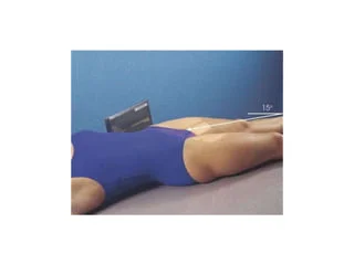
MODIFIED AXIOLATERAL PROJECTION (CLEMENTS-NAKAYAMA MODIFICATION): POSITION OF PART
adjust the grid parallel to the axis of the femoral neck and tilt its top back 15 degrees
MODIFIED AXIOLATERAL PROJECTION (CLEMENTS-NAKAYAMA MODIFICATION): CENTRAL RAY
directed 15 degrees posteriorly and aligned perpendicular to the femoral neck and the grid
MODIFIED AXIOLATERAL PROJECTION (CLEMENTS-NAKAYAMA MODIFICATION): STRUCTURES SHOWN
acetabulum and proximal femur-including the head, neck, and trochanters-in lateral profile
PA AXIAL OBLIQUE PROJECTION (TEUFEL METHOD) RAO OR LAO POSITION: POSITION OF PT
pt lie recumbent in an anterior oblique position on the affected side
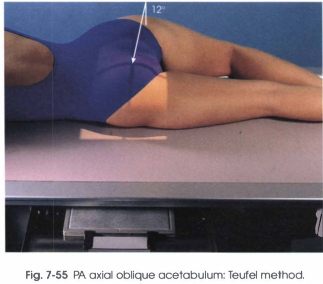
PA AXIAL OBLIQUE PROJECTION (TEUFEL METHOD) RAO OR LAO POSITION: POSITION OF PART
elevate the unaffected side so that the anterior surface of the body forms a 38-degree angle from the table
PA AXIAL OBLIQUE PROJECTION (TEUFEL METHOD) RAO OR LAO POSITION: CENTRAL RAY
directed through the acetabulum at an angle of 12 degrees cephalad; the central ray enters the body at the inferior level of the coccyx and approximately 2 inches lateral to the MSP toward the side being examined
PA AXIAL OBLIQUE PROJECTION (TEUFEL METHOD) RAO OR LAO POSITION: STRUCTURES SHOWN
fovea capitis and the superoposterior wall of the acetabulum
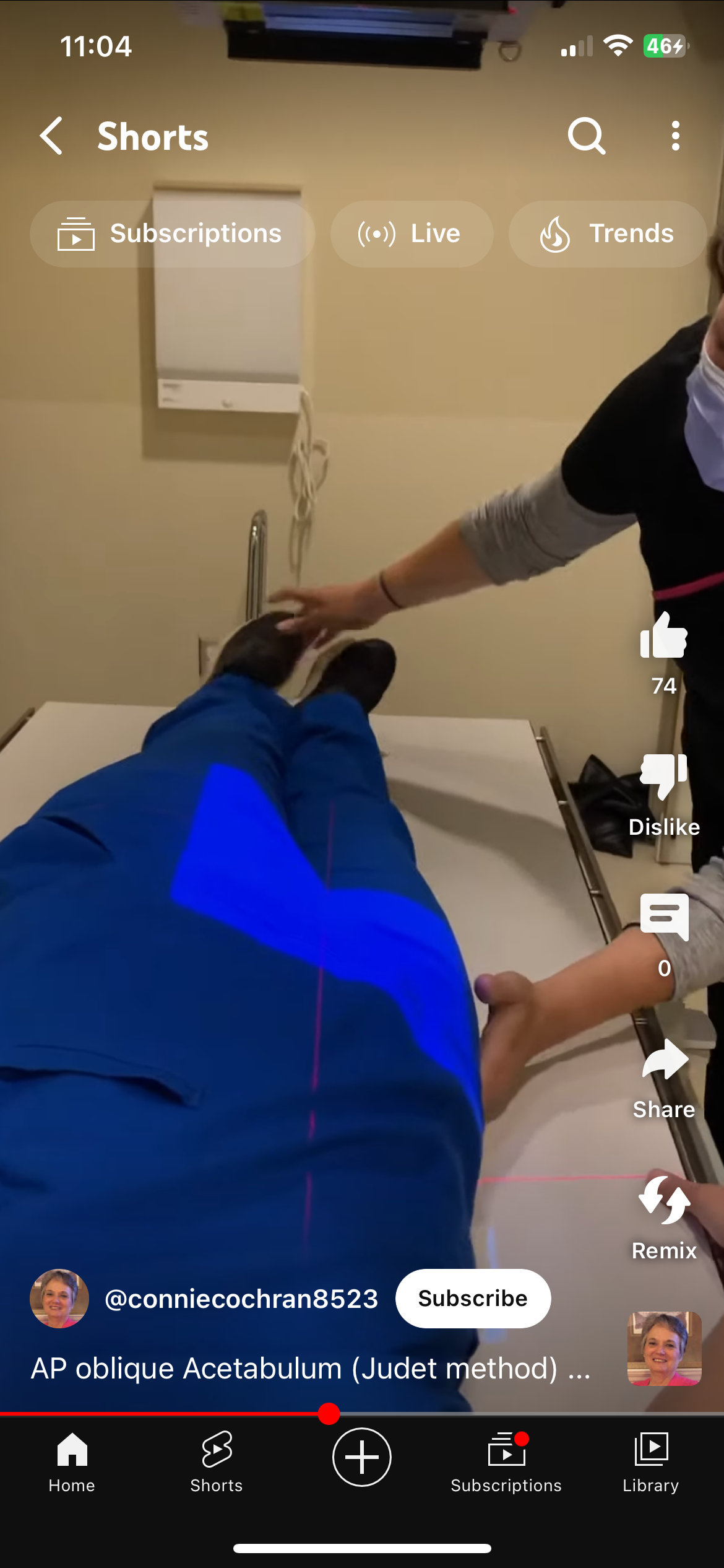
AP OBLIQUE PROJECTION (JUDET METHOD) RPO OR LPO POSITION
JUDET described two 45-degree posterior oblique positions that are useful in diagnosing fractures of the acetabulum: the internal oblique position (affected side up) and the external oblique position (affected side down); both positions must be performed to demonstrate the entire acetabulum, as well as the iliopubic and ilioischial columns of the affected side
AP OBLIQUE PROJECTION (JUDET METHOD) RPO OR LPO POSITION INTERNAL OBLIQUE
used for a pt with a suspected fracture of the iliopubic column and the posterior rim of the acetabulum
AP OBLIQUE PROJECTION (JUDET METHOD) RPO OR LPO POSITION INTERNAL OBLIQUE: NOTE
iliopubic column (anterior), composed of a short segment of the ilium and the pubis, extends up as far as the anterior spine of the ilium and from the symphysis pubis and obturator foramen through the acetabulum to the ASIS
AP OBLIQUE PROJECTION (JUDET METHOD) RPO OR LPO POSITION INTERNAL OBLIQUE: POSITION OF PT
place the pt in a posterior oblique position with the affected hip up
AP OBLIQUE PROJECTION (JUDET METHOD) RPO OR LPO POSITION INTERNAL OBLIQUE: POSITION OF PART
elevate the affected side so that the MCP of the body forms a 45-degree angle from the table
AP OBLIQUE PROJECTION (JUDET METHOD) RPO OR LPO POSITION INTERNAL OBLIQUE: CENTRAL RAY
perpendicular to the IR and entering 2 inches inferior to the ASIS of the affected side
AP OBLIQUE PROJECTION (JUDET METHOD) RPO OR LPO POSITION EXTERNAL OBLIQUE
used for a pt with a suspected fracture of the ilioischial column (posterior) and the anterior rim of the acetabulum
AP OBLIQUE PROJECTION (JUDET METHOD) RPO OR LPO POSITION EXTERNAL OBLIQUE: POSITION OF PT
place the pt in a posterior oblique position with the affected hip down
AP OBLIQUE PROJECTION (JUDET METHOD) RPO OR LPO POSITION EXTERNAL OBLIQUE: POSITION OF PART
elevate the unaffected side so that the MCP of the body forms a 45-degree angle from the table
AP OBLIQUE PROJECTION (JUDET METHOD) RPO OR LPO POSITION EXTERNAL OBLIQUE: CENTRAL RAY
perpendicular to the IR and entering at the pubic symphysis
AP OBLIQUE PROJECTION (JUDET METHOD) RPO OR LPO POSITION EXTERNAL OBLIQUE: STRUCTURES SHOWN
acetabular rim
AP OBLIQUE PROJECTION (JUDET METHOD) RPO OR LPO POSITION EXTERNAL OBLIQUE: NOTE
ilioischial column (posterior), composed of the vertical portion of the ischium and the portion of the illium immediately above ischium, extends from the obturator foramen through the posterior aspect of the acetabulum
UPRIGHT LEG MEASUREMENT PROCEDURE: POSITION OF PT
stand the pt upright with the back against the vertical imaging stand
UPRIGHT LEG MEASUREMENT PROCEDURE: POSITION OF PART
if the two lower extremities are examined simultaneously, separate the ankles 5 to 6 inches and place the specialized ruler between the pt and the IR, with the top at the level of the pelvis and extending down between the legs
UPRIGHT LEG MEASUREMENT PROCEDURE: POSITION OF PART
if the extremities are examined separately, position the pt with the special ruler behind each extremity
UPRIGHT LEG MEASUREMENT PROCEDURE: LOCALIZATION OF JOINTS
localize each joint and mark the central ray centering point
UPRIGHT LEG MEASUREMENT PROCEDURE: LOCALIZATION OF JOINTS
locate the hip joint by placing a mark 1 to 1 ¼ inches laterodistally and at a right angle to the midpoint of an imaginary line extending from the ASIS to the pubic symphysis
UPRIGHT LEG MEASUREMENT PROCEDURE: LOCALIZATION OF JOINTS
locate the knee joint just below the apex of the patella at the level of the depression between the femoral and tibial condyles
UPRIGHT LEG MEASUREMENT PROCEDURE: LOCALIZATION OF JOINTS
locate the ankle joint directly below the depression midway between the malleoli
UPRIGHT LEG MEASUREMENT PROCEDURE: PROCEDURE 1ST EXPOSURE
exposure field is centered to the pt’s hip
UPRIGHT LEG MEASUREMENT PROCEDURE: PROCEDURE 2ND EXPOSURE
exposure field is centered to the pt’s knee joint
UPRIGHT LEG MEASUREMENT PROCEDURE: PROCEDURE 3RD EXPOSURE
exposure field is centered to the pt’s ankle joint
UPRIGHT LEG MEASUREMENT PROCEDURE: CENTRAL RAY
perpendicular to the center of the IR for the manual procedure
*for the automated procedure, there may be a small cephalad angle for the hip exposure and a small caudad angle for the ankle exposure
UPRIGHT LEG MEASUREMENT PROCEDURE: STRUCTURES SHOWN
a composite of the three exposures digitally stitched into one image, which includes anatomy from the hip joints to the ankle joints