PNS Exam 1 Material
1/263
There's no tags or description
Looks like no tags are added yet.
Name | Mastery | Learn | Test | Matching | Spaced | Call with Kai |
|---|
No analytics yet
Send a link to your students to track their progress
264 Terms
The central nervous system includes what 2 structures?
(1) Brain
(2) Spinal Cord
The peripheral nervous system consists of what structures?
(1) Cranial Nerves
(2) Spinal Nerves
What is the nucleus of origin?
Motor Function
Nucleus within the CNS that contains cell bodies of lower motor neurons
What is the nucleus of termination?
Sensory Function
Nucleus that receives information from the PNS and sends it up to higher centers of the brain
How do cranial nerves exit the skull? What is the clinical significance of this?
Exit the skull via patent openings
These openings are FIXED (not dynamic) —> so if there is an issue, more likely to be compromised somewhere else
Compare it to spinal nerves via IVF = changes shape just by moving the neck/spine in the cervicals
How many spinal nerves do we have?
31 pairs (mixed)
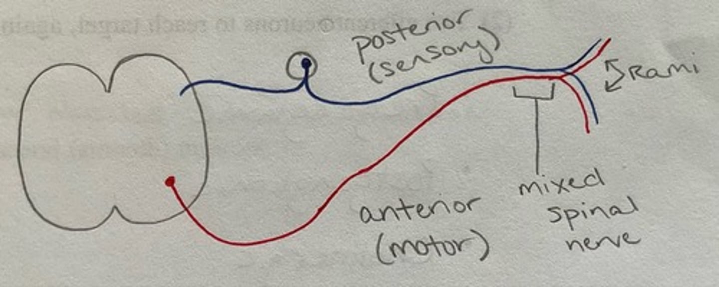
Rootlets vs Rami
Rootlets: spinal nerves attach to the cord via ventral (anterior — motor) and dorsal (posterior — sensory) rootlets
Rami: spinal nerve splits into a ventral (anterior) primary ramus and a dorsal (posterior) ramus
Rami are mixed...rootlets are NOT!
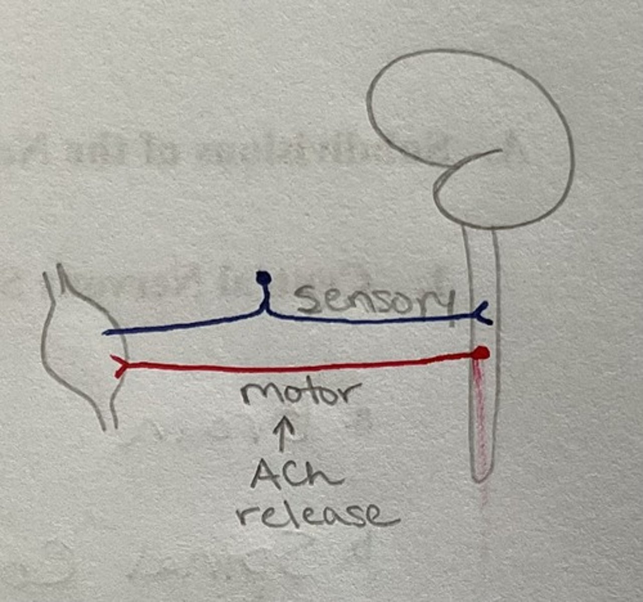
Somatic Subdivision of the Nervous System
Controls skeletal muscle
— sensory (afferent) and motor (efferent)
— all sensory nerves have cell bodies in PNS
— motor nerves release ACh
Visceral (Autonomic) Subdivision of the Nervous System
Controls vital body functions
— glands, smooth muscle, and cardiac muscle
2 Divisions:
(1) Sympathetic
(2) Parasympathetic
Sympathetic Nervous System: function and targets
Fight or flight / Emergency
Targets: heart, lungs, abdominopelvic organs, blood vessels, arrector pili
Sympathetic preganglionic neurons release what neurotransmitter? Postganglionic?
Preganglionic = Ach
Postganglionic = Norepinephrine (NE)
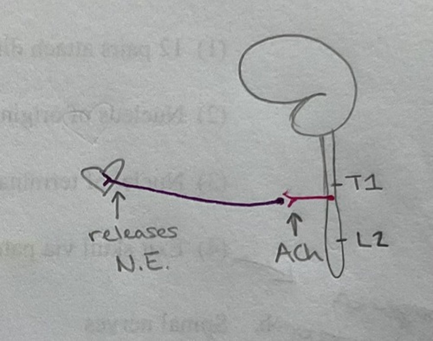
At what level of the spinal cord do we find the sympathetic nervous system?
T1 - L2
Splanchnic nerves are a part of which division of the nervous system? Are they the pre- or pstganglionic neurons?
Sympathetic (greater, lesser, and least) and Parasympathetic (pelvic)
Preganglionic neurons (travels right through the sympathetic trunk in the sympathetic division)
Parasympathetic Nervous System: function and targets
Rest and digest function
— "return to normalcy" = allows small fluctuations (instead of all or nothing between sympathetic and parasympathetic
Targets: essentially the same as sympathetic, but fewer blood vessels
Parasympathetic nerves come from what levels of the spinal cord?
Cranial Nerves 3, 7, 9, and 10
Spinal Cord: S2-S4
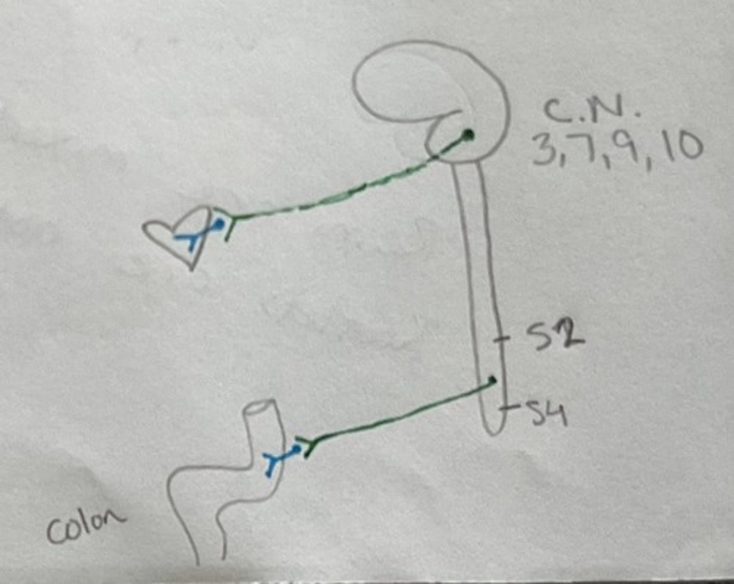
Parasympathetic preganglionic neurons release what neurotransmitter? Postganglionic?
Pre- and postganglionic release Acetylcholine (ACh)
Parasympathetic Division: Visceral Afferents
Senses B.P, toxins, etc.
Cell bodies are located in the PNS (looks just like somatic sensory)
— in the spinal ganglion or ganglion associated with the cranial nerve
Cranial Nerve I (overview)
Olfactory Nerve
— Special Sensory = smell
— NOT through the thalamus
— Fibers in olfactory mucosa traverse the cribriform plate and synapse on the olfactory bulb
Cranial Nerve II (overview)
Optic Nerve
— Special Sensory = vision
— fibers from the retina converge on the optic disc and are carried in the optic nerve, chaisma, and tract to the lateral geniculate bodies (thalamus)
Cranial Nerve III (overview)
Oculomotor Nerve
— Motor (5 somatic muscles), Somatic, and Parasympathetic (2 smooth muscles)
Cranial Nerve IV (overview)
Trochlear Nerve
— Somatic Motor (superior oblique muscle)
— Superior Oblique Muscle action = moves eyes down and out)
— Smallest cranial nerve
— only cranial nerve that leaves the CNS on the posterior side
Cranial Nerve V (overview)
Trigeminal Nerve
— Mixed: somatic motor and sensory
— "Great sensory nerve of the face"
— Largest cranial nerve
— Motor supply to muscles of mastication
Cranial Nerve VI (overview)
Abducens Nerve
— Somatic Motor (lateral rectus)
— Lateral rectus action = moves eyes outward
Cranial Nerve VII (overview)
Facial Nerve
— Mixed: motor ("nerve of facial expression"), somatic sensory, and parasympathetic
— carries taste information from the anterior 2/3 of the tongue
— supplies muscles of facial expression (including stylohyoid and stapedius)
— lacrimal and salivary glands (parasympathetic role)
Cranial Nerve VIII (overview)
Vestibulocochlear Nerve
— Special sensory = hearing and equilibrium (2 nerves carried together, but never mix)
— sensory information from the cochlea and vestibule
Cranial Nerve IX (overview)
Glossopharyngeal Nerve
— Mixed: motor (branchiomotor and parasympathetic) and sensory (somatic, special, and visceral)
— carries taste information from the posterior 1/3 of the tongue
— motor supply to the stylopharyngeus muscle
— Parasympathetic to the parotid gland
Cranial Nerve X (overview)
Vagus Nerve
— Mixed: motor (branchiomotor and parasympathetic) and sensory (somatic, visceral, and special)
— Longest of cranial nerves
— motor supply to the pharyngeal constrictors
— Parasympathetic supply to the bronchi, heart, and GI tract from the esophagus to the distal 1/3 of the transverse colon
Cranial Nerve XI (overview)
Accessory Nerve (Spinal Accessory)
— Motor
— Cranial and Spinal parts
— Supplies muscles of the pharynx, larynx, and palate (cranial part) as well as the trapezius and sternocleidomastoid muscles (spinal part)
Cranial Nerve XII (overview)
Hypoglossal Nerve
— Motor: intrinsic and extrinsic muscles of the tongue
CN I exits the skull via which opening?
Cribriform plate of the ethmoid bone
CN II exits the skull via which opening?
Optic canal
CN III exits the skull via which opening?
Superior orbital fissure
CN IV exits the skull via which opening?
Superior orbital fissure
The 3 divisions of CN V exit the skull via which openings?
V1 = superior orbital fissure
V2 = foramen rotundum
V3 = foramen ovale
CN VI exits the skull via which opening?
Superior orbital fissure
CN VII exits the skull via which opening?>
Internal acoustic meatus
CN VIII exits the skull via which opening?
Internal acoustic meatus
CN IX exits the skull via which opening?
Jugular foramen
CN X exits the skull via which opening?
Jugular foramen
CN XI exits the skull via which opening?
Jugular foramen
CN XII exits the skull via which opening?
Hypoglossal canal
Somatic Motor Pathway: 2 Neurons
(1) Upper Motor Neurons (UMN)
— only in the CNS
— runs from cortex to the spinal cord
(2) Lower Motor Neurons (LMN)
— enters into the PNS
Adjustments directly influence which neuron in the somatic motor pathway?
Directly affects LMN (Indirectly affects UMN)
2 Types of UMNs
(1) Pyramidal
— in the cortex
— conscious control
(2) Extrapyramidal
— mostly in the brainstem
— not conscious control
Pathway of UMNs
(1) Descends through the corona radiata, internal capsule, and crus cerebri
(2) most cross in the pyramids and further descend in the lateral corticospinal tract
(3) synapses in the anterior horn (primarily Rexed lamina VII) of the gray matter
Pathway of LMNs
Cell Body Location = Anterior horn gray nuclei of origin
Exit cord via anterior rootlets and are carried in named nerves to supply somatic (skeletal ) muscles
Somatic Sensory Pathway
3 Neuron Pathway:
(1) Primary
— cell bodies located in the dorsal root ganglion
— convey info from a receptor to the CNS
— synapse in the spinal cord
(2) Secondary
— sensory neurons decussate and convey info to the thalamus
(3) Tertiary
— relay info to the cerebral cortex where info is integrated
The Olfactory Nerve carries what information?
Special Sensory = smell
Where are olfactory neurons found?
Olfactory epithelium lining the superior part of the nasal cavity (covered by a thin layer of mucous)
Pass through the cribriform plate (part of ethmoid bone)
Olfactory neurons are supported by what cells?
"Supporting cells"
What part of the olfactory neurons functions as receptors?
Olfactory "hairs" = telodendria
Are olfactory neurons myelinated?
No! —> unmyelinated (but covered by Schwann cells)
What happens to olfactory neurons after they pass through the cribriform plate?
Enter the olfactory bulb where they synapse on the dendrites of mitral cells
What is formed at the synapse between olfactory neurons and mitral cells?
Synaptic glomeruli
What are the secondary sensory neurons of the Olfactory Pathway? Where are their cell bodies located?
Mitral Cells
Cell bodies located in the olfactory bulb
Where do the mitral cells carry their information?
Travel posterior along the olfactory tract
At the anterior perforated substance the tract fibers split into medial and lateral olfactory striae (singular = striatum)
Where do medial stria fibers travel?
Medial stria fibers cross the midline via the anterior commissures and travel to the opposite olfactory bulb
Where do lateral stria fibers travel?
Carry information to the primary olfactory cortex (periamygdaloid and prepiriform areas, including the uncus = Bdmn area 34) on the medial aspect of the temporal lobe
Then to secondary olfactory cortex (entorhinal area in the parahippocampul gyrus = Bdmn area 28)
Do we segregate smell?
No!
Terminal Nerve (Olfactory Pathway)
Carries info from the nasal septum —> autonomic function
Vomeronasal Nerve (Olfactory Pathway)
Important for tracking prey (poorly developed in humans)
Anosmia
Loss of smell
— rare, but can happen if born without olfactory bulb
Skull fractures and olfaction
If fracture breaks cribriform plate...can tear dura and CSF leaks down into the nasal cavity (halo test)
Can olfactory neurons be replaced/repaired?
YES!
3 Layers (Tunics) of the Eye
Outermost
(1) Fibrous Tunic
(2) Vascular Tunic
(3) Retina
Innermost

Fibrous Tunic of the eye consists of:
(1) Sclera (whites of the eye)
(2) Cornea
Vascular Tunic of the eye consists of:
(1) Ciliary Body
(2) Iris
(3) Choroid (highly vascular)
10 Layers of the Retina (Superficial to Deep)
(1) Pigmented Layer
(2) Photosensitive Outer Segments (of rods and cones)
(3) External Limiting Membrane (ELM)
(4) Outer Nuclear Layer
(5) Outer Plexiform Layer
(6) Inner Nuclear Layer
(7) Inner Plexiform Layer
(8) Ganglion Cell Layer
(9) Nerve Fiber Layer
(10) Internal Limiting Membrane (ILM)
Which layer of the retina is adjacent to the choroid?
Pigmented layer
"Plexiform" means what?
Synaptic activity
"Nuclear" means what?
Cell bodies
Which layer of the retina contains rod and cone cell bodies?
Outer nuclear layer
Which layer of the retina contains the synapse between rods/cones and bipolar cells?
Outer Plexiform Layer
Which layer of the retina contains cell bodies of bipolar cells?
Inner Nuclear Layer
Which layer of the retina contains the synapse between bipolar cells and ganglion cells?
Inner plexiform later
Which layer of the retina contains ganglion cell bodies?
Ganglion cell layer
Which layer of the retina contains retinal ganglion cell axons?
Nerve fiber layer
Are ganglion cell axons myelinated?
NO!
What is the glial boundary separating the retina from the vitreous body?
Internal Limiting Membrane
3 types of cones:
(1) Red
(2) Green
(3) Blue
Hint: think visible light spectrum —> 2 ends plus the middle (ROY G BIV)
Cones require what to function best?
Adequate light!
Where are cones most concentrated?
Towards the center of the retina (fovea)
What do rods sense?
Only light vs dark —> NO COLOR
Where are rods located on the retina?
Periphery of the retina is almost completely composed of rods
No rods in the fovea
Color Blindness
Different types depending on which cones affected
Most sex-linked and recessive
Once depolarized, photoreceptors (rods and cones) send info where?
Relay information to bipolar cells by synapsing in the outer plexiform layer
Bipolar cell bodies are found in the inner nuclear layer
Where do bipolar cells relay their info to?
Relay info to ganglion cells
This synapse occurs in the inner plexiform layer
Where do ganglion cells send their info?
(1) Information carried by ganglion cells travels toward the optic disc in the optic nerve after leaving the eyeball
(2) optic nerve enters the skull through the optic canal and unite to form the optic chiasma
Are the axons in the nerve fiber layer myelinated?
NO —> ganglion cells are unmyelinated
Are the optic nerve axons myelinated?
Yes (if large enough)
Myelin is formed by: oligodendrocytes
What happens in the optic chiasma?
(1) Fibers from the medial (nasal) side of each retina will cross
(2) Fibers from the lateral (peripheral) side of each retina will NOT cross
Fibers then split to form the optic tracts (so now divided into right and left visual fields)
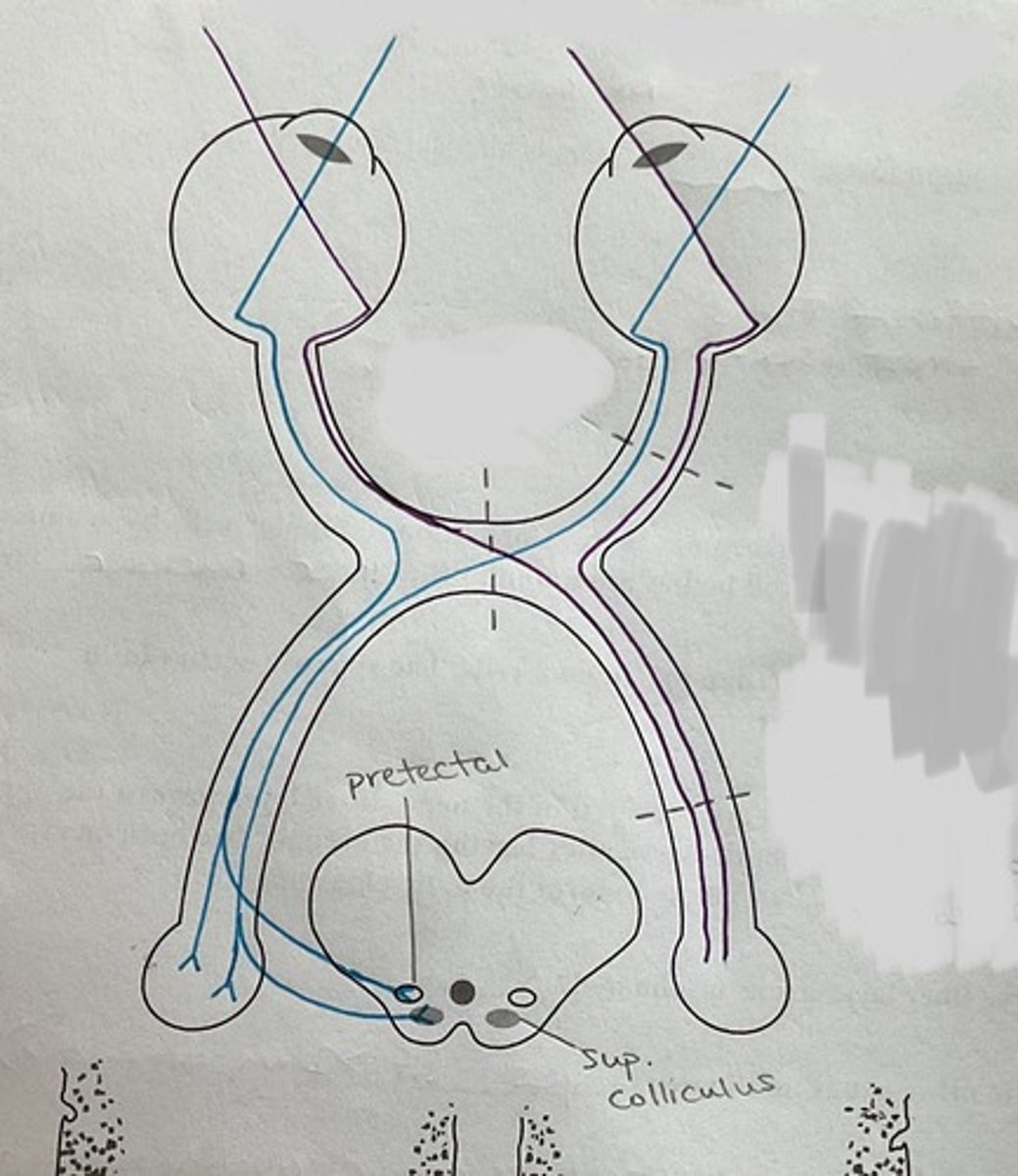
A majority of the fibers of the optic tract synapse where?
Lateral geniculate body of the thalamus —> relayed to the cerebral cortex in the occipital lobe (Bdmn area 17)
OR
Superior Colliculus —> relayed to the tectospinal tract
The tectospinal tract influences what two muscles?
(1) Trapezius
(2) Sternocleidomastoid
Some fibers from the optic tract can also synapse where? What are these fibers involved with?
Pretectal Nucleus of the midbrain (these fibers deal with the light reflex)
What would happen if there was a lesion at the optic nerve?
Smaller visual field
Also issue with depth perception
What would happen if there was a lesion of the decussating fibers of the optic chiasma?
Lose peripheral vision (more narrow field of view)
What would happen if there was a lesion at the optic tract?
Lose half the visual field
Cranial Nerve II is involved with what light reflexes?
(1) Direct (shine flashlight into the eye and pupil constricts) and consensual (indirect; shine flashlight into one eye and the opposite eye should constrict slightly too) light reflexes
(2) Accommodation reflexes (parasympathetic — ciliary muscle makes lens more convex to focus on something closer)
(3) Corneal Reflex (tests more CN 5 and 7; pressure on cornea = slam eye shut)
(4) Convergence ("cross eyes")
NOTE: CN II only can do a sensory component of a reflex!
CN III carries what type of fibers?
MOTOR
Both somatic and parasymapthetic motor fibers
Where is the oculomotor nuclei located?
Located in the periaqueductal gray matter (in midbrain) anterior to the cerebral aqueduct at the level of the superior colliculi