ANT Throrax I Lecture
 Call Kai
 Learn
Learn Practice Test
 Spaced Repetition
 Match
Match Flashcards
 Knowt Play
1/24

There's no tags or description
Looks like no tags are added yet.
Name | Mastery | Learn | Test | Matching | Spaced | Call with Kai | Chat |
|---|
No analytics yet
Send a link to your students to track their progress
25 Terms
What are the functions of the thorax
conduit (passage for things like arteries nerves etc)
protection of vital organs
breathing
What are the compartments of the thorax
mediastinum
L and R pleural cavities
What are the bony parts of the thoracic cage
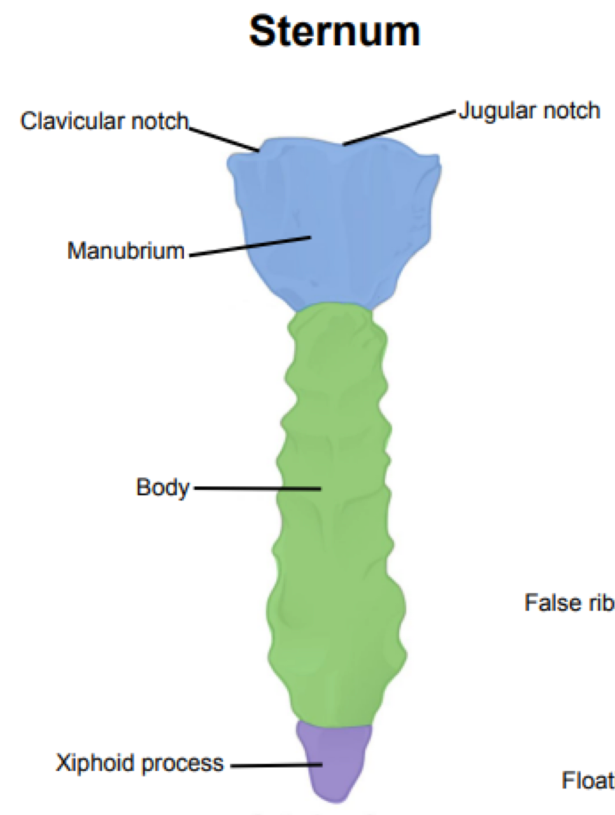
sternum
manubrium
body
xiphoid process
sternal angle (T4-T5)
Ribs (and costal cartilage)
thoracic vertebrae (12)
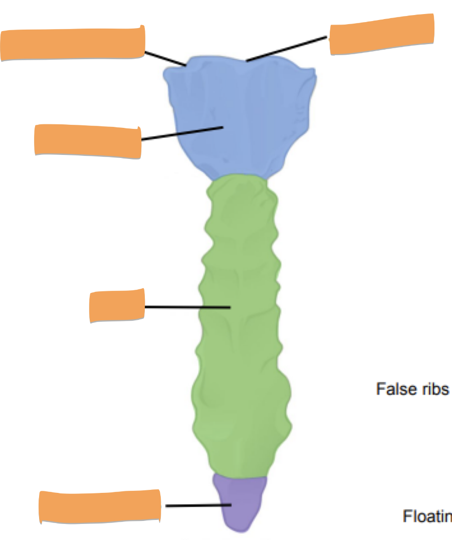
Label the parts of the sternum
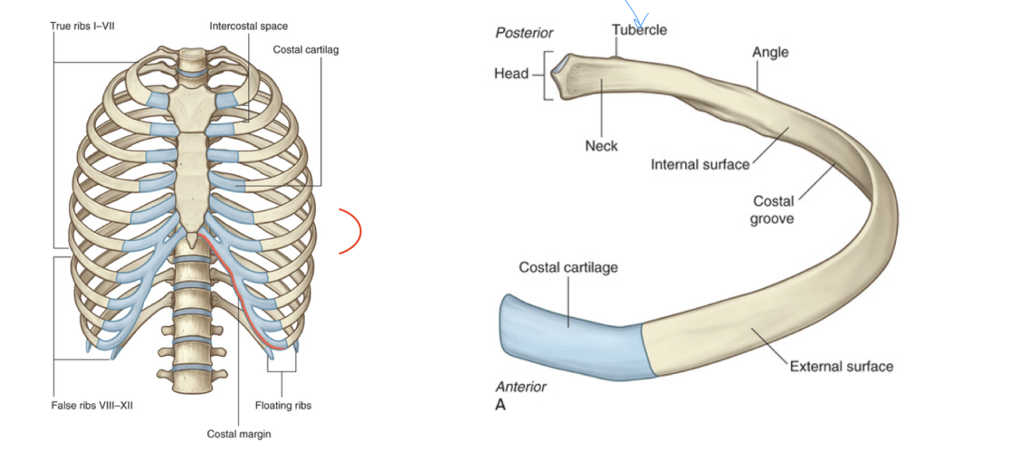
How many true, false and floating ribs do we have
7 true ribs
5 false ribs (2 of which are also floating)
Where is the tubercle of the ribs and why is it important
location: bony portrusion on the side of the rib where it posteriorly is (connects to spine)
function: connects ribs to spine via ligaments at the transverse costal facet on thoracic vertebrae
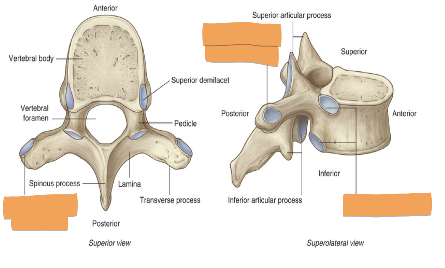
Identify the blanks and list the articulations of where it would connect
demifacets: articulate with head of the ribs
costal facets: articulate with tubercle of ribs T1-10
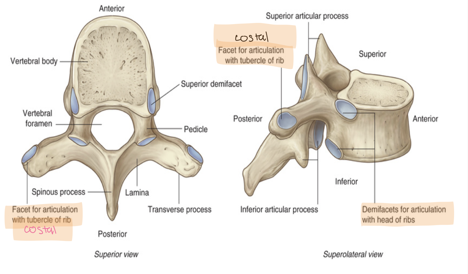
What is the name of the ligament that binds the costal facets to the ribs
radiate ligament of head of rib
What are the features of the superior thoracic aperture
body of T1 posteriorly
medial margin of rib 1 on each side
manubrium anteriorly
open structure
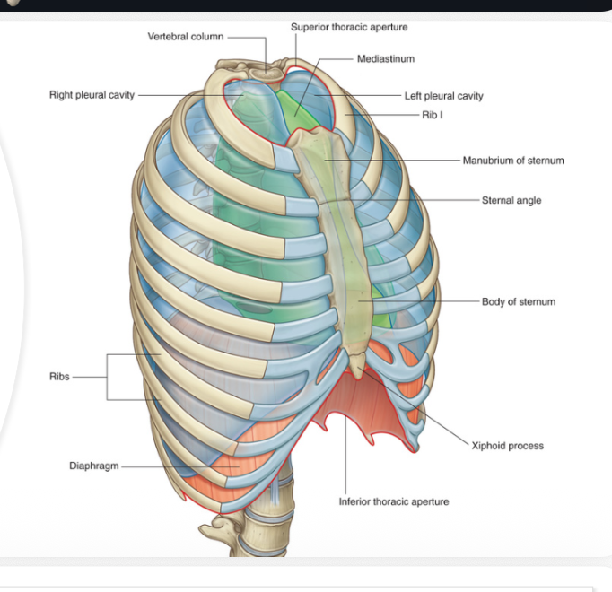
Inferior thoracic aperture features
body of T12 posteriorly
rib 12 and distal end of rib 11 posterolaterally
cartilage of ribs 7-10 anterolaterally
xiphoid process andterior
closed off by the diaphragm
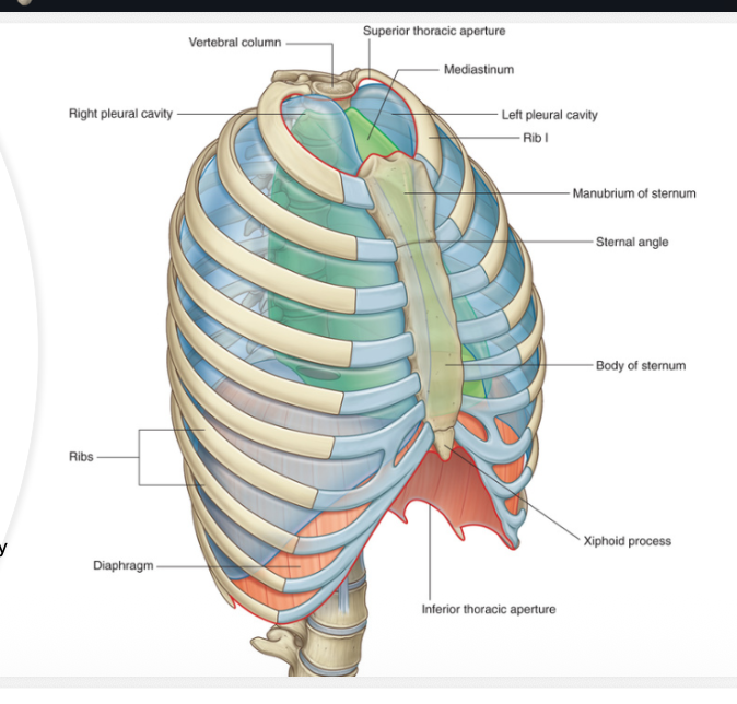
What are the boundaries of the thoracic aperture
body of T1 posteriorly
medial margin of rib 1 on each side
manubrium anteriorly
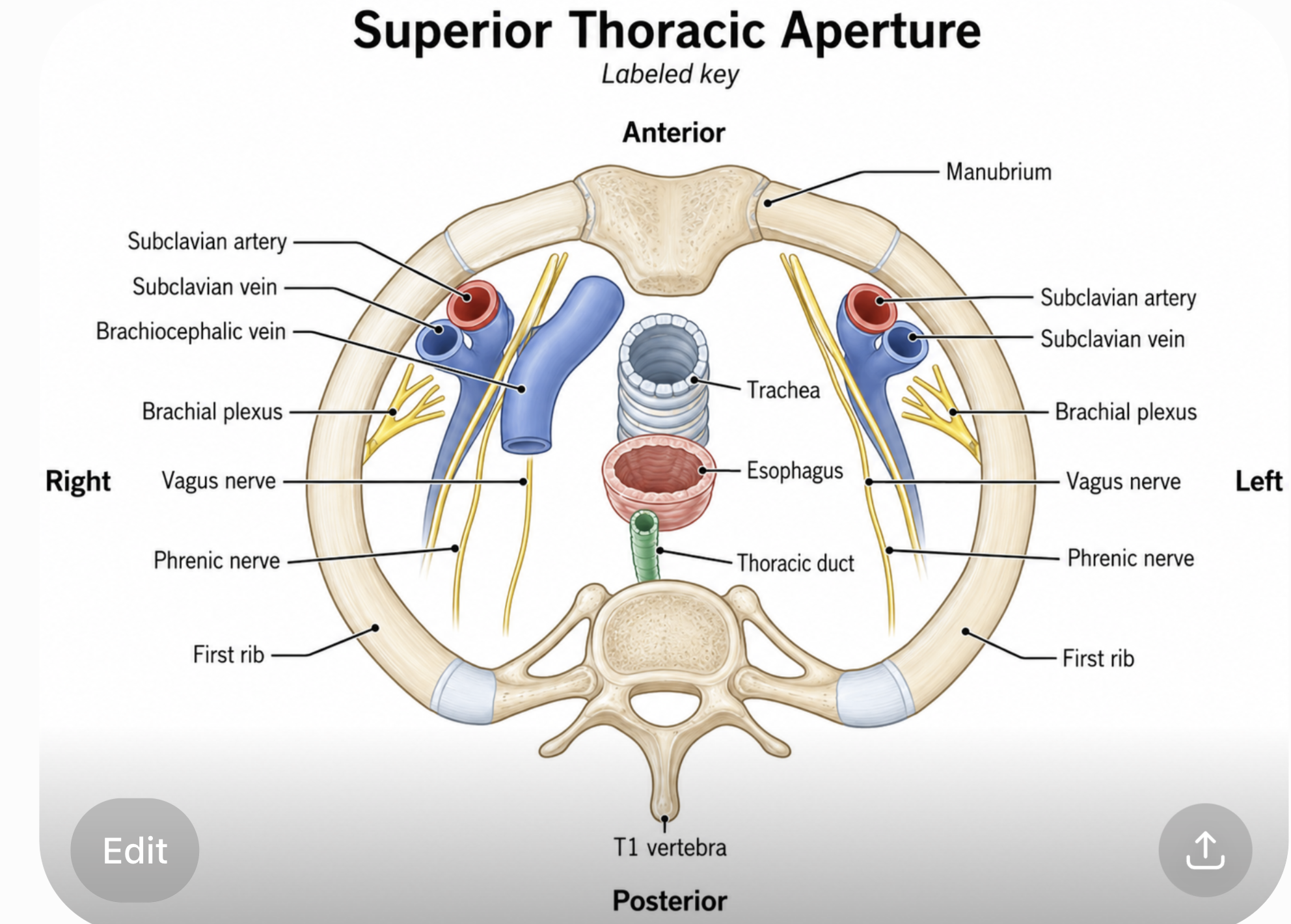
What structures pass through the superior thoracic aperture
vessels
subclavian artery, subclavian vein, brachiocephalic vein
nerves
brachial plexus, vagus nerve, phrenic nerve
organs
trachea, esophagus, thoracic duct
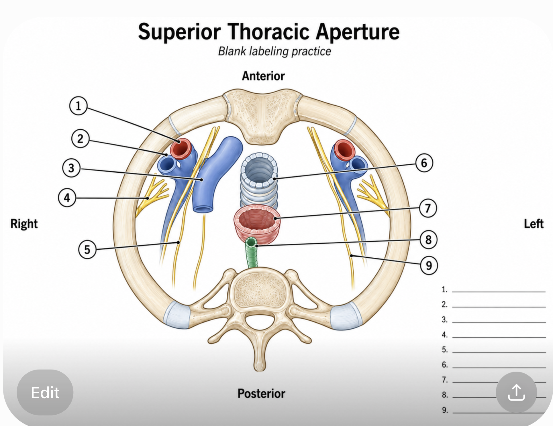
Label each part
What are the three layers of intercostal muscles and their functions
External intercostal muscle
elevates rubs during forced inhalation
Internal
depresses ribs during forced exhalation
innermost intercostal muscles
depresses ribs during forced exhalation
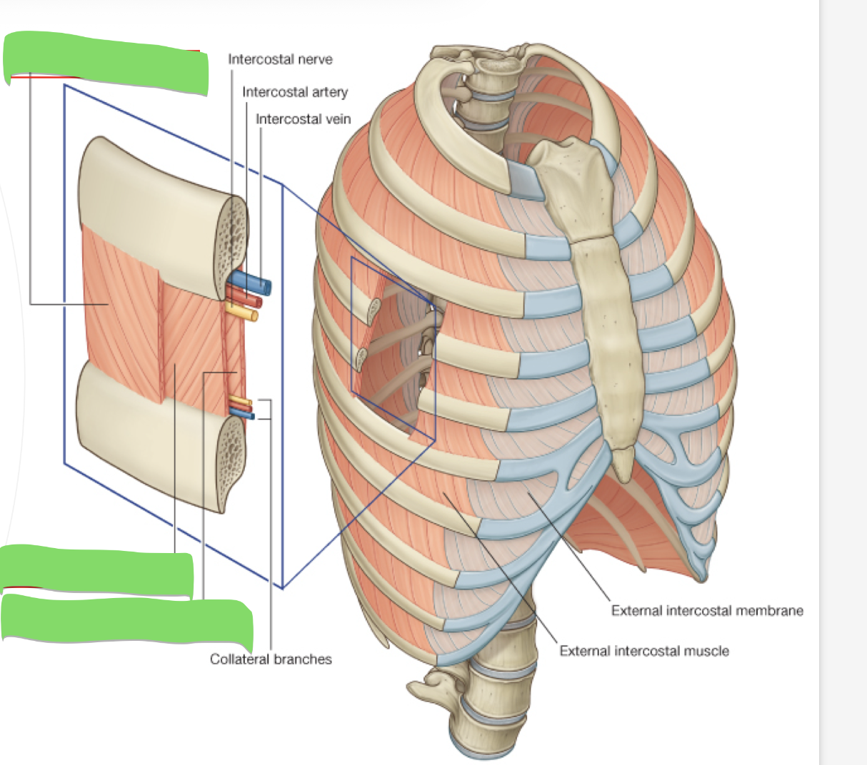
Label
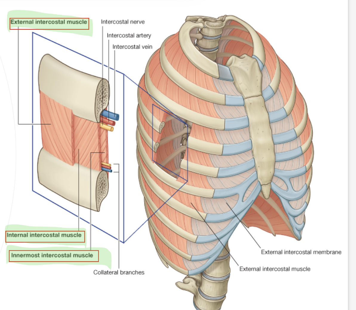
What is the innervation of the intercostal muscles, and what is the arterial supply?
innervation: intercostal nerves
arterial supply: posterior and anterior intercostal arteries
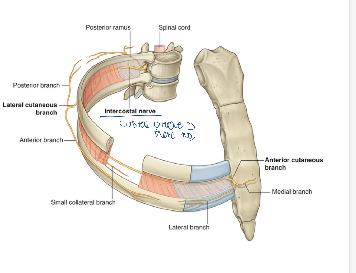
Where is the intercostal nerve located
The costal groove
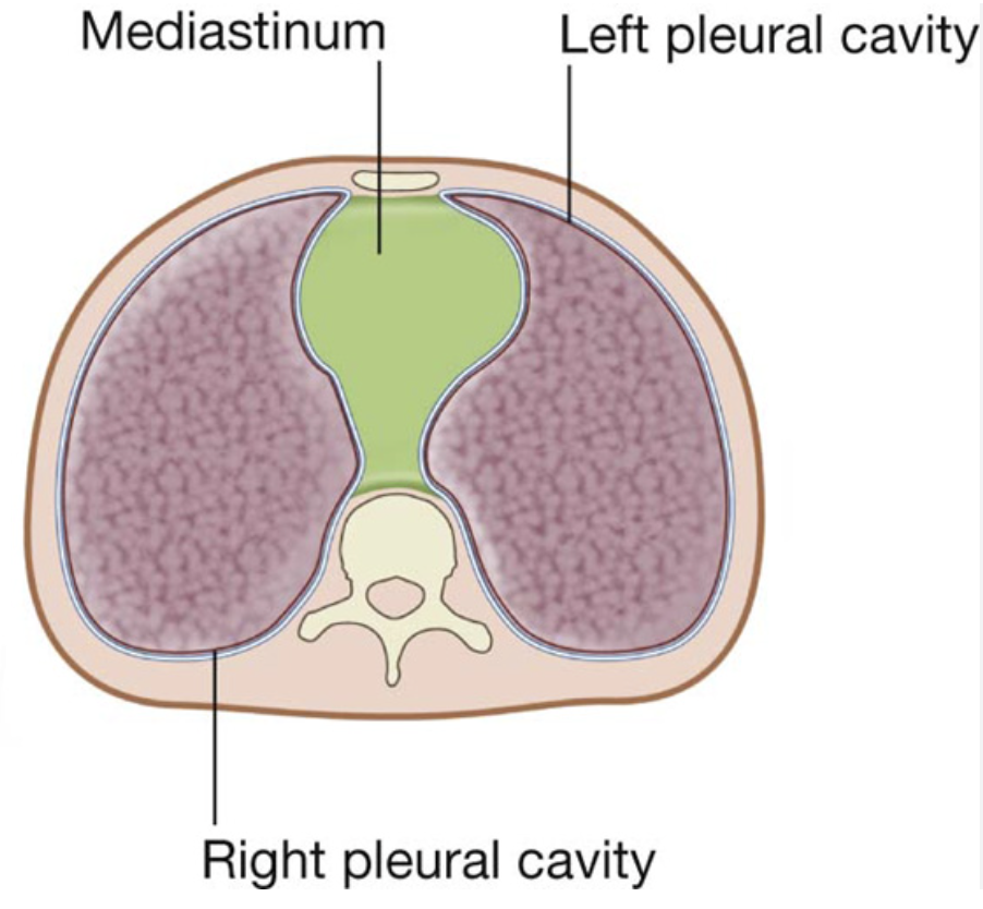
What is the mediastintum, and what does it hold (organs and arteries)
compartment between two lungs that contains heart, esophagus, trachea, thymus gland
and aorta and SVC
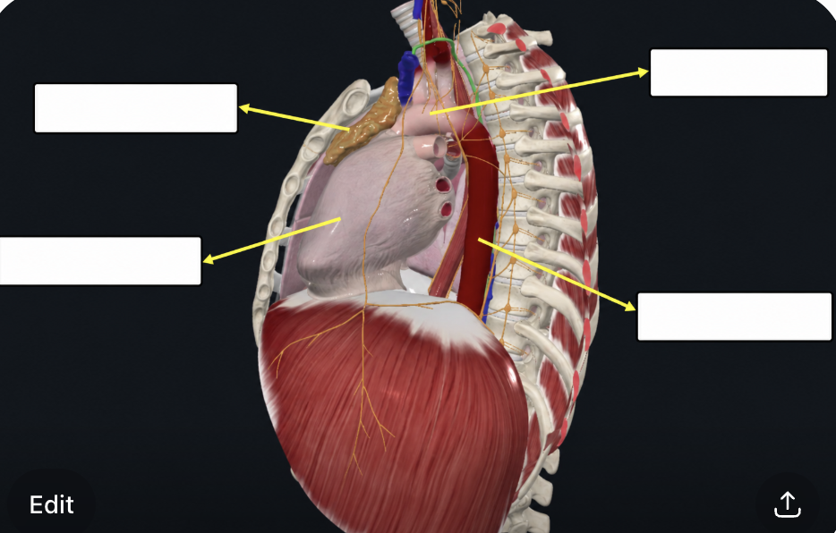
Label
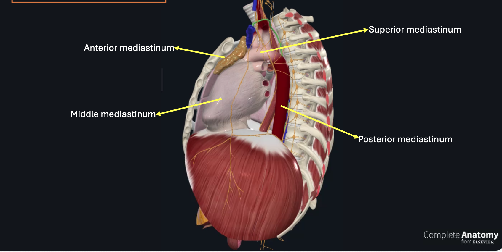
What are the mediastinal divisions
superior: thymus, trachea, esophagus, thoracic duct, aortic arch, veins (SVC, brachiocephalic), nerves (vagus phrenic, left recurrent laryngeal), lympathics
Anterior: remenents of thymus and lymph notes
middle: pericardium, phrenic nerve, heart, pulmonary trunk, svc
posterior: descending thoracic aorta, azygos veins, thoracic duct, esophagus, vagus nerve, splanchnic nerves, lymphatics
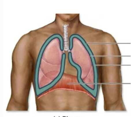
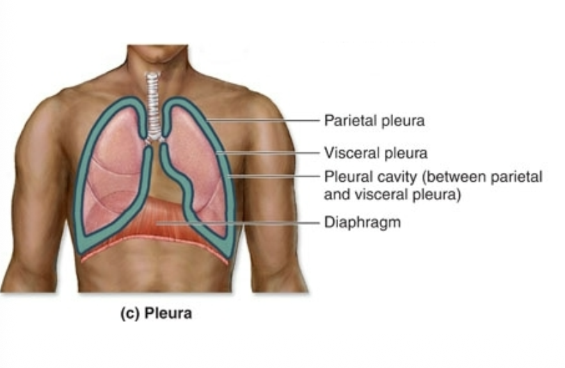
What is pleura (and label this diagram)
lines and protects lungs and chest wall (2 layers) on either side of the mediastinum
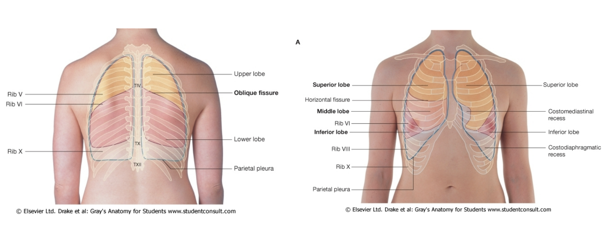
CHECK WITH NOTES AFTER MONDAY What is the difference between the right lung and the left one?
Left: 2 lobes (bc heart), narrower, has the cardiac notch for the heart, has lingula (google idk if you need to know it)
Right: 3 lobes, bigger, heavier, shorts (bc liver under it)
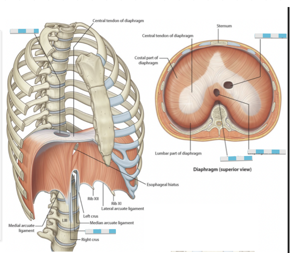
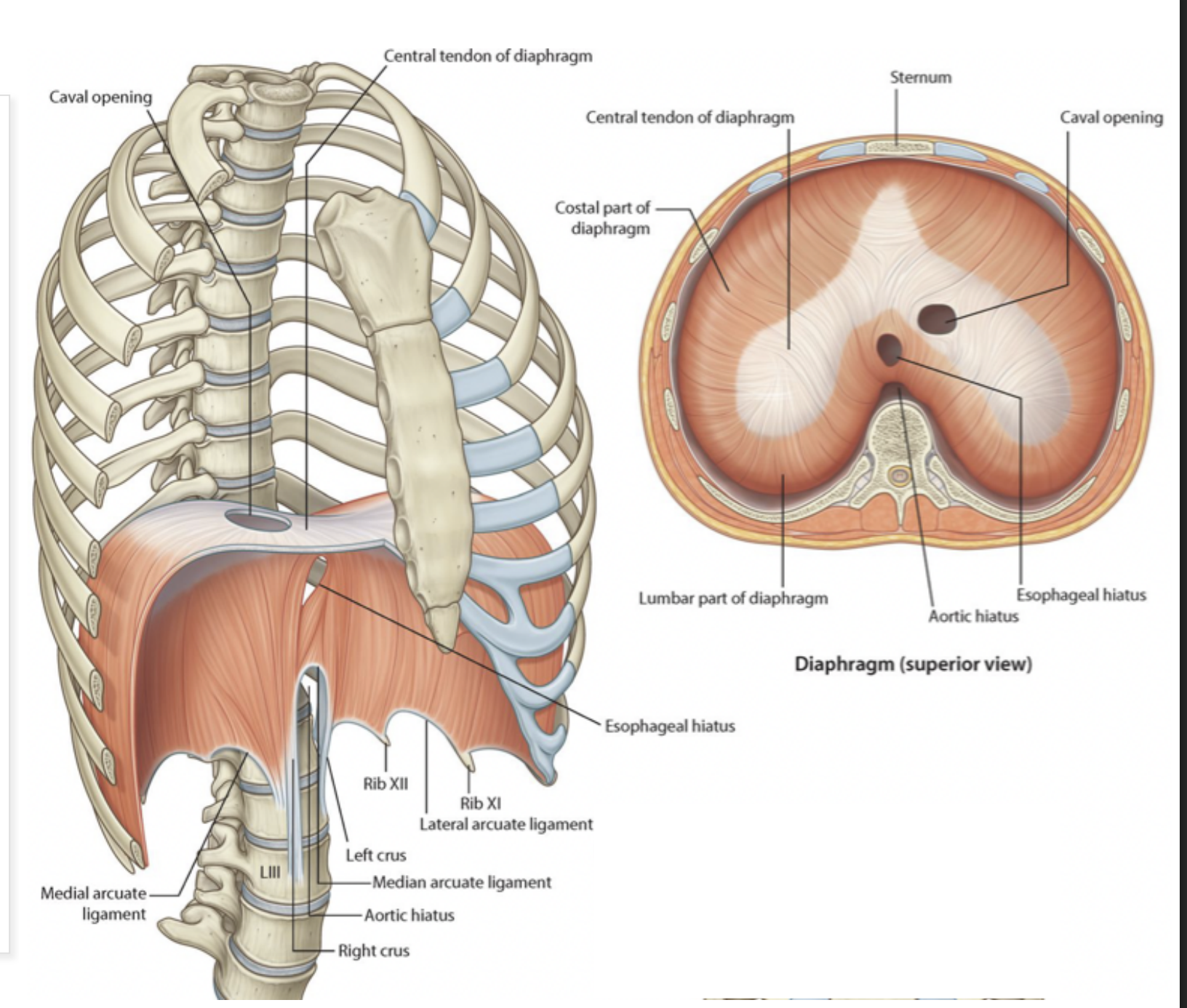
What does the phrenic nerve pass through
C3, C4, C5 keeps the diaphragm alive
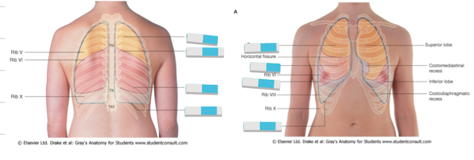
label