MICI 4115 Midterm 1
 Call Kai
 Learn
Learn Practice Test
 Spaced Repetition
 Match
Match Flashcards
 Knowt Play
1/218

There's no tags or description
Looks like no tags are added yet.
Name | Mastery | Learn | Test | Matching | Spaced | Call with Kai | Chat |
|---|
No analytics yet
Send a link to your students to track their progress
219 Terms
What is an immunogen?
substance can elicit an immune response
All immunogens are antigens, not all antigens are immunogenic
What is an antigen?
is defined as any substance that can bind to a specific antibody. All antigens therefore have the potential to elicit specific antibodies, but some need to be attached to an immunogen or adjuvant
All immunogens are antigens, not all antigens are immunogenic
present in different forms (DNA, RNA, Protein, lipid or carbohydrate)
purified or with adjuvant or in live organism
What are the general ways to monitor innate immune responses?
innate immune cells from blood, lymphoid organs and non-lymphoid organs
need to be induced to study
What are the general ways to monitor T cell responses?
lymphocytes from blood, lymphoid organs and non-lymphoid organs
What are the general ways to monitor B cell response?
antiserum (plural: antisera)
What is in vitro vs in vivo?
vitro: glass tube (vitre)
vivo: living body (vivant) need to set up animal by doing additional experiments outside of body to check via ex vivo. Often called immuno assay (often involve the antibodies)
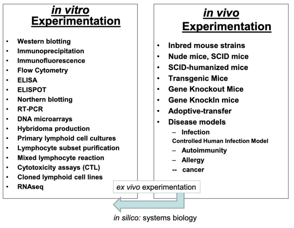
What are transgenic mice?
overexpress of the same gene (gene already have)
What is knockout genes vs knockin genes?
Knockout remove gene
knockin add in the gene (add in foreign protein)
What does in silico mean?
computer done experiments (not covering in class)
Is western blotting and northern blotting in vivo or in vitro?
in vitro
Is immunoprecipiation and Immunofluorescence in vivo or in vitro?
in vitro
Is Flow cytometry in vivo or in vitro?
in vitro
Is ELISA, ELSIPOT in vivo or in vitro?
In vitro
Is RT-PCR in vivo or in vitro?
In vitro
Is DNA microarrays in vitro on in vivo?
In vitro
Is hybridoma production in vivo or in vitro?
In vitro
Is primary lymphoid cell cultures, lymphocyte subset purification, cloned lymphoid cell lines and mixed lymphocyte reaction in vivo or in vitro techniques?
In vitro
Are cytotoxicity assays (CTL) in vitro or in vivo?
In vitro
Is RNAseq in vivo or in vitro?
In vitro
Are inbred mouse strains, nude mice, SCID mice, SCID-humanized mice, trangenic mice, gene knockout mice, gene knockin mice in vitro or in vivo?
In vivo
Is adoptive transfer in vivo or in vitro?
In vivo
Are disease models (infection- controlled infection human model, autoimmunity, allergy and cancer) in vivo or in vitro?
In vivo
What is ex vivo?
Ex vivo (Latin for "out of the living") refers to procedures or experiments performed on tissues, cells, or organs outside the body, in a controlled lab environment, but still maintaining them in a living state
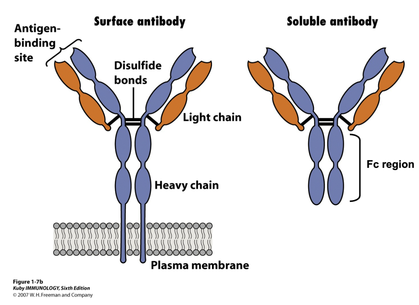
What are the physical features of an antibody?
2 heavy 2 light chain
antibody often referring to soluble
surface= BCR
antigen binding site is fundamental characteristic for research
Fc region: determines isotype and it mediates antibody function
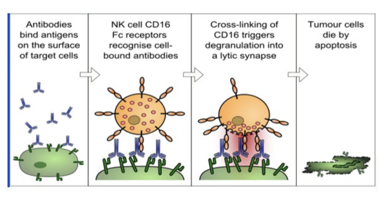
What is antibody dependent cell-mediated cytotoxicity (ADCC) assay?
Fc region allows for the antibody to bind to CD16 receptor of NK
binding is critical in ADCC function
crosslinking allows NK to degranulate
What is an epitope?
The site on an antigen to which each distinct antibody molecule binds is called antigenic
determinant or epitope
Different antibodies bind to distinct epitopes on an antigen
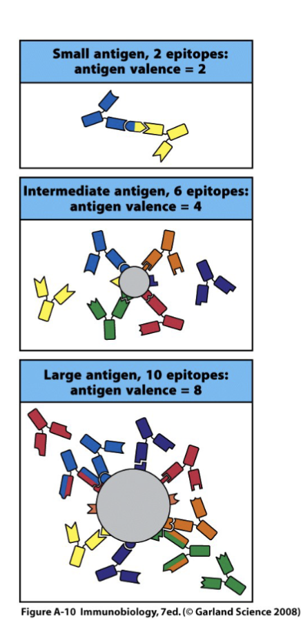
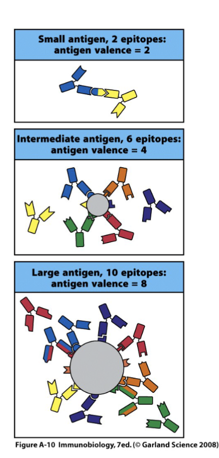
What is valence?
The number of antibody molecules that can bind to an antigen at one time defines the antigen’s valance. Similarly, the number of binding sites per antibody molecule refers to the antibody’s valence. The valence of an antibody depends on its structure class (IgG, IgA, IgM)
focus on this: antigen binding site
IgA valence of 4 bc works as dimer
IgM valence of 10
Antigen valence (or valency) refers to the number of epitope sites available on an antigen for binding with antibodies. While some antigens are monovalent, most are multivalent, having multiple, sometimes repeating, epitopes that increase binding strength (avidity) and enable cross-linking.
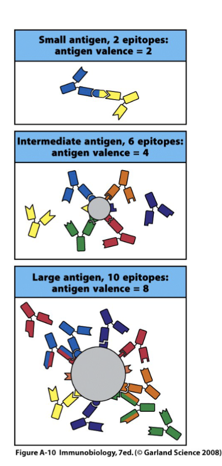
What limits the amount of antibodies that can bind to to the surface of an antigen?
Steric considerations can limit the number of different antibodies that bind to the surface of an antigen at any one time so that the number of epitopes on an antigen is always greater than or equal to its valence.
What is cross-reactivity?
antibody elicited by one antigen can cross-react with an un-related antigen due to shared epitope or very similar epitope
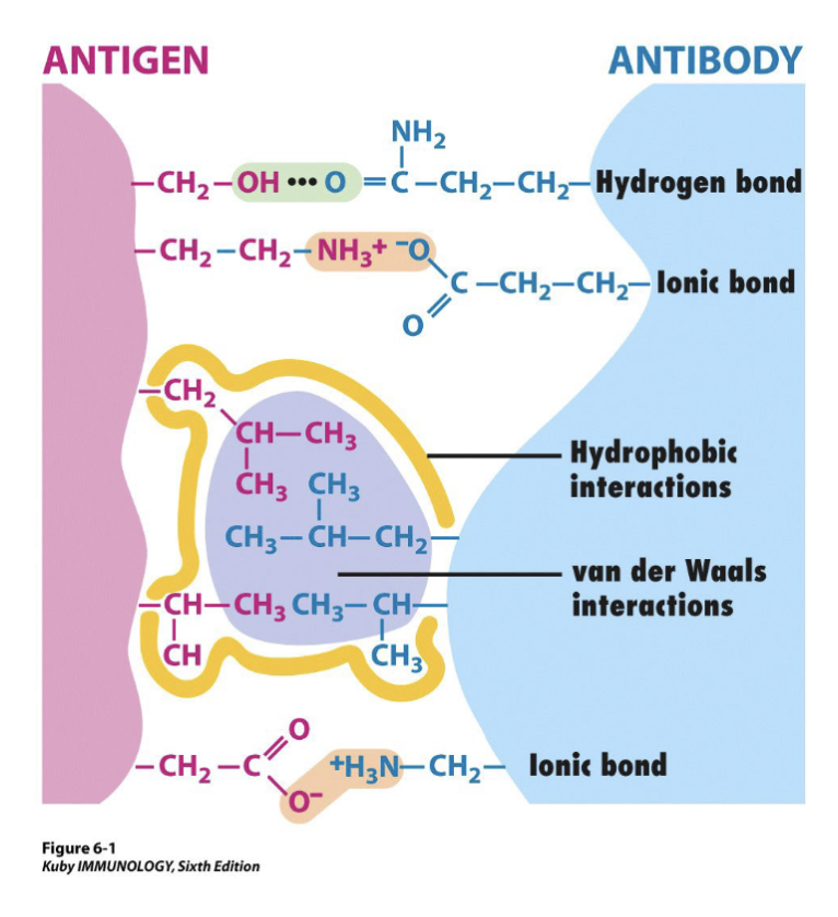
The interaction between an antibody and an antigen depends on _
Four types of non-covalent forces
noncovalent is the nature of antibody and antigen binding
doesn’t share e-
What is specificity, affinity, avidity and titer?
Specificity:determines the ability of the antibody to distinguish the immunogen from other antigens (1 out of more than 10^8 molecules)
Affinity: the strength of binding of the antibody to its antigen in terms of a single antigen-binding site binding to a monovalent antigen. Antibody affinity is a quantitative measure of binding strength
Avidity: the total binding strength of a molecule with more than one binding site
Titer: 50% of the maximum binding
What is the Conventional Method for Antibody Production?
conventional way to create antibody
is produce antigen in natural form, can buy antigens (prepared in vivo)
Ab antibody containing serum
polyclonal creates higher coverage
monoclonal more specific
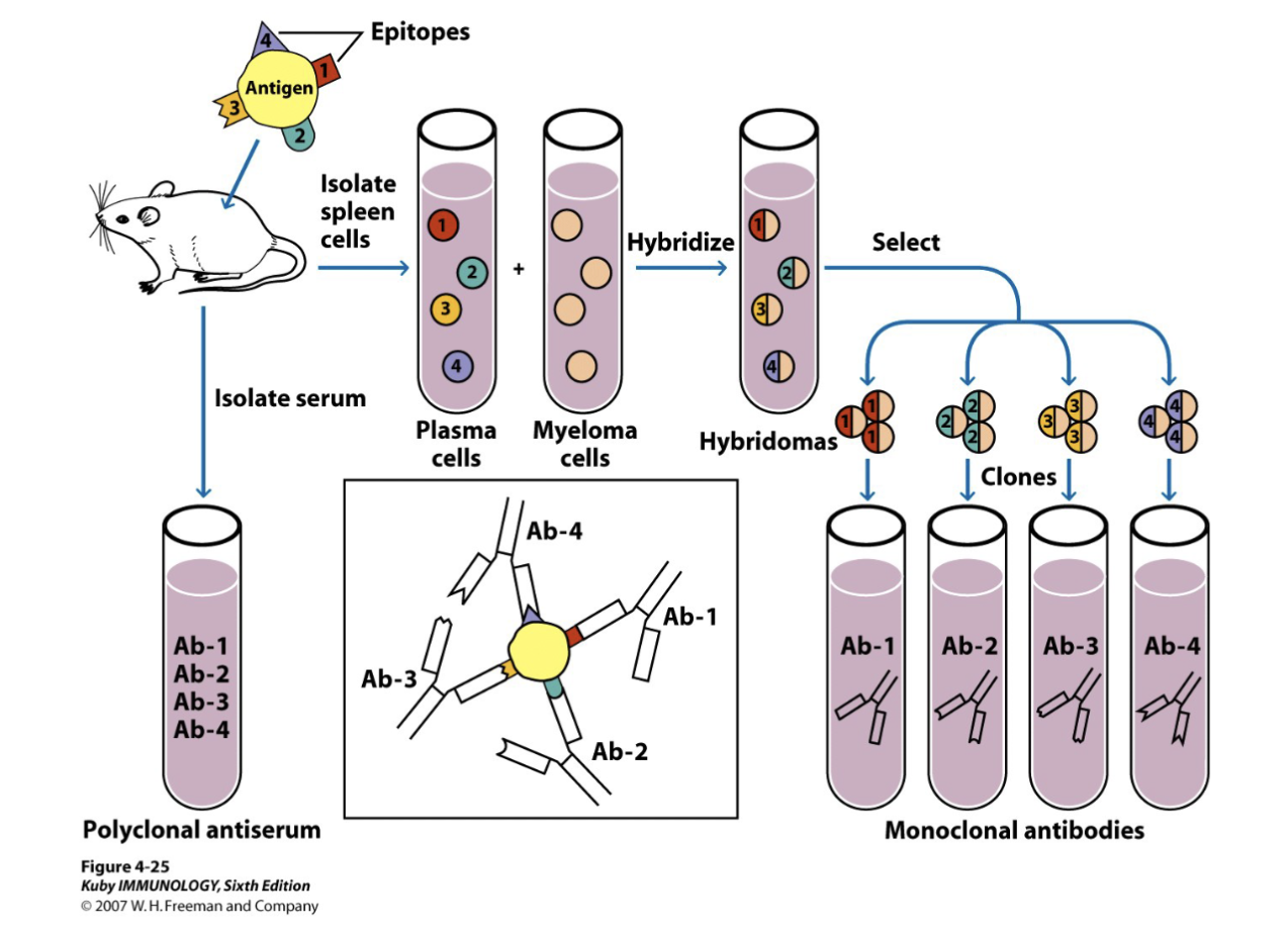
What are the features of The Production of Monoclonal Antibodies by
Hybridoma Technique?
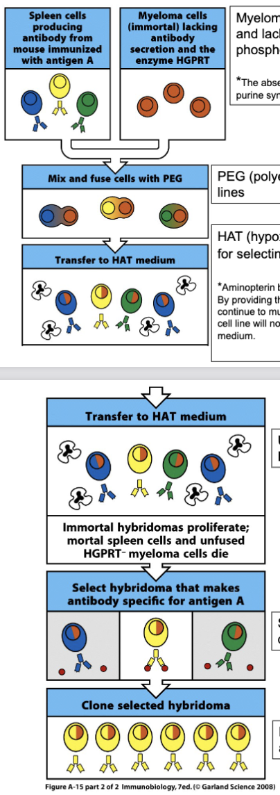
Myeloma cells are pre-selected, they do not secret Ab, and lack the enzyme HGPRT (Hypoxanthine:guanine phosphoribosyl transferase) or Thymidine kinase
-*The absence of HGPRT is not a problem for these cells unless the de novo purine synthesis pathway is also disrupted
PEG (polyethylene glycol) is used to form the hybrid cell lines
HAT (hypoxanthine-aminopterin-thymidine) medium is used for selecting the hybrid cell lines
Unfused spleen cells and myeloma cells die in HAT medium; and fused myeloma cells proliferate
Select different hybridoma cell lines by limiting dilution assay
Each hybridoma cell line only produces one antibody
-Mouse mAbs are routinely produced by hybridoma, yet, this approach is not very successful in making hmAbs
spleen cells that react to antigen and make antibody
myeloma cells (B cell + mast cell)- pre screened, cannot make antibodies themselves, naturally deficient in enzymes involved in RNA DNA synthesis
two different cell types, once the two cell cultures are put together with strong chemical PEG forces merging
primary cells (spleen and myeloma will die in the culture)
one cell put in separate well to grow culture that recognizes single epitope
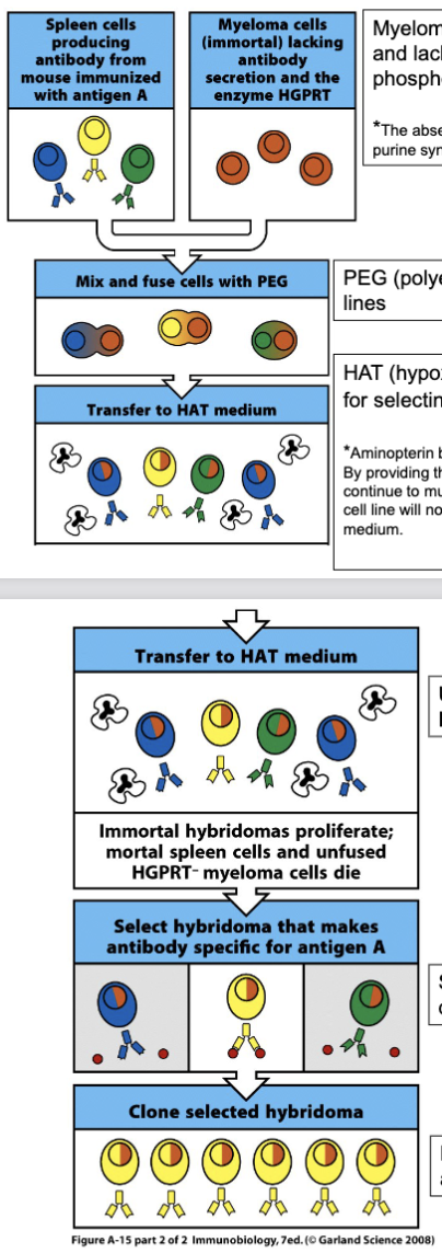
What does aminopterin do? (Production of monoclonal antibodies by hybridoma technique)
Aminopterin blocks the main biosynthetic pathways for DNA and RNA synthesis.
By providing thymidine and hypoxanthine in the growth medium, cells can usually continue to multiply by using the salvage pathway. However, a HGPRT- myeloma cell line will not grow, since the cell cannot utilize hypoxanthine supplied with the medium
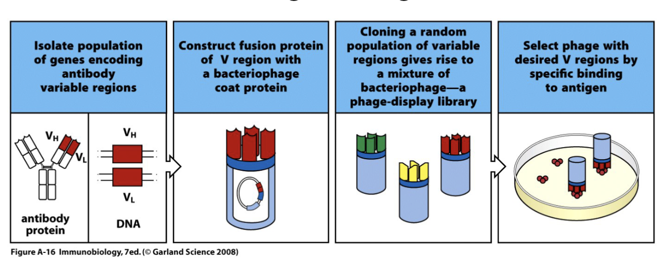
What are the features of The Production of Antibodies By Genetic Engineering?
Phage display library: a collection of recombinant phage, each displaying a
different antigen-binding domain on its surface
fuse variable region and generate a library
What needs to be determined once an antibody is created?
The affinity
antibodies can be made in bulk via Bioreactors Or Ascites Mice
to determine affinity:
equilibrium dialysis (old method)
surface plasmon resonance (new method)
What are the features of Determination of antibody affinity by traditional
equilibrium dialysis?
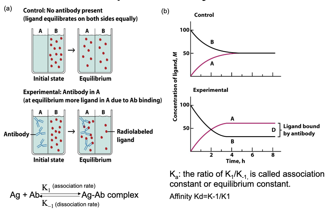
Ka : the ratio of K 1 /K-1, is called association
constant or equilibrium constant
Affinity Kd=K-1/K1
(old method)
need to understand principle
need an apparatus w/ semi permeable membrane in middle
capture radioactivity w/out antibody A and B are the same
antibody on one side will be more with A than B bc some are anchored by antibody
repetition of antibody w/ increased antibody and ligand (tedious= no longer used)
keep detecting antigen antibody speed, two constant numbers being received, association rate and the dissociation rate
they don’t bind and stick there, influenced by surrounding environment
repeating assay can determine association and dissociation rate
Kd: measures dissociation over association
What are the features of surface plasmon resonance?
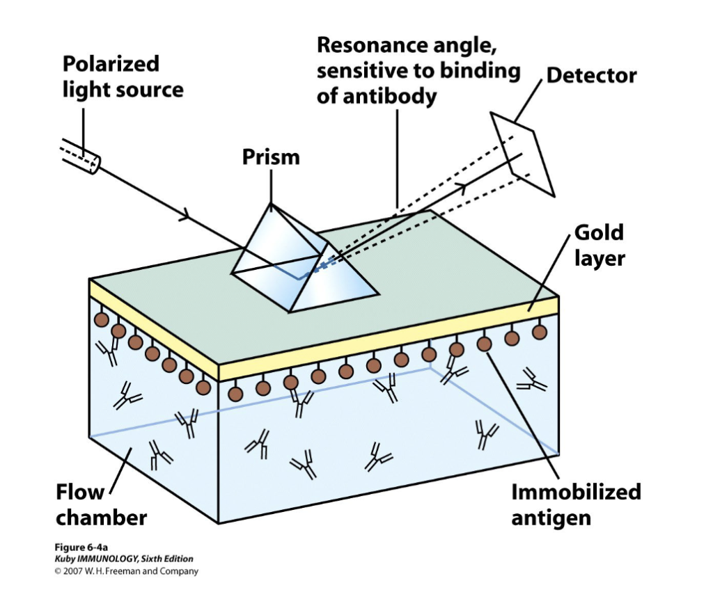
this is the main method
gold chip, immobilized antigen on the chip
the chip will be sensitive to light source and when there is more antigen it will react
change in antigen antibody binding will be detected by the machine
What are the features of the interpretation of a sensogram?
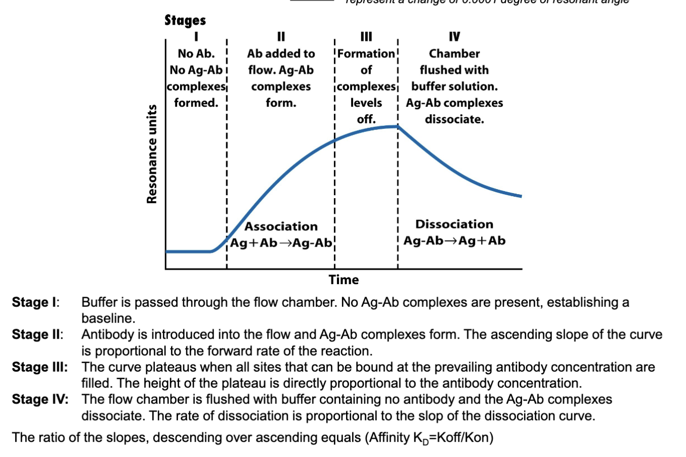
Look at image and the stages
have a baseline formed
inject antibody, more antibody injected= more Ab-Ag association
plateau forms system is separated Ab-Ag is maximized
flush chamber with buffer: some Ab (low affinity Ab will dissociate)
can create two slopes
association slope and dissociation slope
Kd (ratio of the slope) =(how quickly it dissociates/ how quickly antibody can bind to antigen) Koff/Kon
How is antibody affinity commonly expressed?
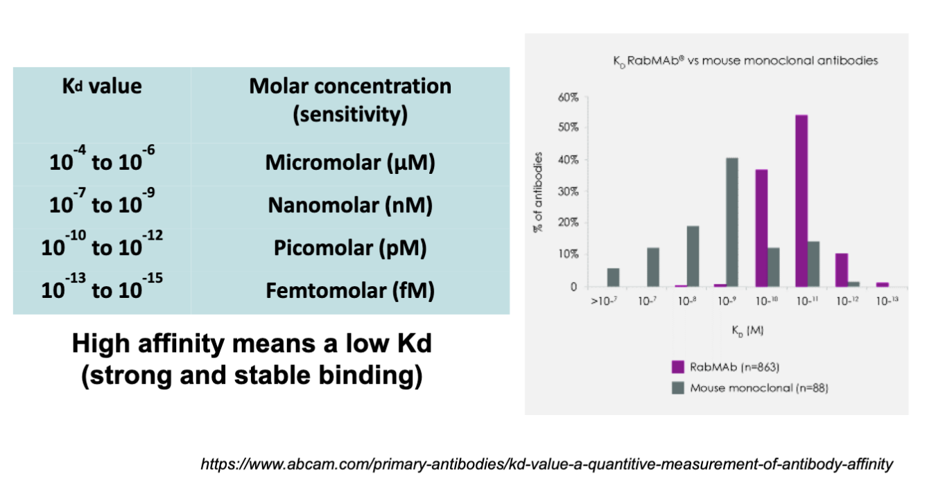
by Kd value
High affinity means a low Kd (strong and stable binding)
antibody affinity gives low number (Femtomolar, v. small= antibody high affinity, binding is strong and stable)
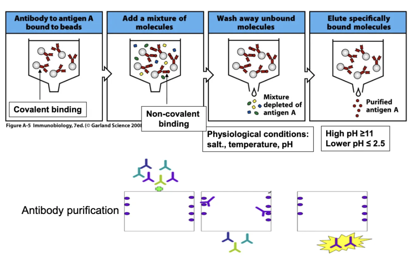
What is Affinity Chromatography?
can purify antigen or antibody
antigen bind covalently to beads, binding needs to be covalent, antigen won’t be able to separate
non-covalent binding
elute: change pH or salt concentration
non-covalent binding is explore with this method
What is ELISA?
Enzyme-linked Immunosorbent Assay
Indirect ELISA
Sandwich ELISA
Competitive ELISA
ELISPOT Assay
What enzymes are used for ELISA?
Alkaline phosphatase (AP) or horseradish peroxidase (HRP)
5-bromo, 4-chloro, 3-indolylphosphate (BCIP)-AP
p-Nitrophenyl Phosphate –AP
3,3’,5,5’-tetramethylbenzidine (TMB)-HRP
ELISA relies on two types of enzymes AP or HRP
they can catalyze a chromogenic substrate no colour→ to colour (chromogenic reaction)
know which substrate for which enzyme
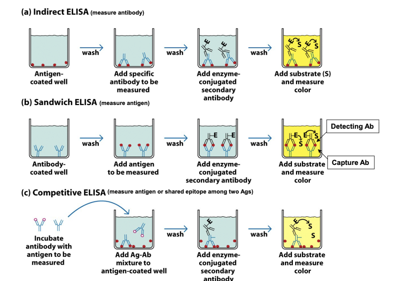
What are the features of the types of ELISA (indirect, sandwich, competitive)?
indirect: can coat the culture plate w/ antigen, add specific primary antibody, add enzyme conjugated 2nd antibody
Sandwich: use capture antibody on bottom, add Ag, add enzyme 2nd Ab
Competitive: preformed product prevents binding to coated, competition creates signal induction
What are the features of the sandwich ELISA format?
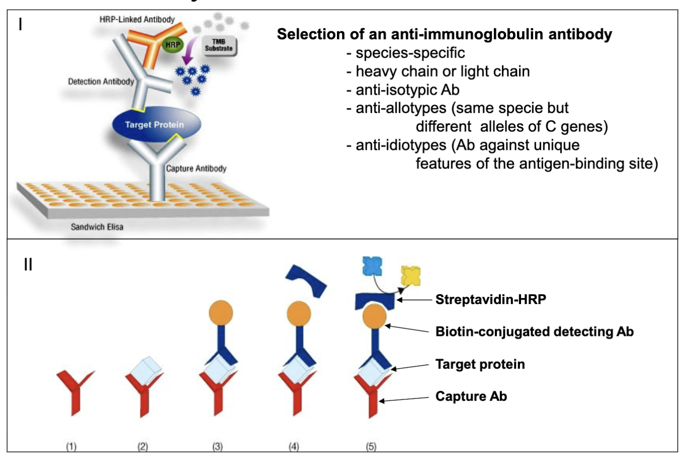
look at image details
commercial purpose not easy to conjugate
capture antibody, protein, 2nd antibody, HRP
biotin: conjugates with streptavidin-HRP (very high binding affinity)
What are the standards of the ELISPOT assay?
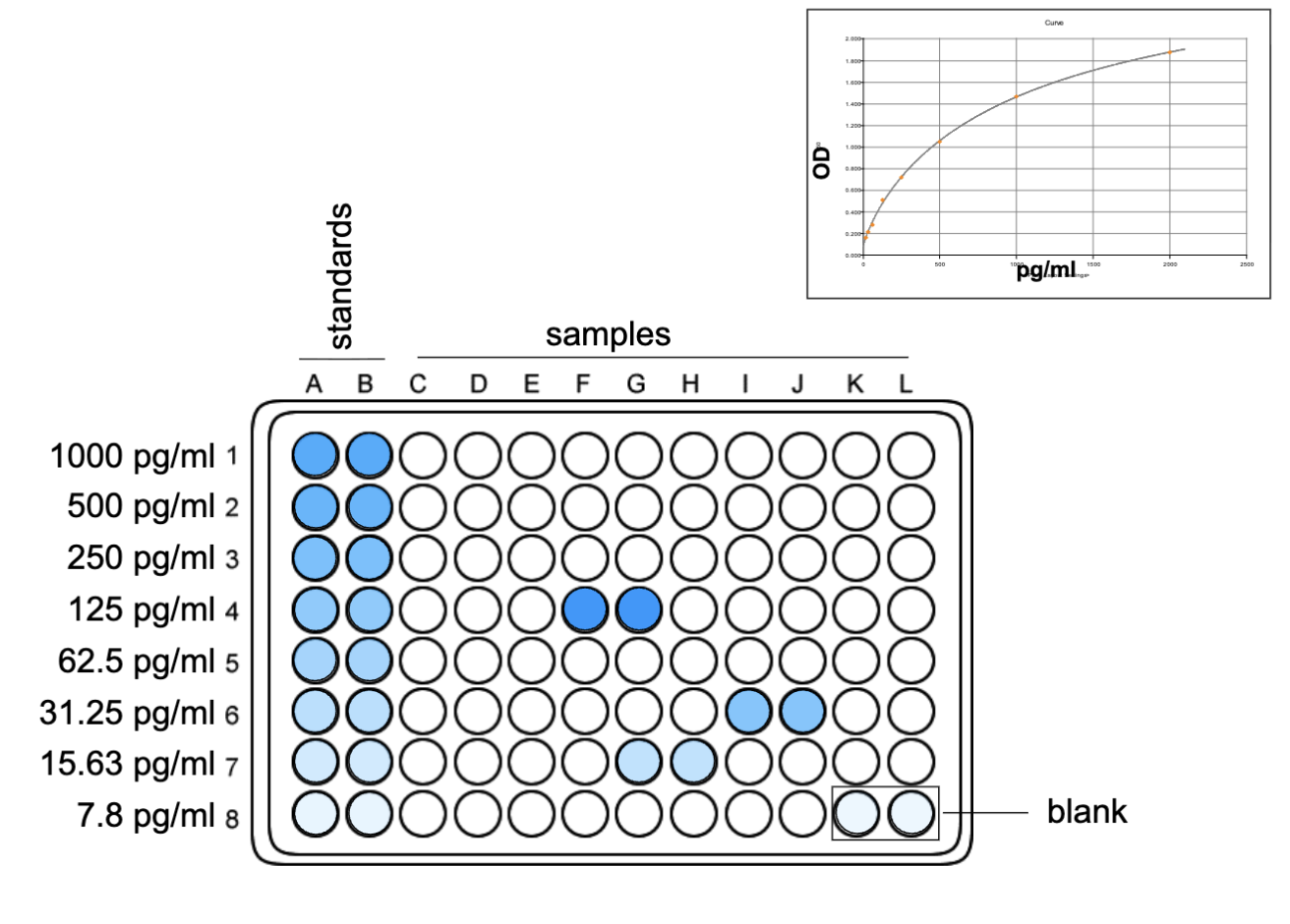
run the assay, need standards: fixed amount and blanks (gives background reading)
based on readings can generate the curve
What are the features of the ELISPOT assay?
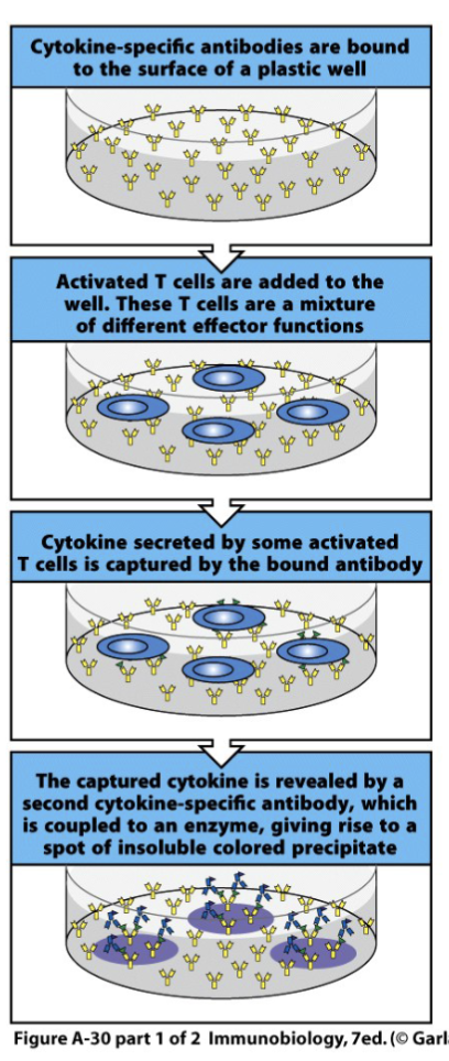
The frequency of cytokine-secreting T cells can be determined by the ELISPOT assay. Antibodies bound to a plastic surface are used to capture cytokines secreted by individual T cells.
Usually, cytokine specific antibodies are bound to the surface of a plastic tissue-culture well and the unbound antibodies are removed.
Activated T cells are then added to the well and settle onto the antibody-coated surface. If a T cell is secreting the appropriate cytokine, this will then be captured by the antibody molecules on the plate surrounding the T cell.
After a period of time the T cells are removed, and the presence of the specific cytokine is detected using an enzyme-labeled secondary antibody specific for the same cytokine.
Where this binds, a colored reaction product can be formed. Each T cell that originally secreted cytokine gives rise to a single spot of color, hence the name of the assay.
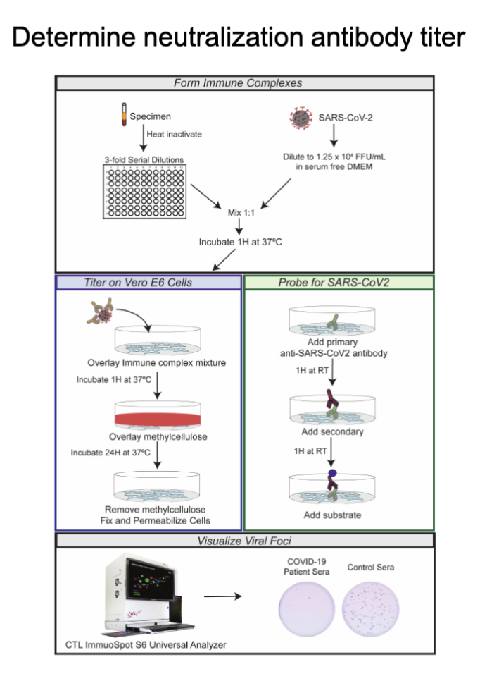
How do you determine the neutralizing antibody titer? What is the neutralizing antibody titer?
A neutralization antibody titer is a blood test measuring the concentration of antibodies that can block a virus or pathogen from infecting your cells, indicating your level of immunity from past infection or vaccination, showing protection, and guiding vaccine booster needs. It quantifies how effectively these specific antibodies can neutralize, or stop, the germ from causing disease, with higher titers generally meaning stronger protection
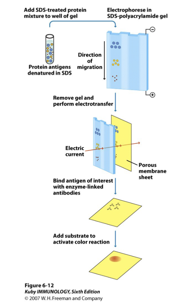
What are the features of Western Blotting?
antigen separated based on size
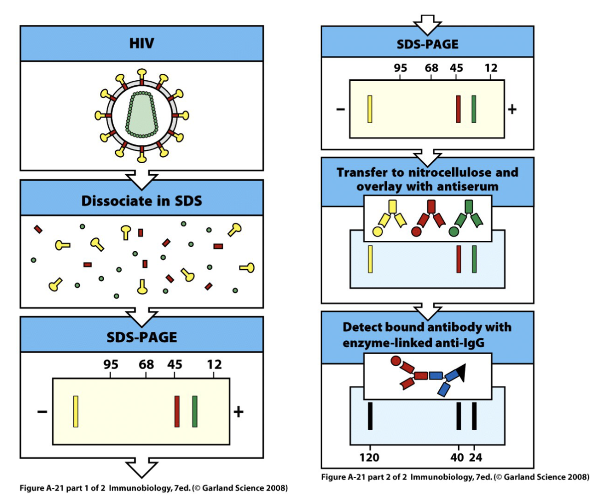
How can western blotting be used with virus and antibodies?
can determine if the antibodies bind to part of dissociated virus the bands would appear in a different location if bound
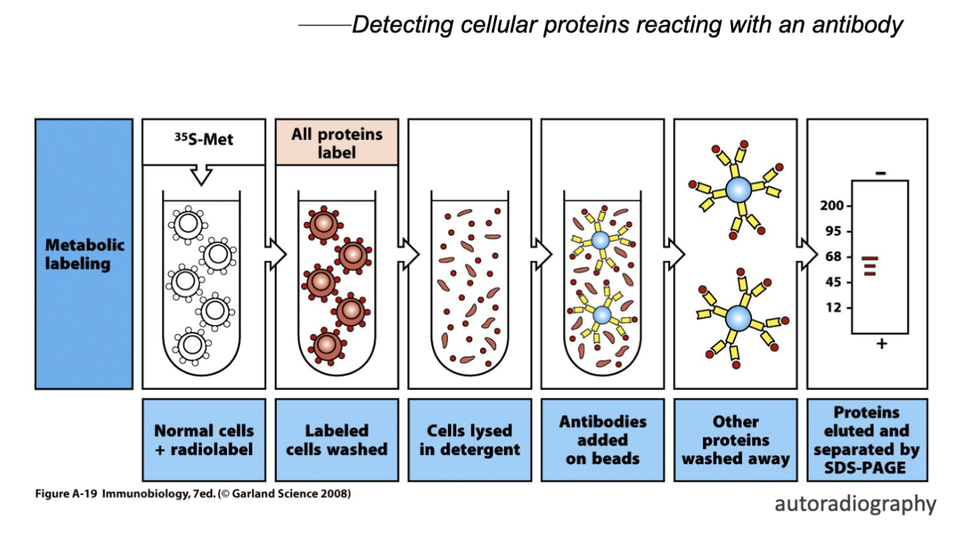
What are the features of Immunoprecipitation and Co- Immunoprecipitation?
visualize protein
detecting cellular proteins reading with an antibody
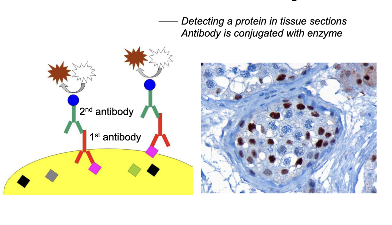
What are the features of immunohistochemistry?
Detecting a protein in tissue sections antibody and conjugate enzyme
don’t need to separate via western blot can generate tissue section and probe antigen w/ particular antibody
can see antibody expression in anatomical location (membrane, cytoplasm, ER, …)
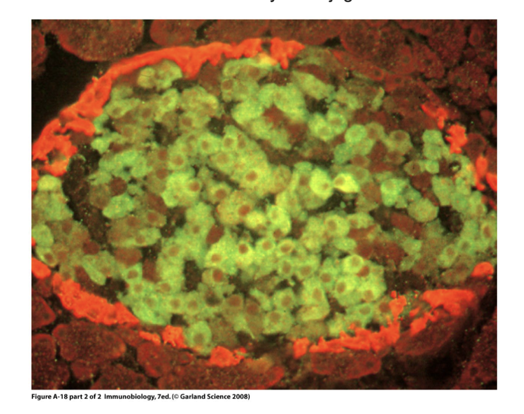
What are the features of immunofluorescent staining?
Antibody is conjugated with a fluorescent probe
change detection method but everything dependent on antibody antigen interaction, can conjugate the antibody w/ fluorochrome
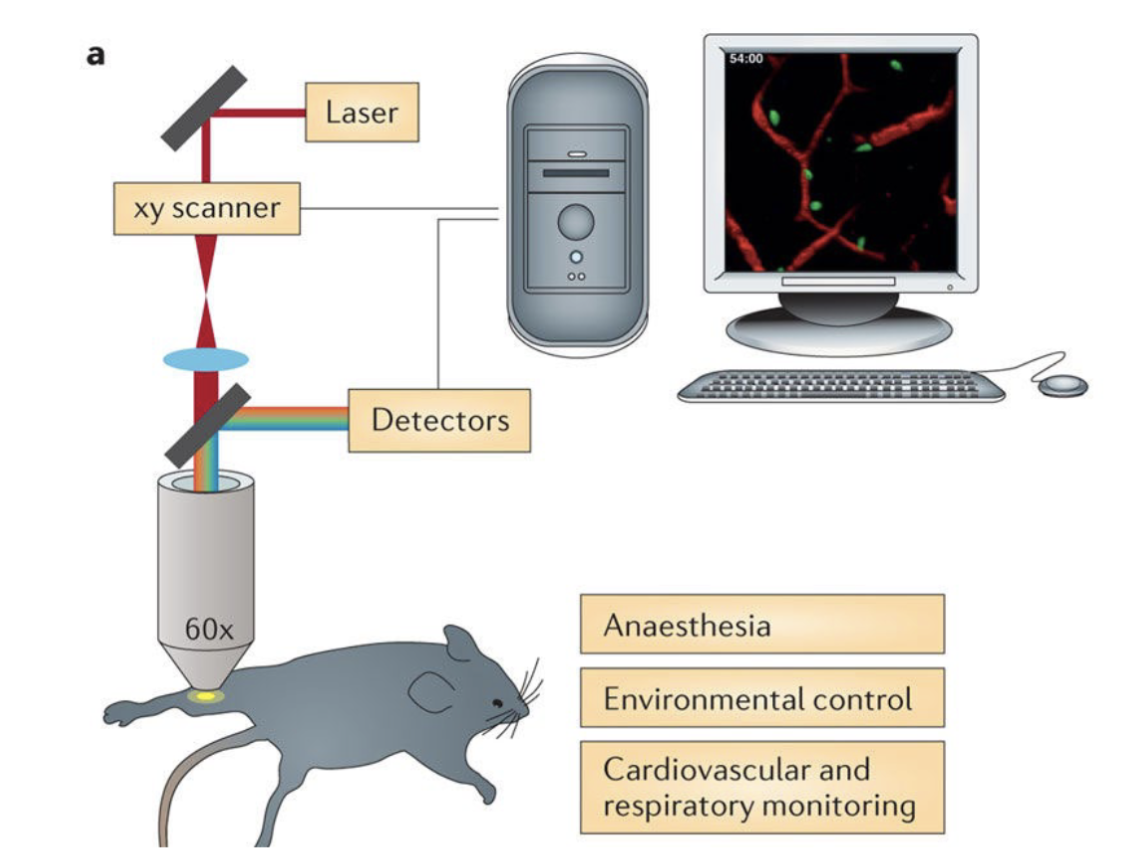
What are the features of multiphoton intravital microscopy?
can follow the molecule or cell type change in live animal
produces real time observations
What does bacterial agglutination diagnose?
Infection
when a spot appears= infection
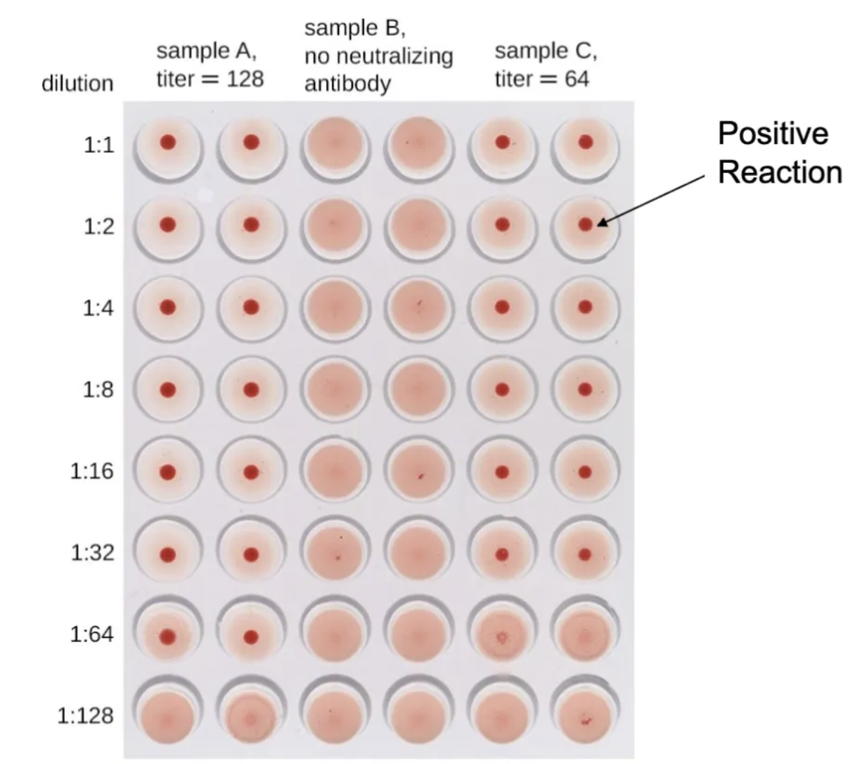
What are the features of Hemagglutination and blood typing?
Based on the ability of antibody binding to alter the physical state of the antigen it binds to
O type no A antigen
antigens are sugars (genetic predetermined) highly homologous to bacteria in gut flora
if B antigen is not self will create antibody
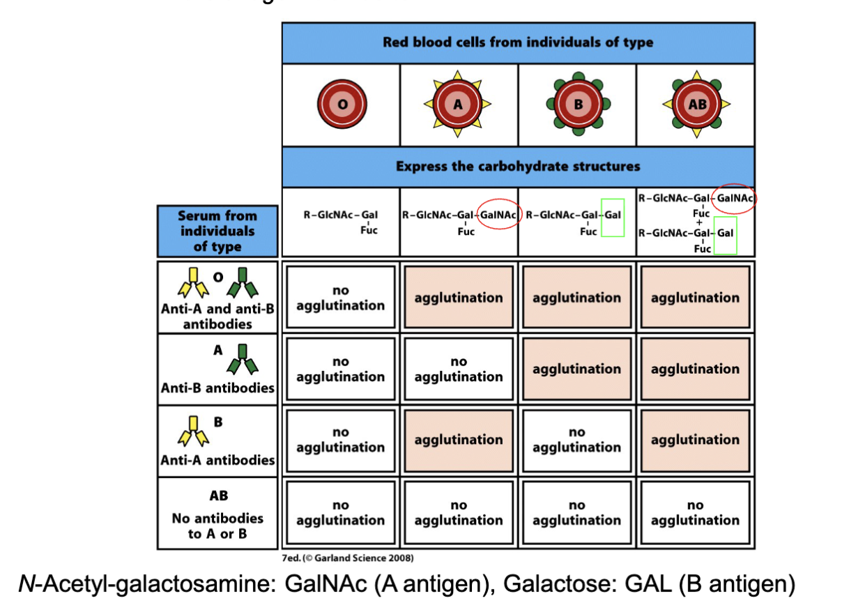
What are the features of a Hemagglutination Inhibition Reaction?
Used to detect the presence of viruses and of the antiviral antibodies
virus expresses surface molecule hemagglutinin and binds to sialic acid on RBC
mix them: creates aggregate (hemagglutination)
can use it to test of serum sample contains antibody, it will bind to virus and prevent formation
can be used for other viral infections but need to modify reagent
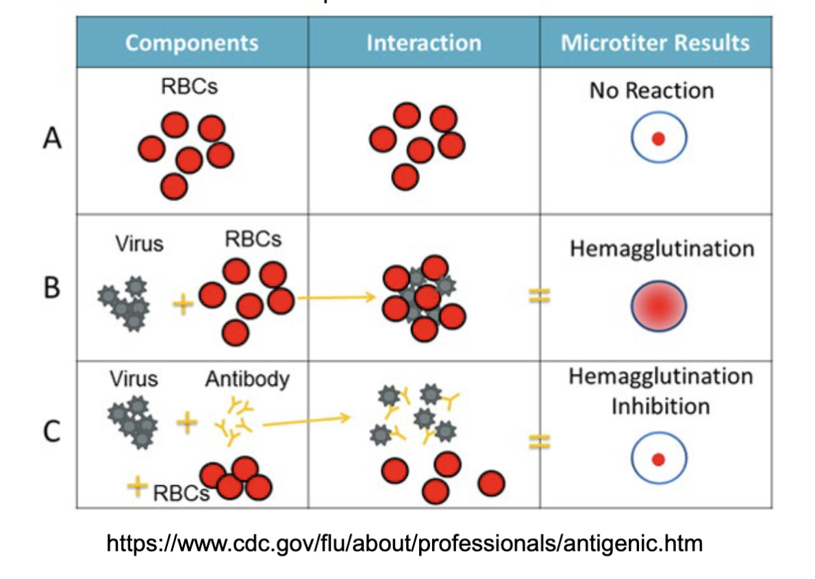
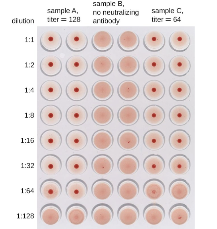
What are the results of this test?
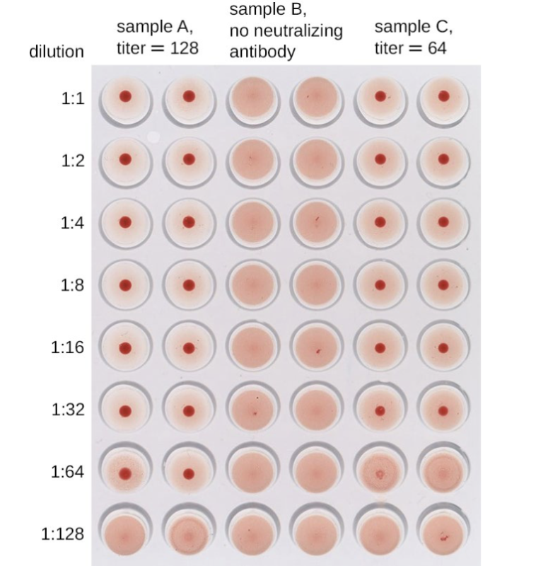
In this HIA, serum containing antibodies to influenzavirus underwent serial two-fold dilutions in a microtiter plate.
Red blood cells were then added to the wells. Agglutination only occurred in those wells where the antibodies were too dilute to neutralize the virus. The highest concentration at which agglutination occurs is the titer of the antibodies in the patient’s serum. In the case of this test, Sample A shows a titer of 128, and Sample C shows a titer of 64. (credit: modification of work by Evan Burkala)
What is Antigenic characterization?
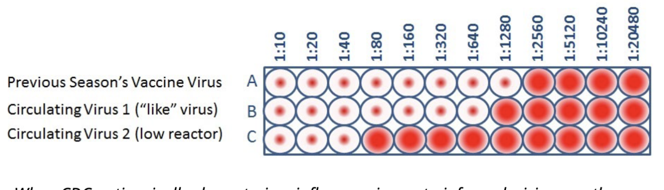
When CDC antigenically characterizes influenza viruses to inform decisions on the formulation of the seasonal flu vaccine, the HI test is used to compare currently circulating viruses (B&C) with vaccine viruses (A). This allows scientists to quickly determine if a virus used in the seasonal flu vaccine is antigenically similar to circulating influenza viruses and therefore capable of producing an immune response against them
test if the vaccine will match to circulating virus
use this assay to test
if full red circles not really working
What is the sensitivity of various immunoassays?
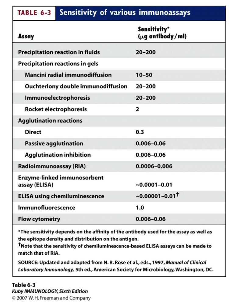
flow cytometry is quite accurate
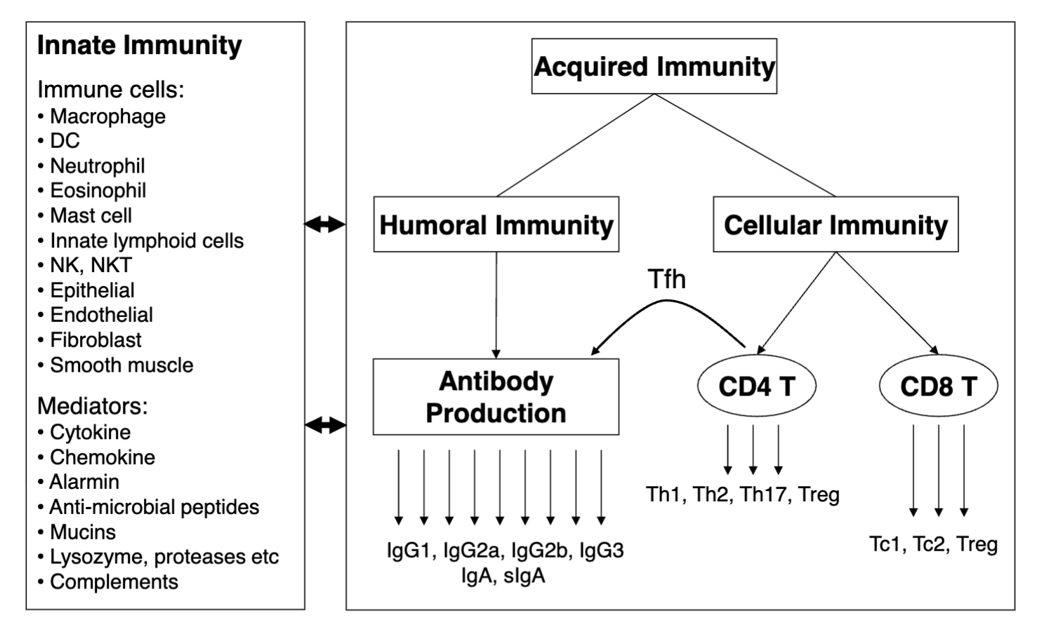
What is acquired vs innate immunity?
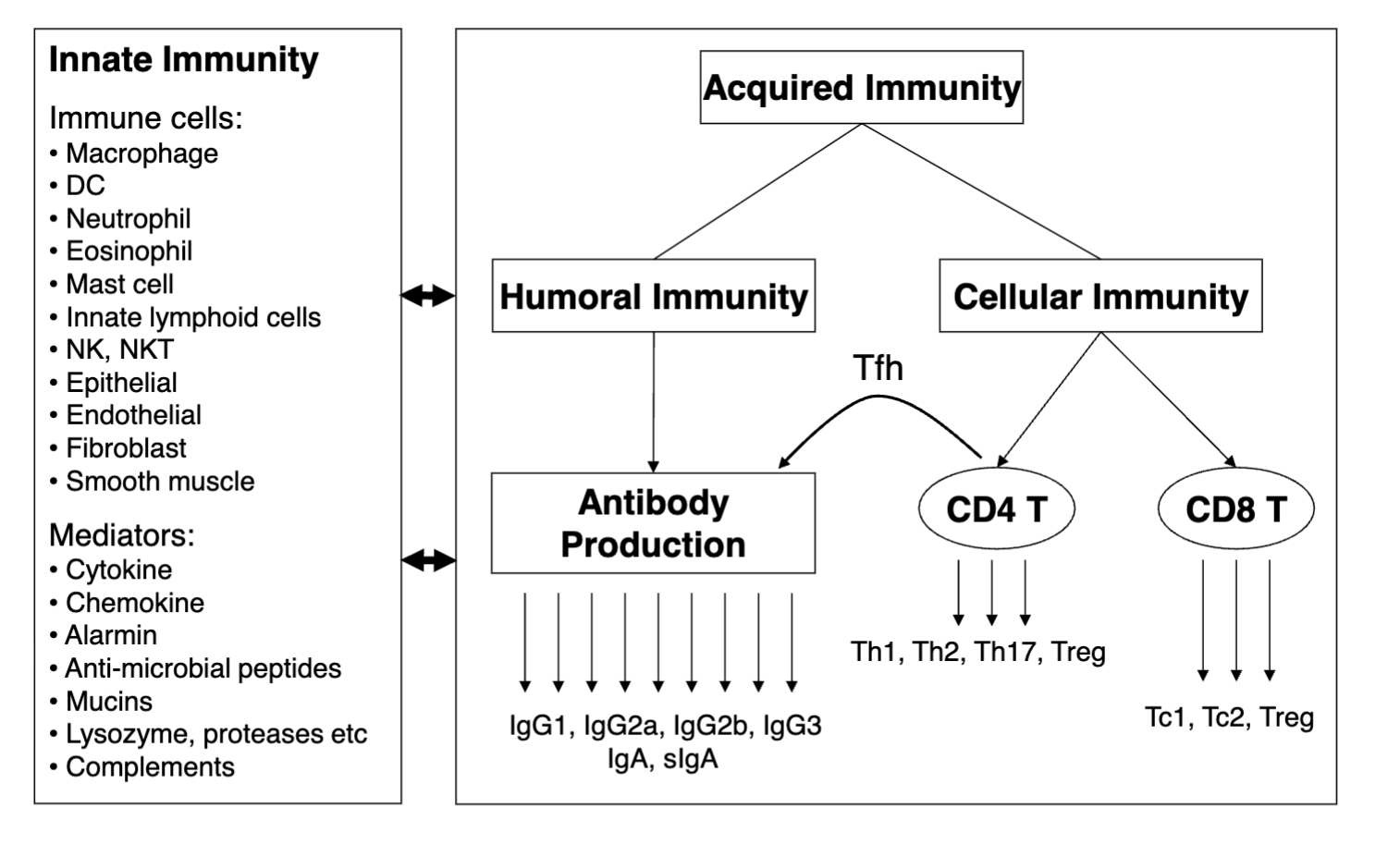
acquired: T and B cells (develops over time) slower reaction
innate: fast reaction immune cells and mediators
What are the immune cells of innate immunity?
• Macrophage
• DC
• Neutrophil
• Eosinophil
• Mast cell
• Innate lymphoid cells
• NK, NKT
• Epithelial
• Endothelial
• Fibroblast
• Smooth muscle
What are the mediators of innate immunity?
• Cytokine
• Chemokine
• Alarmin
• Anti-microbial peptides
• Mucins
• Lysozyme, proteases etc
• Complements
What are the two types of acquire immunity?
Humoral (B) and cellular (T)
What are the general features/ what is Flow Cytometry And Fluorescence-activated
Cell Sorter (FACS) Analysis?
FACS : fluorescence-activated cell sorting
Flow cytometry is a laser-based, biophysical technology
employed in cell counting, cell sorting, biomarker detection
by suspending cells in a stream of fluid and passing them
by an electronic detection apparatus. It is used to study
cells as they move in fluid suspension, allowing multiple
measurements to be made per cell
What are the systems of Flow Cytometry And Fluorescence-activated Cell Sorter (FACS) Analysis?
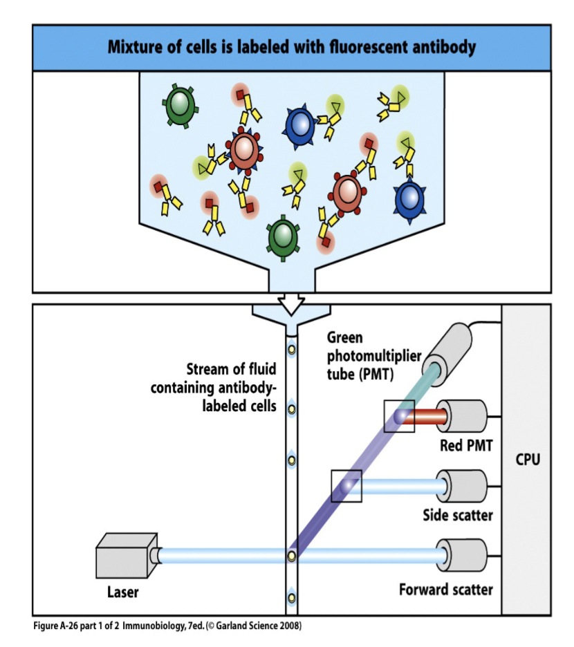
A flow cytometer is made up of three main systems: fluidics, optics, and electronics
The fluidics system transports particles in a stream to the laser beam for interrogation.
The optics system consists of lasers to illuminate the particles in the sample stream and optical filters to direct the resulting light signals to the appropriate detectors called photomultiplier tubes (PMTs).
The electronics system converts the detected light signals into electronic signals that can be processed by the computer (CPU).
every drop one cell →liquid handling system
laser: when liquid passes through it will be activated by laser bc labelled by fluorescent marker = emit signals and collected by the PMTs (different for different colours) and is converted by digital signal for the computer
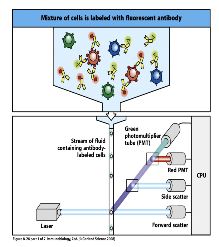
What is the optical layout that FACS creates?
creates graph of the colour wavelengths, machine will only collect a fraction of each signal (fraction controlled by filter to tell the machine which signal to collect needed to separate diff. signals that overlap)
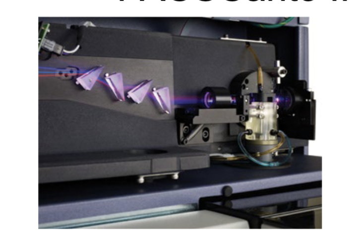
What are the features of the FACSCanto II machine?
The optics of the BD FACSCanto II system consist of an excitation source with
up to three lasers:
• blue (488-nm, 20-mW solid state),
• red (633-nm, 17-mW HeNe),
• violet (405-nm, 30-mW solid state).
FACSCanto II is able to detect 8 parameters simultaneously.
What are the features of the BD LSR Fortessa machine?
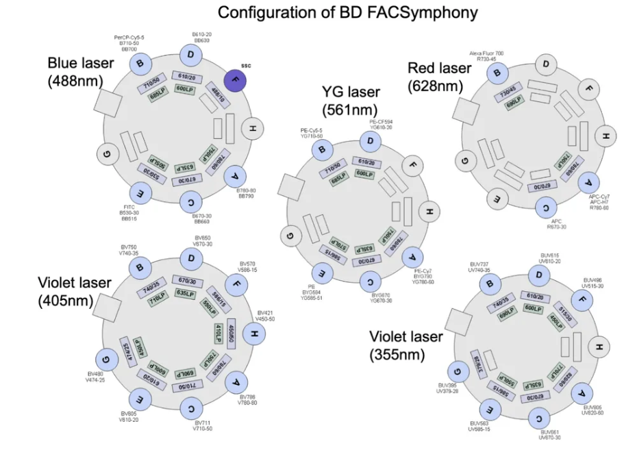
The Fortessa is a 4 laser flow cytometer capable of detecting up to 18 fluorescence parameters simultaneously with state of the art optics to ensure maximal excitation, and collection of signals.
• 50mW 488nm (blue),
• 50mW 532nM (green),
• 200mW 628nm (red),
• 100mW 402nm (violet) laser.
blue laser: different lasers in different filters in the different channels
What measurement can be made with FACS?
Forward light scatter (FSC): proportional to cell size (measurement of mostly diffracted light)
Side light scatter (SSC): proportional to cell granularity (measurement of mostly refracted and reflected light)
Fluorescent light:
– Binding of fluorescent-labeled antibodies
– Ca ++-sensitive dyes within cells
– Fluorescent proteins expressed by cells
– Binding of DNA dyes
– Cellular protein binding dyes (fixable viability dye)
NK can be labelled w/ Ca+ bc they utilise this element
DNA dyes mostly when cell is dead
What needs to be compatible with the fluorochrome in FACS?
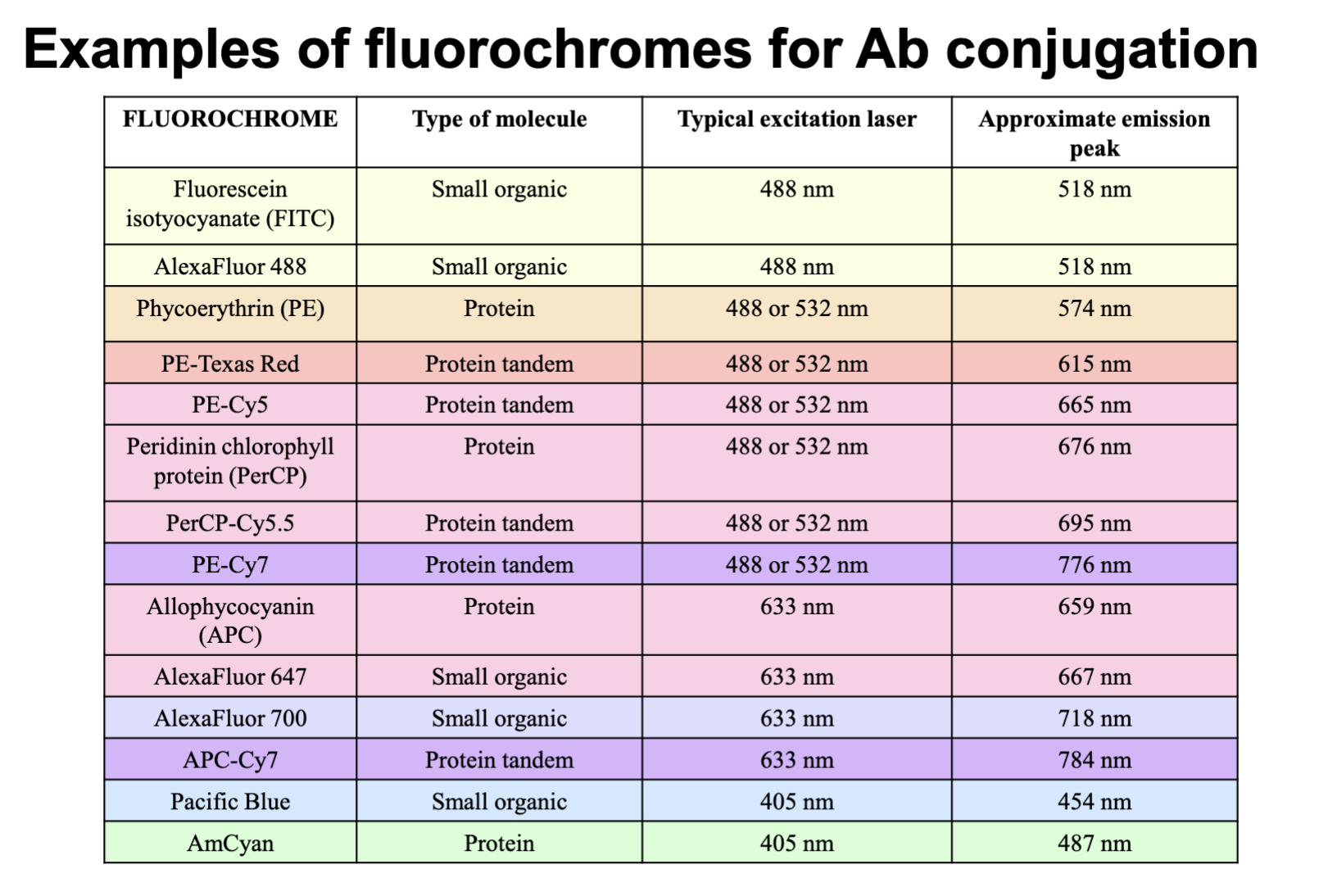
Antibody conjugation
important parameters: every antibody look for the excitation laser (one dose of fluorochrome will be activated at that point) need compatibility of laser and fluorochrome. the emission peak number identified for PMT (which channel it goes to)
What are some issues (outliers that need to be corrected) with FACS method?
All cells have a certain level of background fluorescence, due to:
– Autofluroescence: from pigments and fluorescent moieties on cellular proteins
– Non-specifically bound antibodies, and free antibody in the sample stream
The level of autofluorescence varies with the wavelength of excitation and collection:
– Highest in FITC, PE detectors; lowest in far red (APC, Cy7) detectors
most challenging part is the background and spillover
spectrum, trie to connect only peak signal there is level of spillover
need to titrate antibody make sure there isn’t too much
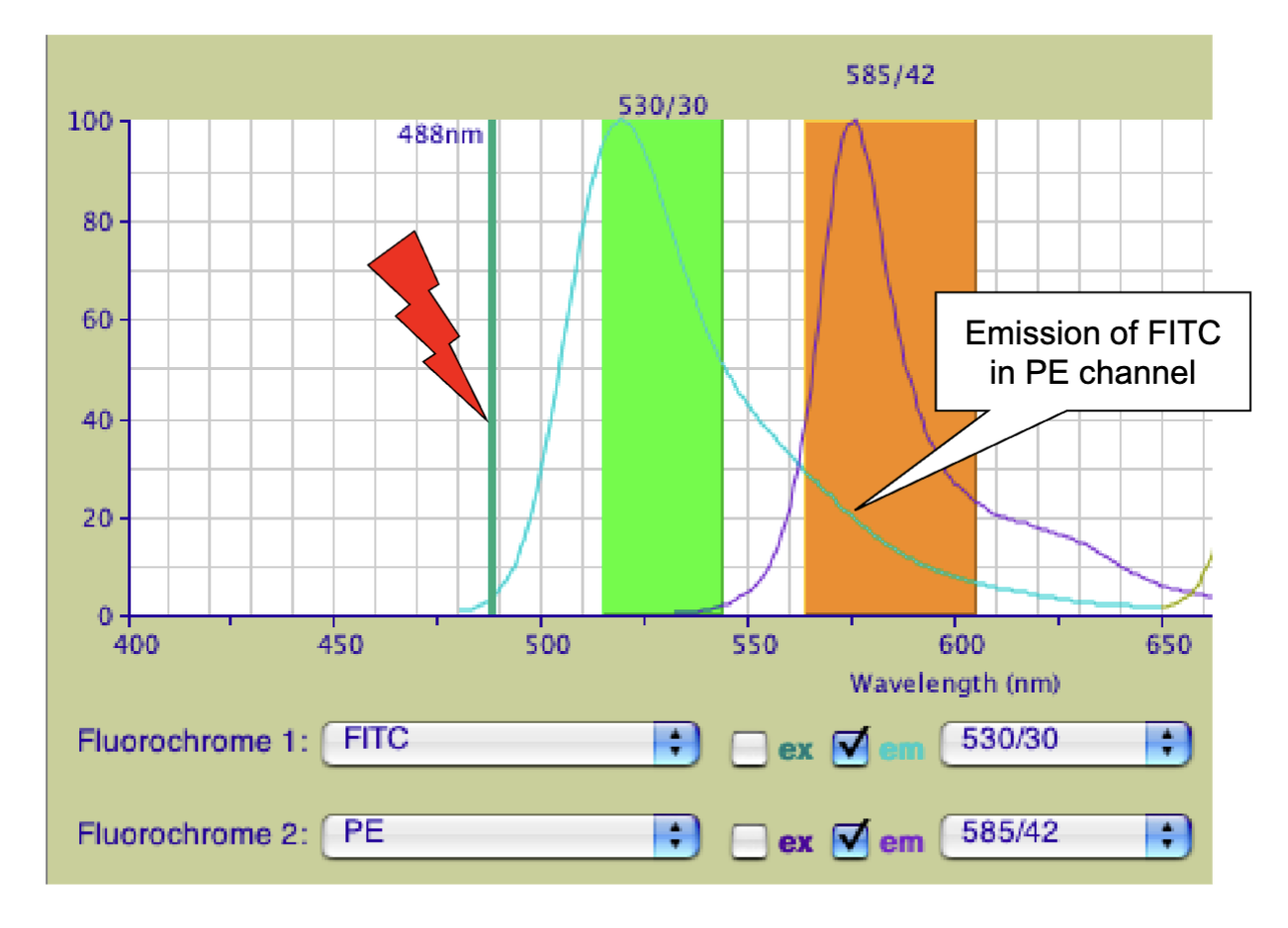
What are the features of fluorescent spillover?
ex: 2 antibody and both are activated by 488 laser
peak level proportional to amount of antibody used want to reduce the peak level to the extent of control spillover into neighbouring channel (blue goes into orange)
white arrow= spillover point
can control spillover w/ amount of antibody and the laser/ voltage
most important control is the titration of antibody (want the minimum sufficient amount less is better) needs high affinity antibody to reduce amount
What are the features of FACS Staining Setup?
• Un-stained samples
• For multiple-color FACS staining, make sure to also have single color staining samples for machine compensation
• Fluorescent-minus-one (FMO) controls for detecting the background signals.
• Cocktail with antibodies that are conjugated with different fluorescent dyes.
4 tubes needed, need un-stained (control, autofluorescences of the cell) regular population has a signal,
need single colour control: used for machine compensation (need it to set up machine and understand spillover when using more than one colour)
FMO control to test and make sure there is no signal for specific fluorophore if it shows up means wrongly calibrated
What are the Parameters of FACS Analysis (when plotted on graph)?
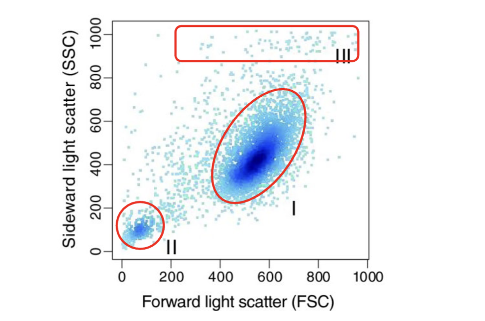
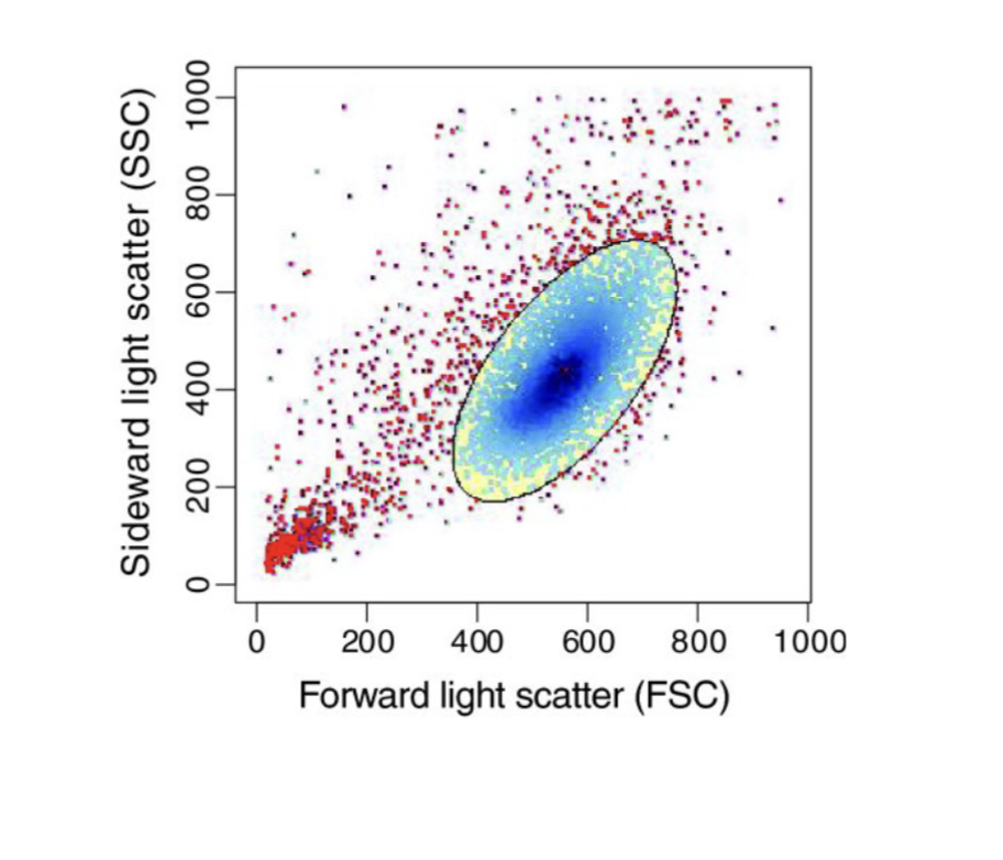
Scatterplot of FACS data showing typical properties of morphologic parameters.
FSC corresponds to cell size and SSC to cell granularity.
Several subpopulations can be distinguished: (I) healthy and well measured cells, (II) cell debris, and (III) cell conjugates and air bubbles.
when looking at file look at forward scatter and side scatter -need to make sure voltage set ups is correct to see the signals
healthy cells in the large dark blue region
low scatter= cellular debris bottom left (some RBCs)
air bubbles (try to avoid) or cells stuck together can appear here
What is gating in FACS?
interested cell population can be specifically selected by gating
step 1 gating cells of interest (look at interested population & program discards remaining cells)
Gating in flow cytometry (FACS) is the process of selecting specific subsets of cells from a larger, heterogeneous population for detailed analysis. By drawing boundaries on 2D plots (using scatter or fluorescence parameters), researchers isolate populations of interest—such as single, viable, or antibody-stained cells—while excluding debris, dead cells, and doublets
What are the basics of doublet discrimination?
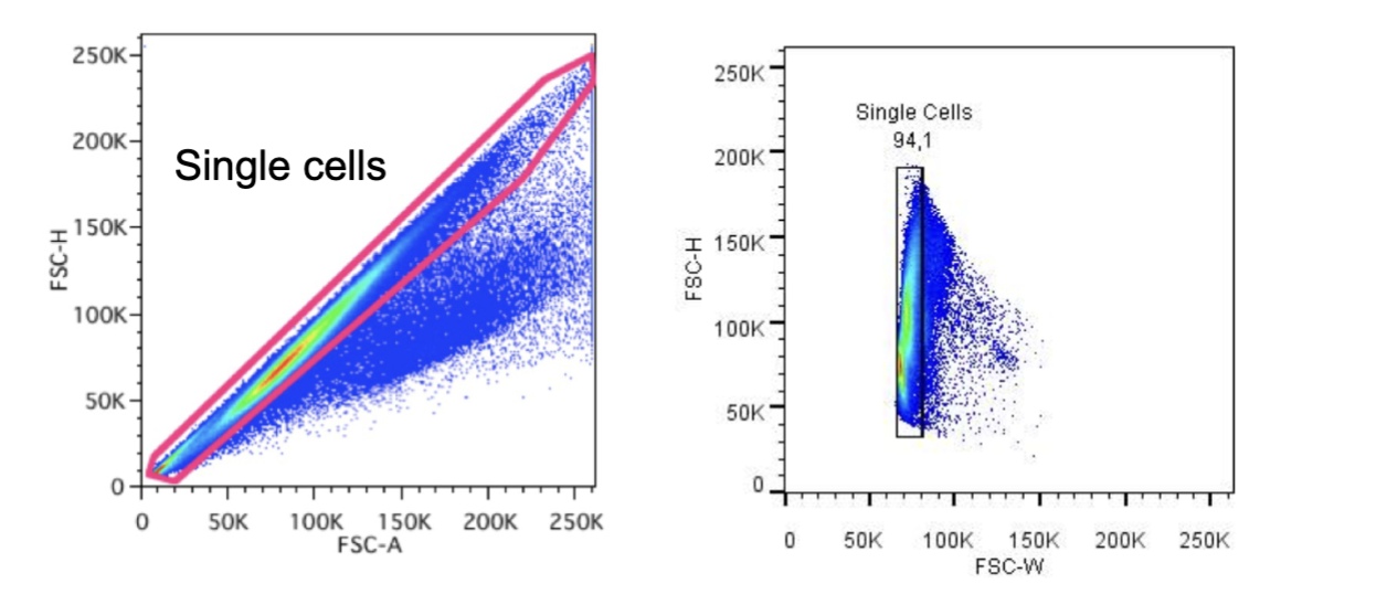
is to detect disproportions between cell size vs. cell signal
Height (H), cell's maximum signal, is directly impact by any changes in PMT voltage;
Width (W), represents time duration of the signal, not impacted by PMT voltage, directly correlates with cell size;
Area (A), calculated from all H measurements during the time (W) the cell passes through the laser beam.
The most common one uses FSC-A vs. FSC-H. But you also can use FSC-H vs FSC-W and SSC-H vs. SSC-W
Forward scatter height and area on same cells, most of them should stay in position, if doublet height and area changes
this method locates single cells first
How are live and dead cells distinguished from another in FACs?
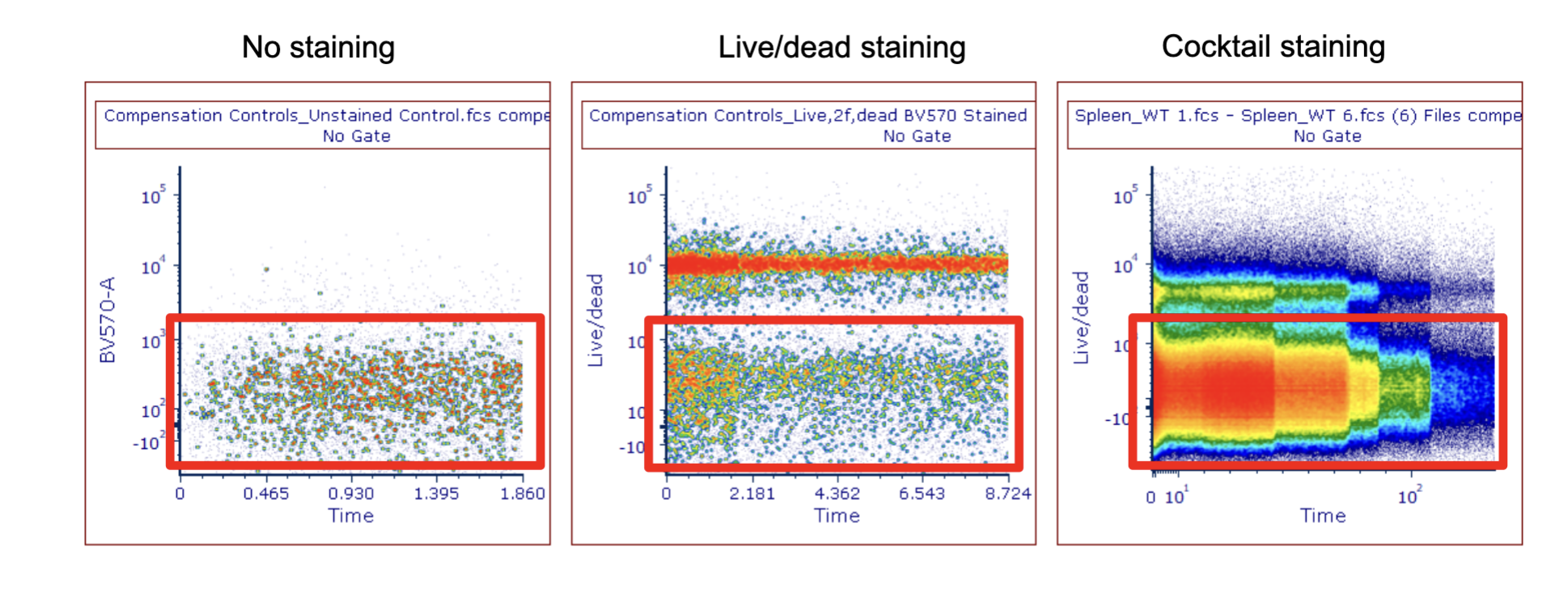
Distinguish live/dead cells using positive and negative controls
remove dead cells from analysis (tend to be sticky and non-specifically stick to antibody)
understand where the gait to look at
no staining: what the background signal are
top middle: cells w/ high fluorescent intensity= dead
remove the dead cells and focus on the live cells
What is the process of sequential gating strategy in FACs?
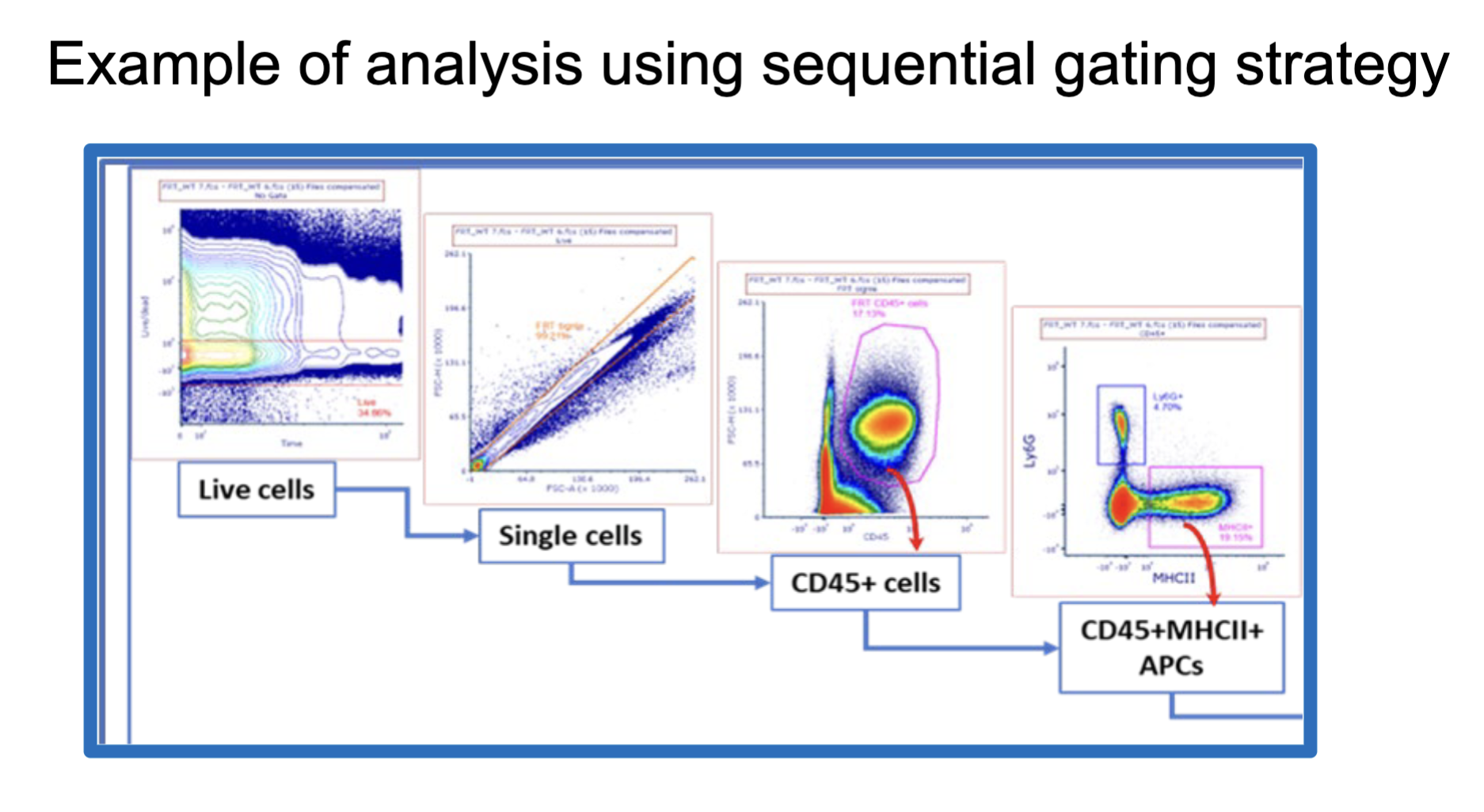
can do live cells or single cells first
fluorescent marker= CD45+ (marker for all leukocytes all of them express it)
What are the features of two colored plots for FACs analysis?
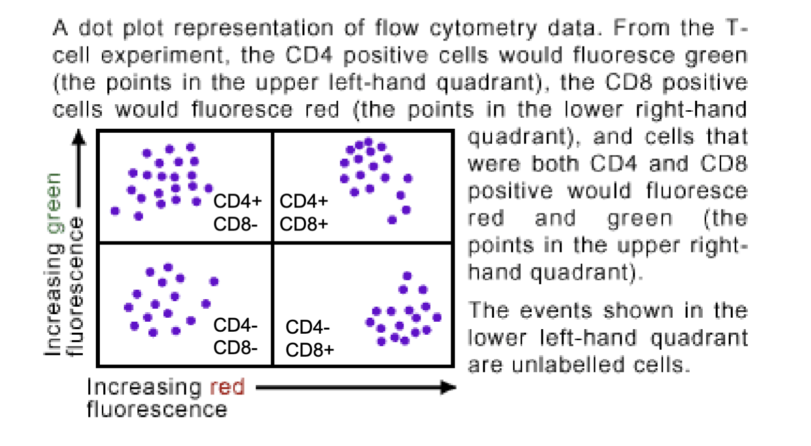
when looking at data might be presented with two fluorescent signal intensities
when the cells are found in these positions need to understand the meaning
Red= CD8 marker
Green= CD4 marker
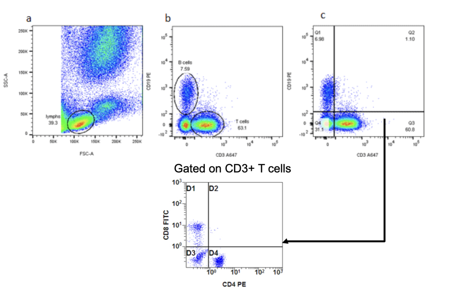
What is the process of two coloured plots and gating?
top left: start gaiting then look at CD3 and CD19 then set the gait for specific population then CD4 and CD8 cells
interpretation doesn’t change
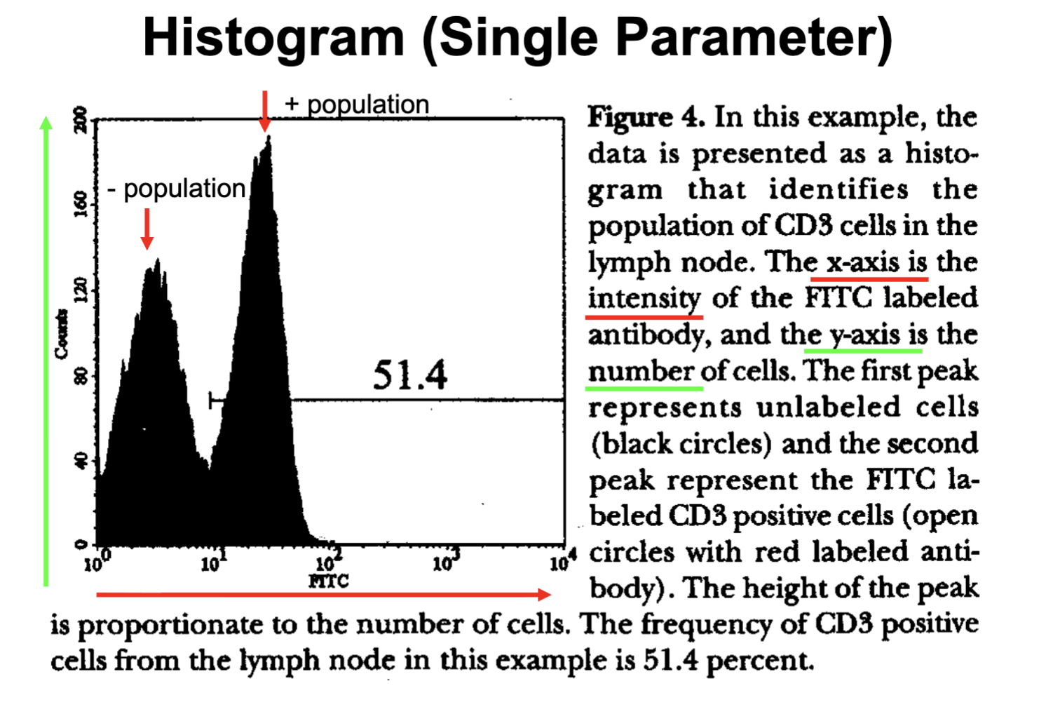
What are the features of histogram plots for FACs data analysis?
x axis identical to 2 color plot= intensity
y axis is different it is the number of cells
have to draw plot in midterm (maybe not this way but indicate the double or single positive point)
What are in-vivo models that are used in FACs?
each mouse has unique traits and immune characteristics, that might be suitable for certain types of research common used are BALB/C
C57BL/6
BALB/C C57BL/6 often used for infections
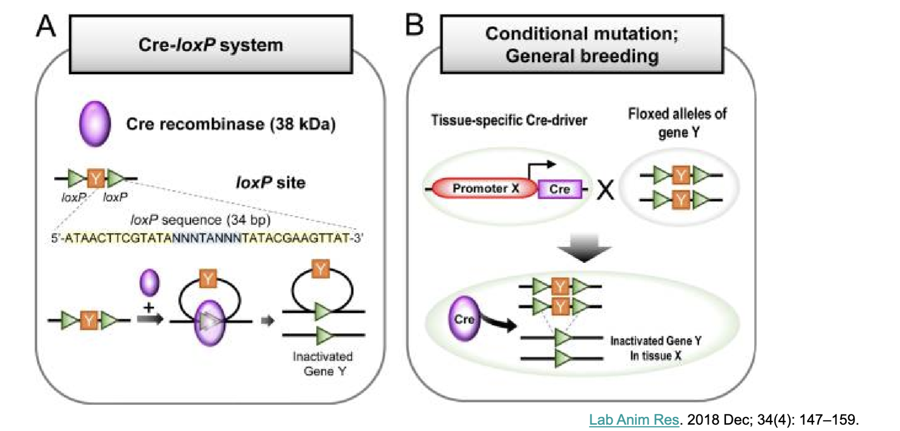
What is the Cre-LoxP system? What is it used for?
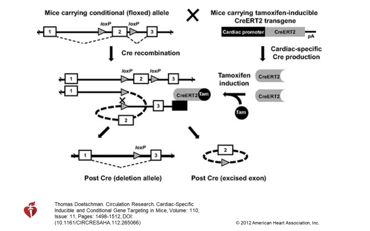
Cre-LoxP system to generate tissue-specific or cell-specific gene knockout mice
Cre-Lox system discovered
before using whole body knockout mice not give enough information
but examining gene deletion in a type of cell type, liver, CD4 cells, heart, … understand the function better
A:
Cre-lox Cre recombinase (bacteria protein) generate mouse and over express Cre which recognizes specific sequence the loxP and catalyse recombination if Y gene (the flanked region gene will be deleted bc of Cre activity)
B:
other strands insert Lox P site and flank the gene for it to be removed, generate Cre in cell and cause gene deletion in that particular cell type of tissue but other parts are not affected= precise examination how that gene is expressed by certain cells ex: CD4 cell
What are the features of Tam-Inducible conditional gene targeting?
Control Cre expression, common construction is the promoter is connected to construct estrogen receptor allows cells responding to tamoxifen and bind to ERT2 to stimulate Cre expression (can control Cre expression at different time points- can delete certain oncogenes at different age )
What are the features of Hu-PBL-scid model?
injecting human PBMCs into immune deficient mice (quick and easy, mainly on T cell memory response)
SCID mouse human like model
What are the features of Hu-SRC-scid model?
Engrafting CD34+ human hematopoietic stem cells into immune deficient mice. The cells are obtained from human fetal liver, bone marrow or from the umbilical cord. (multilineage development of hematopoietic cells, naïve immune system)
SCID mouse human like model
What are the features of BLT (bone marrow/liver/thymus) model?
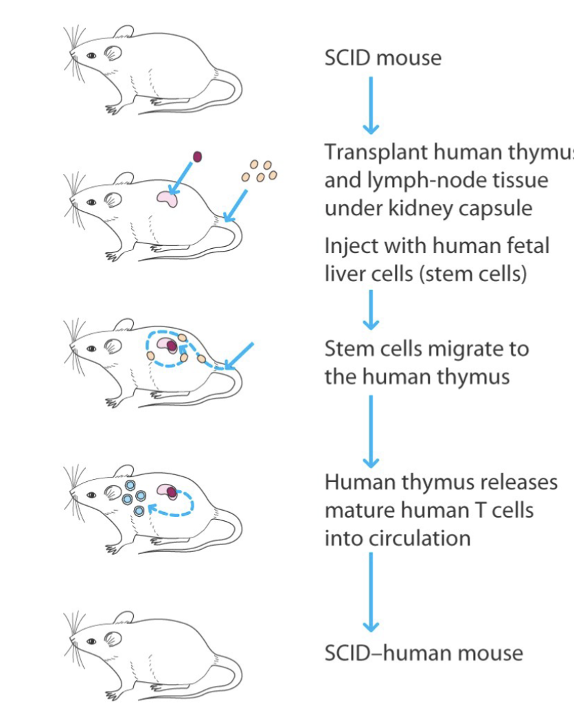
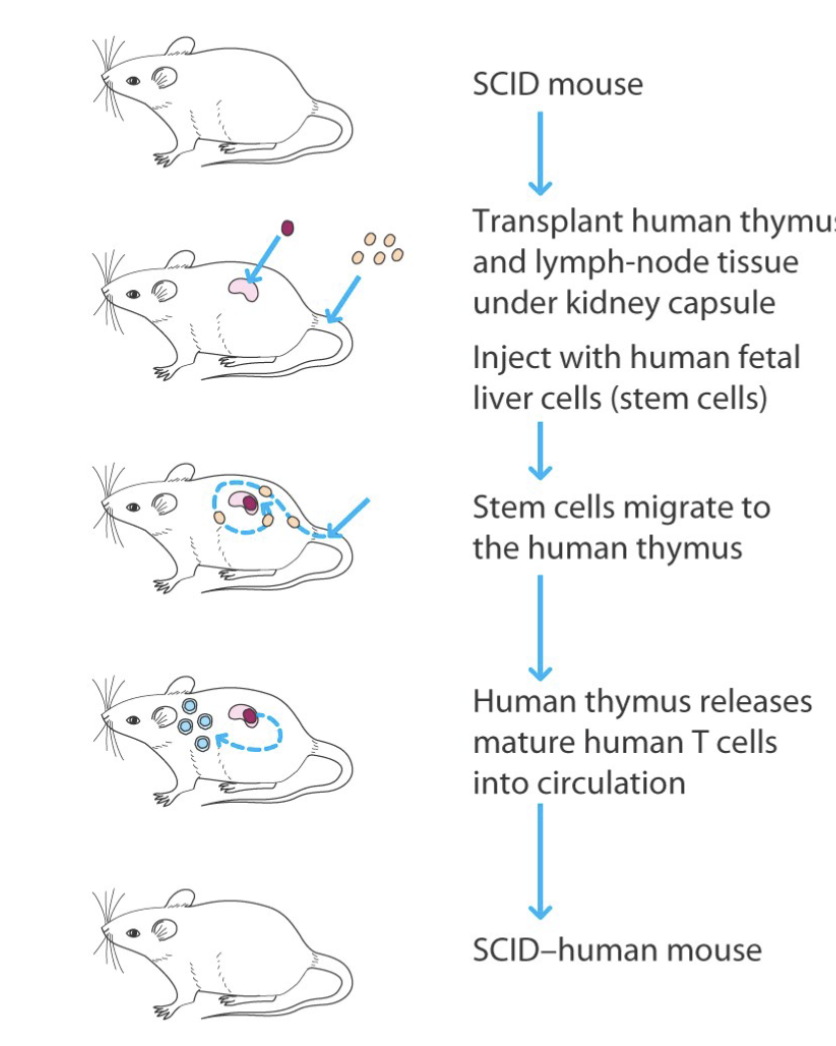
The engraftment is carried out by implantation of liver and thymus under the kidney capsule and by transplantation of HSCs obtained from fetal liver.
(highest human cell reconstitution but difficult to do)
SCID mouse human like model
What are the features of the isolation of blood lymphocytes by Ficoli-Paque Density Gradient?
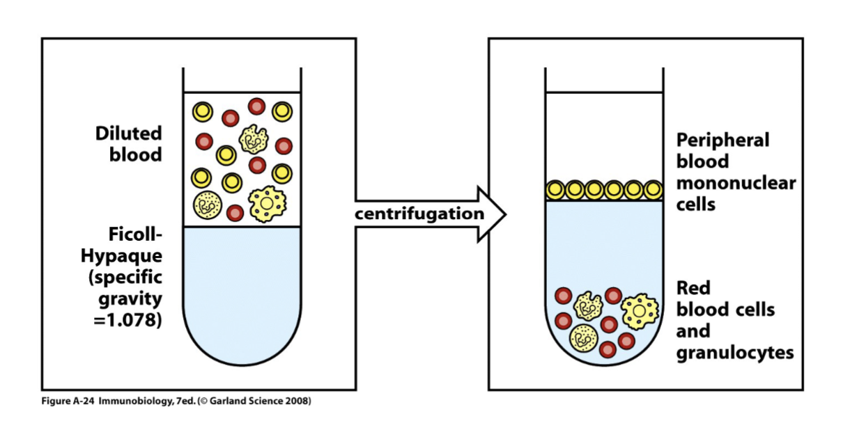
Diluted anticoagulated blood (left panel) is layered over Ficoll-Hypaque and centrifuged. Red blood cells and polymorphonuclear leukocytes are more dense and centrifuged through the Ficoll-paque, while mononuclear cells consisting of lymphocytes together with some monocytes band over it and can be recovered at the interface (right panel)
usually use peripheral blood
need to use gradient with Ficoll (high density) used to separate the cells that have high vs low density centrifuge and heavy density penetrate and remain at bottom of the tube
those with lower density remain on top (these are collected= peripheral blood)
What is the distribution Of Lymphocyte Subpopulations In Human Peripheral Blood Mononuclear Cells (PBMC)?
Majority T cells
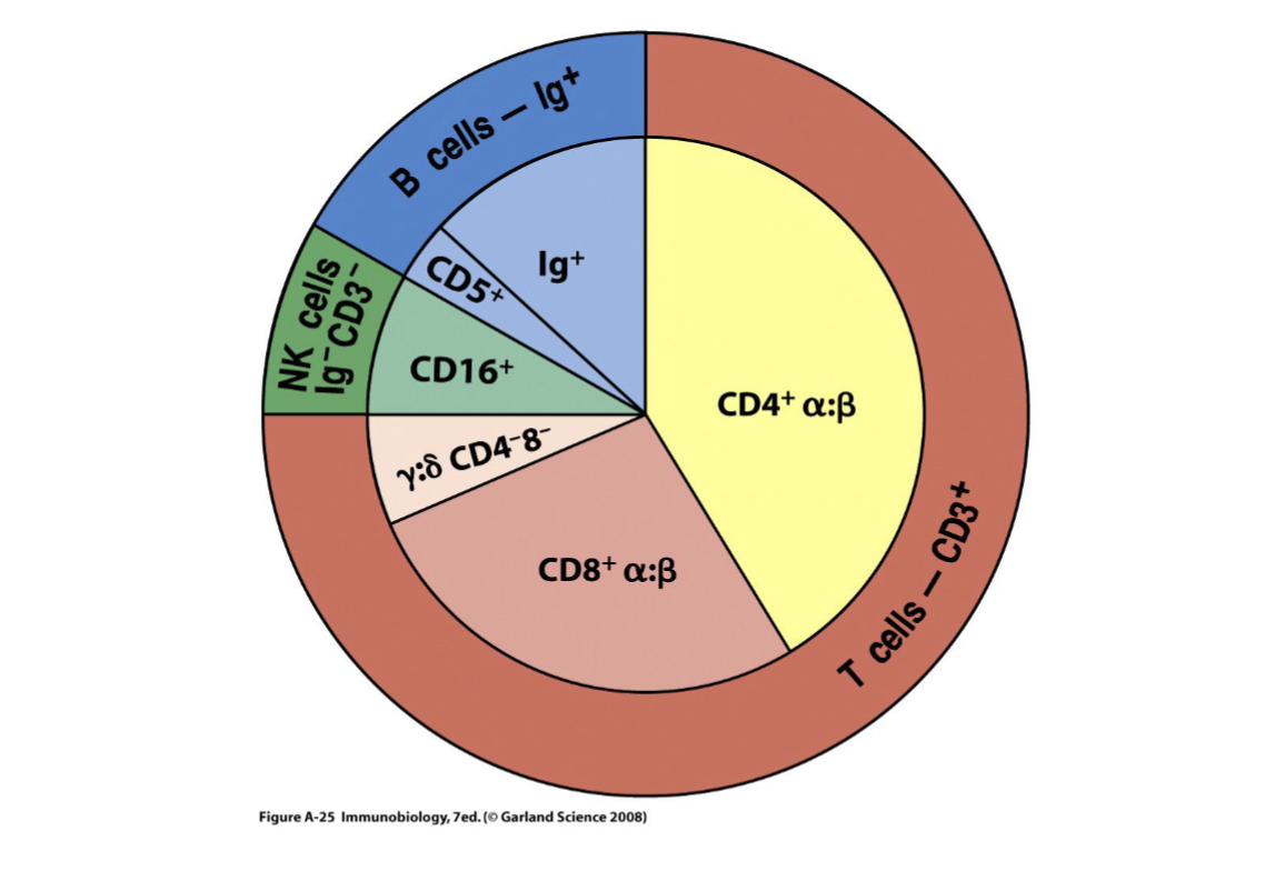
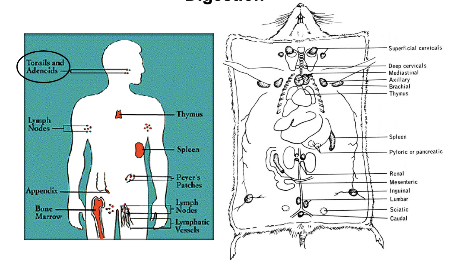
What are the features of Lymphocyte Isolation From Lymphoid Organs
By Mechanical Detachment and Enzymatic Digestion (in humans and mice)?
normally tonsils and adenoids
or taken from deceased or organ donations
blood is the common one used
animal model: all accessible lymphoid organs, spleen is common, draining lymph nodes, chop the lymph nodes to smaller pieces and enzymatically digest to increase recovery
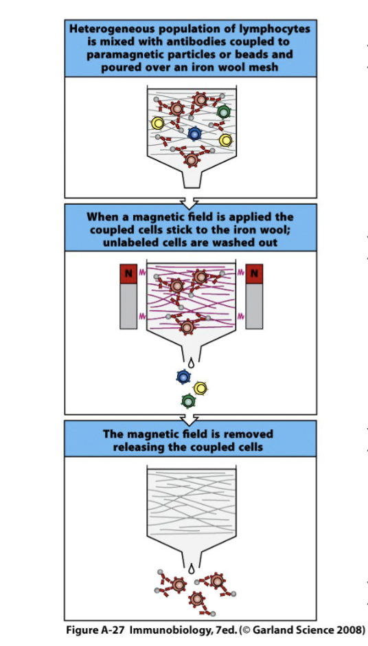
What are the features of Lymphocyte Subpopulation Isolation Using
Antibody-coated Magnetic Beads?
A mouse monoclonal antibody specific for a particular cell-surface molecule is coupled to paramagnetic particles or beads. It is mixed with a heterogeneous population of lymphocytes and poured over an iron wool mesh in a column.
A magnetic field is applied so that the antibody-bound cells stick to the iron wool while cells which have not bound antibody are washed out; these cells are said to be negatively selected for lack of the molecule in question.
The bound cells are released by removing the magnetic field; they are so called positively selected for presence of the antigen recognized by
the antibody.Purity: 85-95%
can use negative of positive fragment
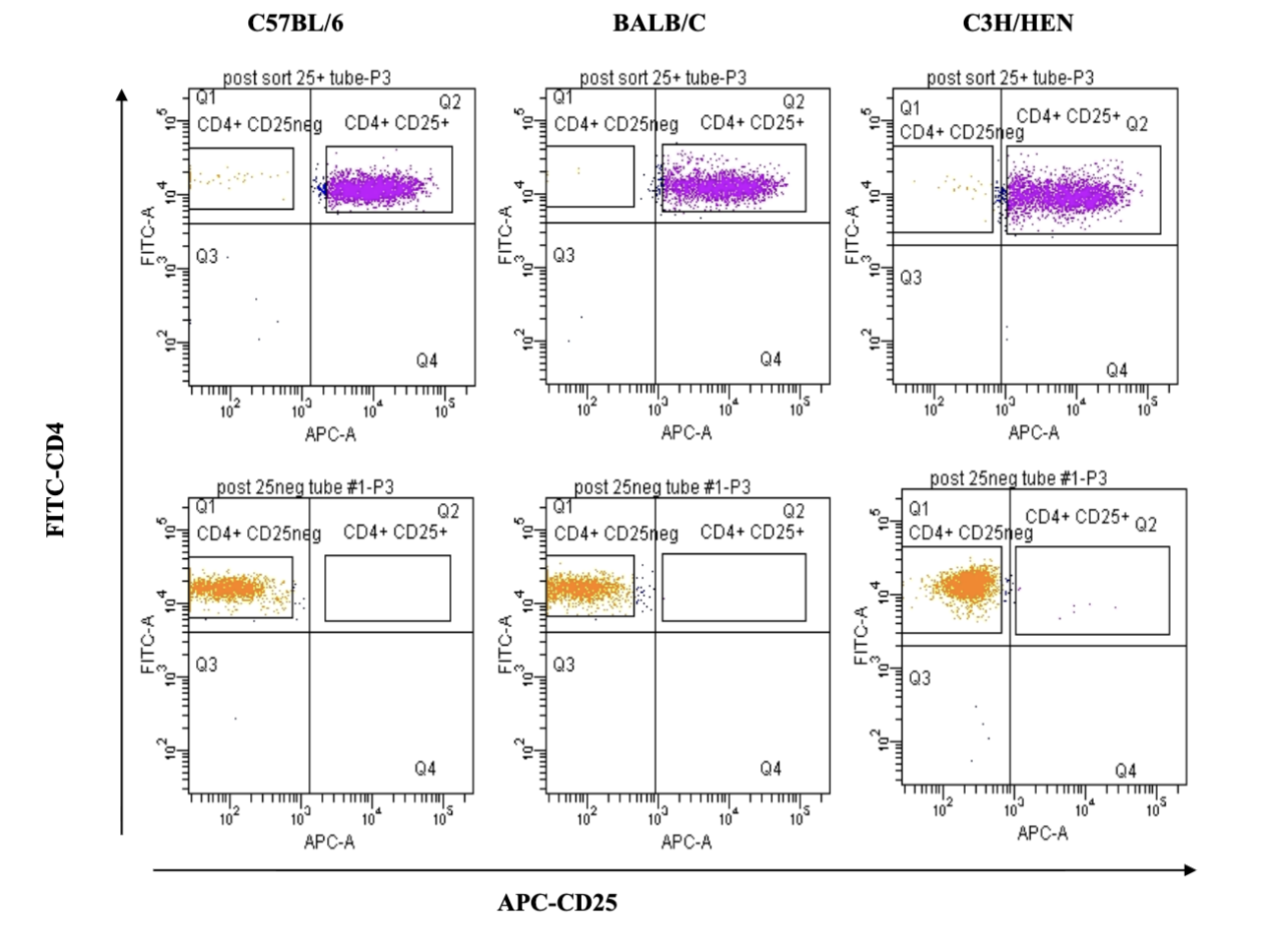
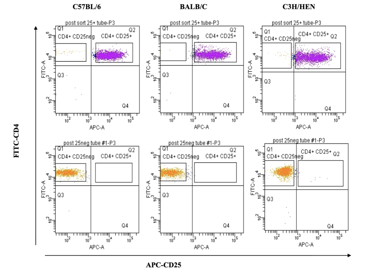
What does this demonstrate?
Cell Isolation By FACS Sorting
3 diff mouse strands
positive and negative fractions
testing CD4 and CD25 (both bound to individual fluorophores) (yellow is CD25 negative)
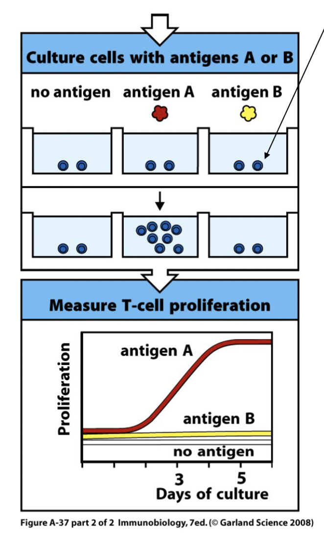
How can the characterization Of Lymphocyte Frequency, Specificity, And
Function be done?
Via humoral responses
Via cellular responses
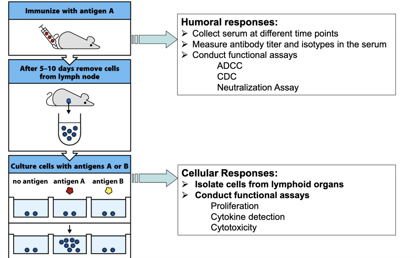
What are the humoral methods to characterize Lymphocyte
Frequency, Specificity, And Function?
Collect serum at different time point
Measure antibody titer and isotypes in the serum
Conduct functional assays
ADCC
CDC
Neutralization Assay
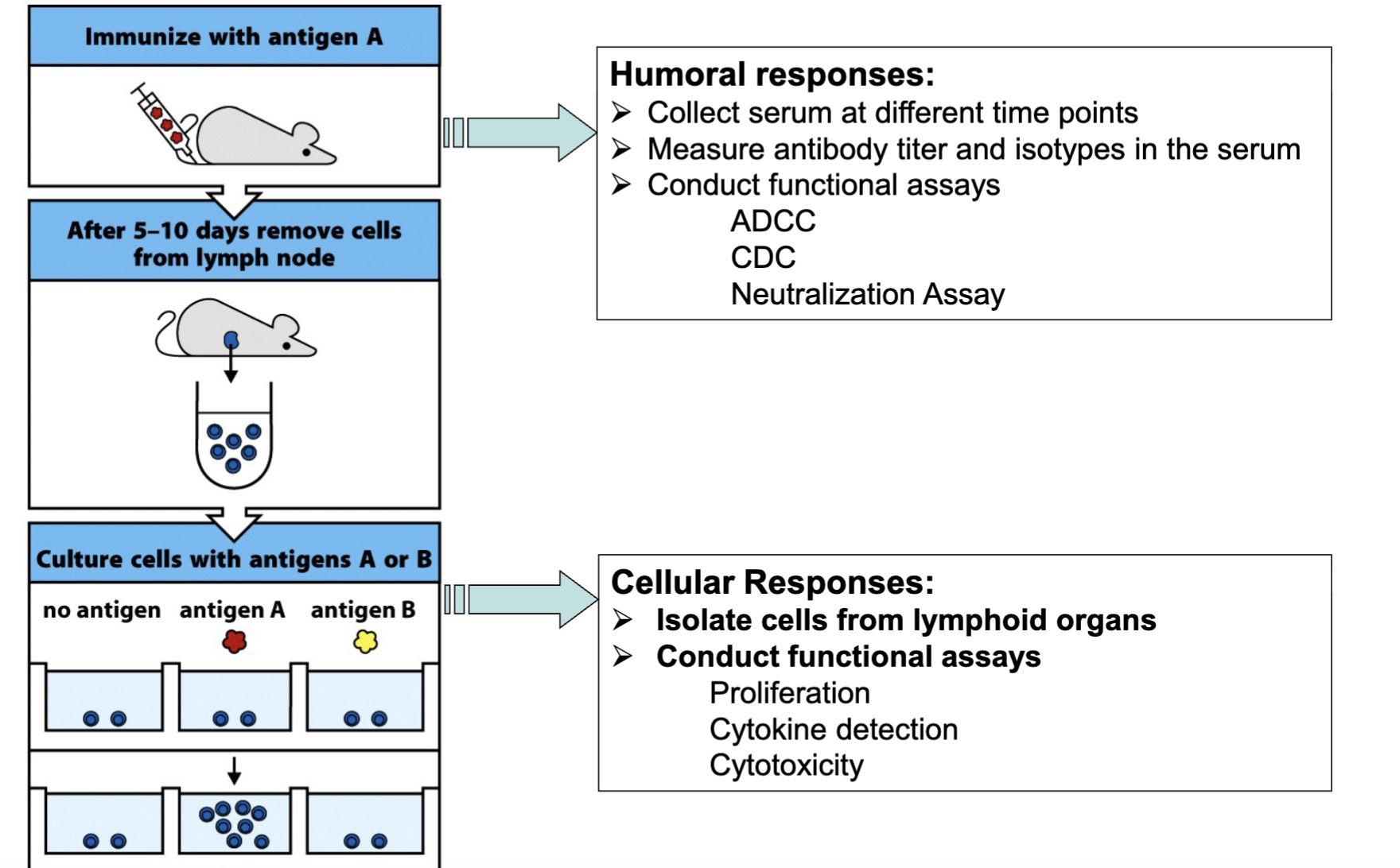
What are the cellular methods to characterize Lymphocyte
Frequency, Specificity, And Function?
Isolate cells from lymphoid organs
Conduct functional assays
Proliferation
Cytokine detection
Cytotoxicity
What are the features of Thymidine incorporation assays?
It relies on the fact that dividing cells synthesize DNA at a rapid pace, and radioactive thymidine in the culture fluid will therefore be quickly incorporated into DNA
measuring T cell proliferation in past used thymidine incorporation assay: type of reagent that imitates DNA synthesis and it gets incorporated into DNA and it gives radioactivity to determine if cell proliferates
What are the features of MTT proliferation assay?
The tetrazolium compound is reduced by metabolically active cells to form insoluble, purpule formazan dye crystals, which can be directly read by
spectrophotometer
MTT depositing compound can be incubated and picked up by live cells and undergo high levels of metabolism
(modern method of measuring T cell proliferation)
What are the features of Brdu proliferation assay
When introduced into cells, bromodeoxyuridine (BrdU) is rapidly phosphorylated to bromodeoxyuridyl triphosphate (an analogue for deoxythymidine triphosphate) and is incorporated in its place into newly
synthesized DNA . Cells that divide following BrdU incorporation can then be identified using antibodies to BrdU.
BrdU molecule being introduced during DNA synthesis and they get incorporated
(modern method of measuring T cell proliferation)
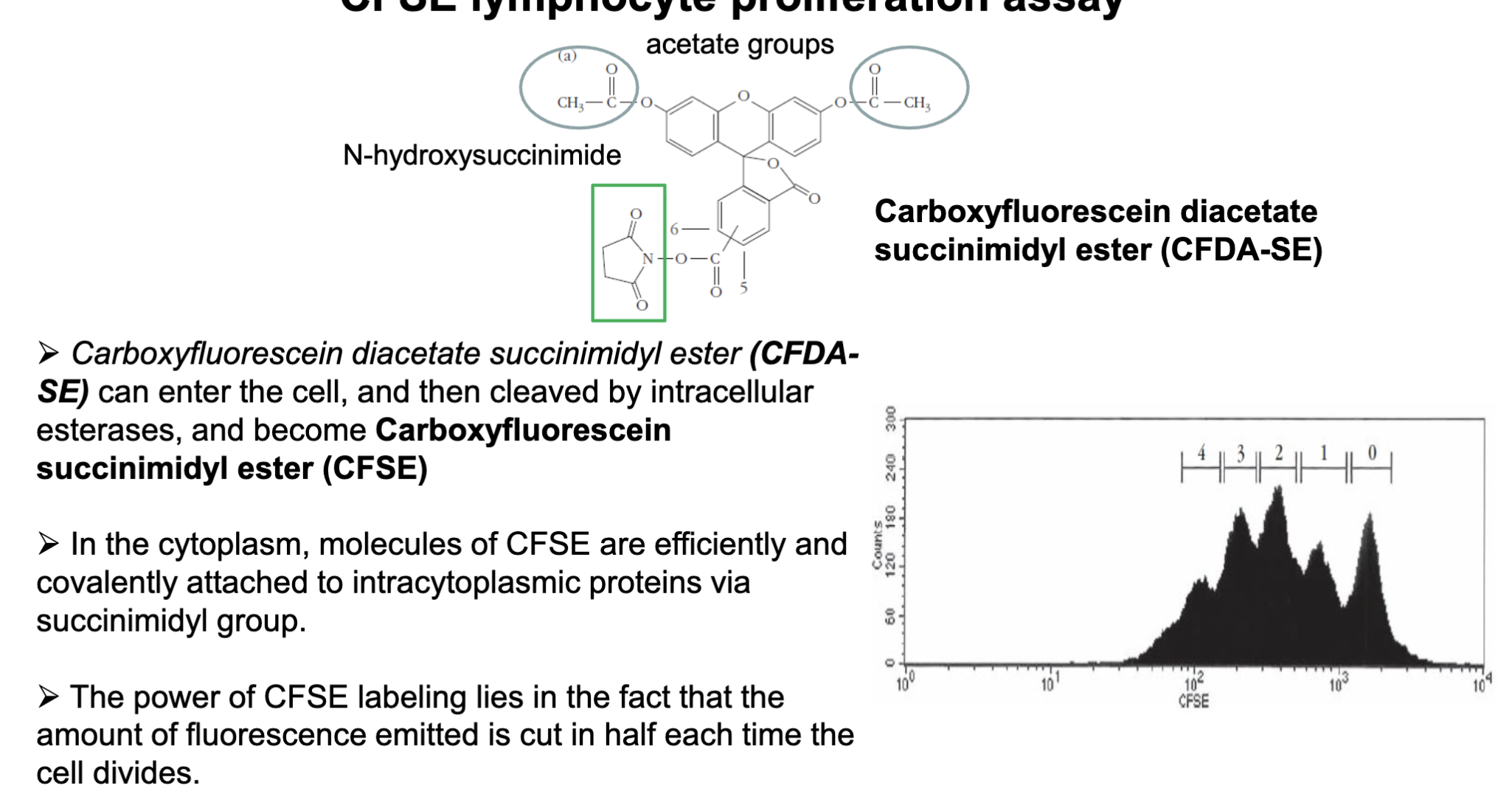
What are the features of the CFSE lymphocyte proliferation assay?
Carboxyfluorescein diacetate succinimidyl ester (CFDA-SE) can enter the cell, and then cleaved by intracellular esterases, and become Carboxyfluorescein succinimidyl ester (CFSE)
In the cytoplasm, molecules of CFSE are efficiently and covalently attached to intracytoplasmic proteins via succinimidyl group.
The power of CFSE labeling lies in the fact that the amount of fluorescence emitted is cut in half each time the cell divides.
common method today is CFSE
-chemicals has two functional acetate groups (allow the cells to be highly permeable to cell membranes) can enter the cells
-once entered cell the acetates are cleaved and the chemical becomes fluorescent converts itself to fluorescent dye
has a hydroxysuccinimide which has high affinity to lysine and amine (covalent binding to cytoplasmic proteins)
able to detect the different intensity in proliferating cells: idea is 0 is parental cell and then 1 (daughter cell) fluorescences reduced by half 4 (is the 2nd division)