BIO 251 Final Exam Study Points
1/48
There's no tags or description
Looks like no tags are added yet.
Name | Mastery | Learn | Test | Matching | Spaced |
|---|
No study sessions yet.
49 Terms
CH 2. Understand the Gram stain, including Gram stain reagents, procedure, outcomes, the principles of the stain.
What is it?
★ A differential stain that allows you to classify bacteria as either gram-positive or gram-negative
Reagents
★ REMEMBER VIDS
★ Crystal VIOLET: stains cells purple
★ Iodine: cells remain purple
★ Decolorizer (alcohol): washes away stain from GRAM - cell wall or remains purple in gram +
★ Safranin: counterstains and makes gram - PINK or gram + remains purple
Outcomes
Purple: gram + bacteria (thick peptidoglycan wall)
Pink: gram - bacteria (thin peptidoglycan + and outer LPS membrane)
Reveals cell morphology (rod or cocci)
CH 2. Explain staining techniques
★ Basic stain (simple stain): + charge, stains CELLS
★ Acidic stain (simple stain): - charge, stains the background
★ Endospore stain: endospore present or not present
★ Acid-fast: used to identify mycolic-acid outer layer → present= mycobacterium
CH 2. Visualizing microbes, microscopy, light microscopy size limitations
Compound Light Microscope
★ invented by Hooke
★ has magnification up to 1000x
★ placing stained microbes (to improve contrast) on bright background to identify morphology
Limitations
★ Bacteria can be seen with light
★ Generally viruses are too small to be seen with light
CH 3. Understand the structure and components of Gram-positive and Gram-negative cells walls, and compare and contrast bacterial cell wall structures.
Gram Positive Bacteria
★ Thick wall of peptidoglycan (stains purple) and teichoic acid (more simple and porous)
★ No outer membrane
Gram Negative Bacteria
★ Thin layer of peptidoglycan (stains pink)
★ Outer membrane of endotoxin lipopolysaccharides (LPS)
★ More resistant to antibiotics due to LPS
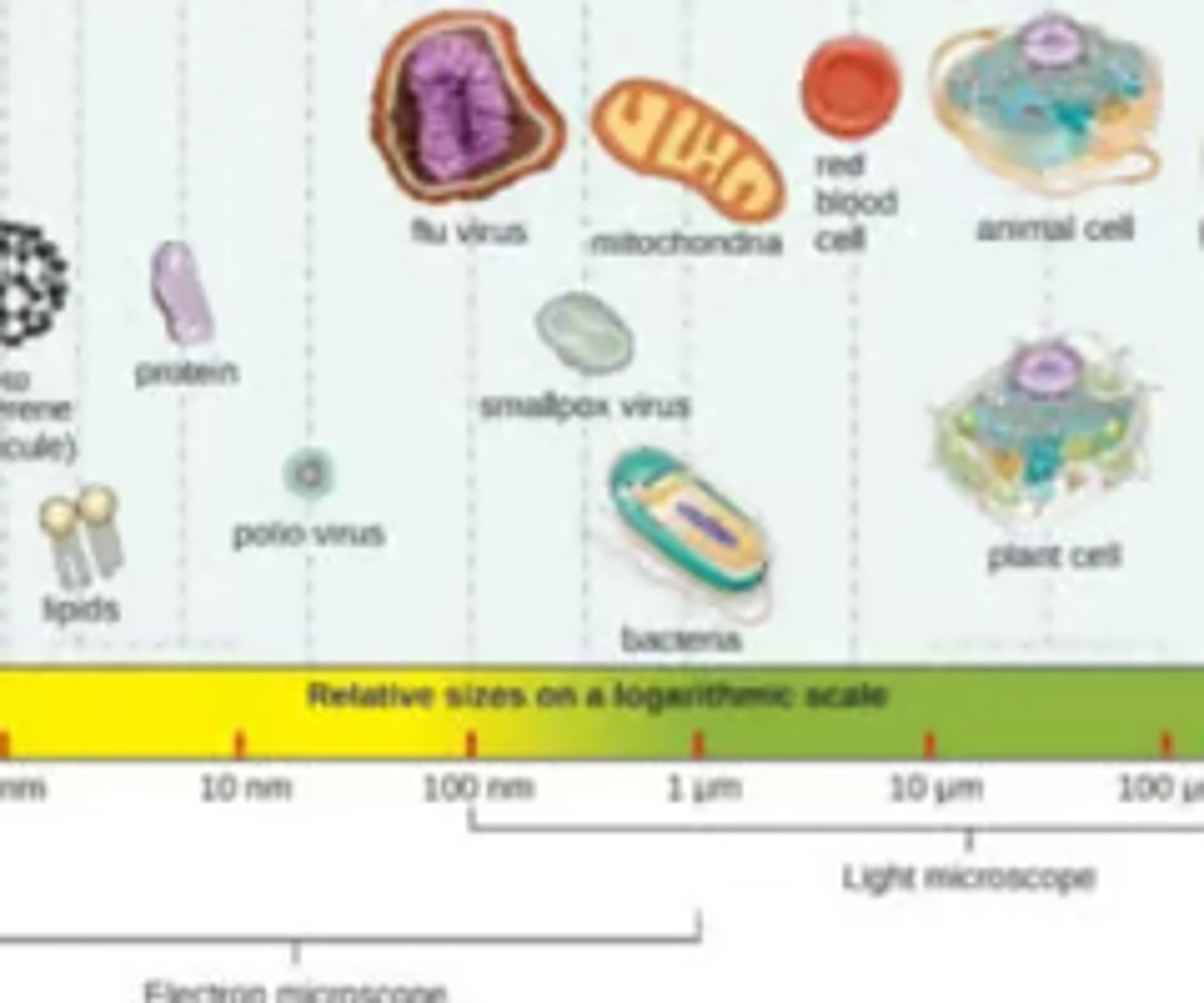
CH 1. Relative Sizes
Lipid→ virus→ bacteria→ yeast→ red blood cell
CH 1. Members of the Microbial World
Three domains of Life (Carl Woese)
★ Bacteria, Archaea, Eukarya
★ built off comparisons of DNA sequences of 16s rRNA genes
★ NOTE THAT VIRUSES ARE NOT ON TREE BECAUSE THEY ARE ACELLULAR (NON LIVING)
CH 1. Understand species vs. strains of bacteria
★ Species: basic unit of classification of related strains or isolates
★ Strain: generic variant of a species
CH 1. Taxonomy: Identification and naming of Microorganisms
★ Est. by Carolus Linnaeus
★ (broadest) Domain→ Kingdom→ Phylum→ Class→ Order→ Family→ Genus→ Species (specific)
★ Binomial system (naming system): Genus then species (ex: Clostridium difficile--> C. difficile
CH 3. Bacterial cell structure (structures and their functions)
★ Cell wall: peptidoglycan cell wall
★ 70S Ribosome
★ Plasmids: extra DNA that can code for helpful, but nonessential traits
★ Motility/adhesion: flagella, pili, fimbriae, capsule
★ Endospores: resistant to heat, drying, and numerous chemicals
★ Glycocalyx: jelly-like substance secreted out of cell that allows capsule to attach to cell wall
CH 3. Compare/contrast prokaryotic and eukaryotic cells (PROKARYOTIC DESCRIPTION)
Prokaryotic cells (bacteria cells)
★ no nucleus
★ non membrane-bound organelles
★ Bacteria and Archaea ONLY
★ Peptidoglycan cell wall
★ 70S ribosome and 16S rRNA
★ Bacteria (prokaryote): DOUBLE STRANDED, SINGLULAR CIRCULAR MOLECULE OF DNA
CH 3. Compare/contrast prokaryotic and eukaryotic cells (EUKARYOTIC DESCRIPTION)
Eukaryotic (Human cells)
★ membrane-bound organelles
★ Animal, plant, fungi, etc.
★ No cell wall in animal cells, but cell wall in plants and fungi
★ 80S ribosomes and 18S rRNA
★ more complex structure
★ MULTIPLE rod shaped chromosomes
CH 6. Viruses - Explain structure and composition, replication
Structure
★ Acellular (non-living)
★ either DNA or RNA NOT BOTH
Composition
★ nucleic acid surrounded by capsid
★ either naked or enveloped
★ If enveloped → virus had protein/glycoprotein spikes rising from capsid allowing for better attachment to specific host cells
CH 6. Steps of Viral Replication
1. attachment: Virus binds specifically to one or more receptor sites on host cell
2.Virus enters target cell (penetration): ENTIRE virus enters (unlike bacteriophage)
Via endocytosis (of enveloped or naked) or fusion with host cell membrane (envelope only)
3. Biosynthesis of viral nucleic acids and proteins
4. Assembly and maturation:
Spontaneous self-assembly when nucleic acid and capsid proteins accumulate in host cell
5.Release: Most enveloped virus leave via budding
★ Non-enveloped viruses released when host cell dies, often by apoptosis initiated by virus or host
CH 10 & 11. Know the details of plasmids, including structure, genes on plasmids, plasmid replication and how they are transferred.
Structure
★ NON-ESSENTIAL circular DNA that can code for antibiotic resistance, pilus synthesis, toxin production, capsule, and biofilms
Plasmid replication
★ self-replicating but have a narrow-host range
★ recipient must be similar and have double-stranded DNA
★ often transferred via conjugation, but also thru transformation and transduction
CH 13. What are endospores are, why they are difficult to kill/eradicate, bacteria genera that are endospore formers
★ Bacterial endospores: protective structures made by certain bacteria that keep them safe until conditions are livable again
Resistant to: heat, drying, and numerous chemicals
Endospore formers
★ Bacillus (think B. anthracis → anthrax)
★ Clostridium → C. botulinum, C. tetani, C. difficile, and C. perfringens
CH 10 & 11. Explain the three mechanisms of gene transfer in bacteria; know the details of each, how they differ from each other; what can be transferred; and the
significance of gene transfer
★Gene transfer: where new donor genes are introduced into a recipient cell
★ Transformation: cells TAKES UP DNA directly from environment
★ Transduction: bacteriophage INJECTS a HYBRID of viral DNA and DNA from other infected bacterial cells
★ Conjugation: DNA is TRANSFERRED between cells via conjugation pilus bridge
❤︎only between two living cells
Vertical: genes shared from generation to generation (mother to daughter cells)
Horizontal: genes received and shared from cells of SAME generation
CH 10 & 11. Explain lysogenic conversion and know the examples discussed in class of toxin genes that are transferred to bacteria via transduction; how this contributes to strain variation
★ when a virus (called a bacteriophage) infects a bacterium, inserts its DNA into the bacterial chromosome, and gives the bacterium new abilities -- like producing toxins.
★ Prophage: phage DNA becomes part of bacterial genome
Explain the cholera toxin example of transduction
❤︎ Patient diagnosed with cholera
❤︎ Prescribed Doxycycline but does not work
❤︎ Bacteria is found to undergone lysogenic conversion and now carries cholera toxin gene due to horizontal gene transfer (transduction)
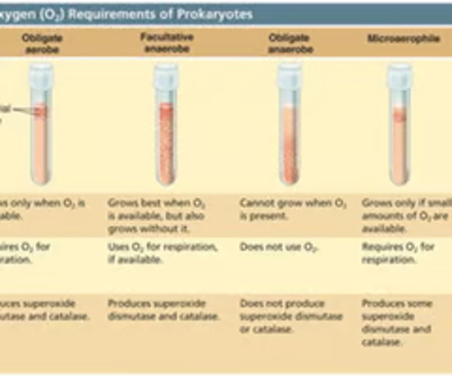
CH 9. Microbial Growth - Know the different categories for grouping microbes according to oxygen requirements/utilization
★ Obligate aerobes: requires oxygen, grows top of tube
★ Obligate anaerobes: can't grow in presence of oxygen, grows in bottom of tube
★ Facultative anaerobes: grows in aerobic or anaerobic conditions but prefers oxygen, grows throughout tube but more towards the top
★ Microaerophiles: require little to no oxygen (not in room air), growth towards top of tube
★ Aerotolerant anaerobes: grows with or without oxygen, equal growth throughout tube
CH 9. Microbial Growth - How are microbes grouped according to acid/base tolerance and practical applications
★ Neutrophiles: pH range 5-8 (neutral)
★ Acidophiles: pH below 5.5 (acidic)
★ Alkaliphiles: pH above 8 (basic)
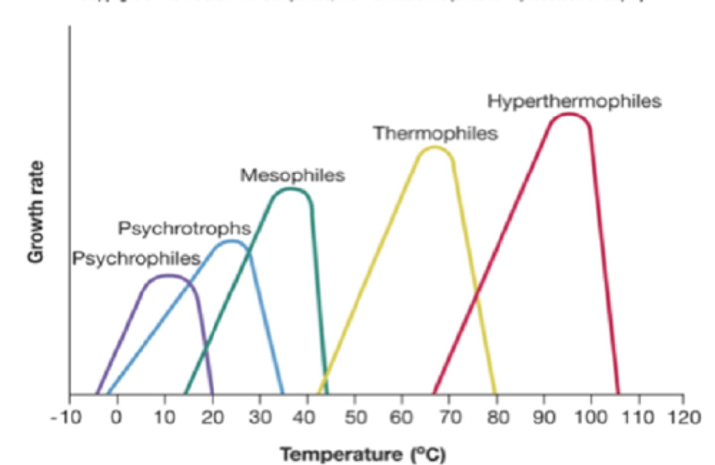
CH 9. Microbial Growth - Understand temperature requirements/tolerance of microorganisms
Optimum temperature depends on species but have a range of what temp it can grow within → Outside of temperature= still grows but slowly
★ Psychrophiles: extremely cold environments
★ Psychrotrophs: refrigerator temperature
★ Mesophiles: body temperature environments (pathogenic bacteria)
★Thermophiles: hot tub temperature
★ Hyperthermophiles: extremely hot temperature
CH 9. Understand solute (salt) concentration and food preservation
★ Halotolerant bacteria: can tolerate higher concentrations of solute
★ Halophilic bacteria: require a higher concentration of solute in their environment (think sea water)
❤︎ Bacon is preserved with high salt concentration since bacteria undergoes plasmolysis
❤︎ Other examples: jams, honey, mustard, ketchup
CH 9. What is a bacterial growth curve, and explain the phases of growth
❤︎ 1) Lag Phase: cells acclimate
❤︎ 2) Exponential phase: more growth than death
❤︎ 3) Stationary phase: equal rate of death and growth
❤︎ 4) Death phase: more death than growth
❤︎ 5) Phase of decline
★ Used to determine the growth rate of bacterial species
❤︎ Often helpful to know how different environmental and nutritional factors affect rate of growth
CH 13. Control of Microbial Growth – what are the most difficult microbes to kill/eradicate
Hardest to kill
★ Bacterial endospores – highly resistant to heat, drying, and chemicals.
★ Protozoan cysts/oocysts – survive harsh conditions.
★ Mycobacterium species – waxy mycolic acid resists disinfectants.
★ Pseudomonas species – chemical-resistant, versatile.
★ Naked viruses – lack envelope; more resistant.
CH 13. Control of Microbial Growth – How methods of control are selected and aseptic technique
★ Based on level of risk (BSL 1–4), item use (critical vs non-critical), and setting (daily life, hospitals, labs, industries).
★ Choose sterilants for critical items, high-level disinfectants for semi-critical, and low/intermediate-level for general surfaces.
Aseptic Technique:
★ Prevents contamination in labs/clinical settings.
★ Maintains sterility of tools, surfaces, and patient areas.
CH 13. Control of Microbial Growth – Understand sterilization methods
Sterilization Methods:
★ Autoclaving (121°C, 15 psi, 15 min),
★ Incineration,
★ Ethylene oxide gas,
★ Radiation,
★ Filtration (air/liquid).
★ Commercial sterilization for food (kills C. botulinum without ruining food quality
CH 13. Know and explain all the different chemicals that are used for sterilization and what they would be used for.
Sterilants (e.g., glutaraldehyde, ethylene oxide):
★ Glutaraldehyde (2%): Sterilizes heat-sensitive medical tools (endoscopes).
★ Ethylene oxide gas: Sterilizes packaged items, fabrics, electronics, artificial joints.
★ Aldehydes: Inactivate proteins/DNA; used in medical equipment sterilization (not skin).
★ Halogens (e.g., chlorine dioxide gas): Used to fumigate/sterilize large enclosed spaces.
CH 14. Antibiotics – know the major categories
cells
β-lactams (e.g., Penicillin, Cephalosporins, Carbapenems):
★ Inhibit cell wall synthesis
Protein synthesis inhibitors (e.g., Azithromycin):
★ Target 70S ribosome
Nucleic acid inhibitors (e.g., Fluoroquinolones, Rifamycin):
★Inhibit DNA/RNA replication
Metabolic inhibitors (e.g., Sulfonamides, Folate inhibitors):
★ Block folic acid synthesis
Cell membrane disruptors (e.g., Polymyxin B):
★ Damage bacterial membrane integrity
CH 14. Antibiotics – what do antibiotics target in prokaryotic cells
Targets in Prokaryotes:
★ Peptidoglycan cell wall
★ 70S ribosomes
★ Bacterial enzymes for DNA replication/transcription
★ Unique metabolic pathways like folate synthesis
CH 14. Antibiotics – what are the features of effective antibiotics
★ Selective toxicity (harms bacteria, not host)
★ Stable and long-lasting in body (e.g., azithromycin has long half-life)
★ Different spectrums (narrow vs. broad) based on infection
★ Low resistance potential, or combined with other agents to reduce resistance (e.g., Augmentin)
CH 14. Antibiotics – How do bacteria develop resistant to antibiotics
★ Intrinsic resistance: naturally occurring antibiotic that was not curated or exposed to a clinical antibiotic that gained resistance due to chromosomal genes
❤︎ (Think of naive bacteria gaining plasmids in a natural environment becoming resistant)
★ Acquired resistance: resistance gained by Human development and use of antibiotics
❤︎ Resistance transferred via horizontal gene transfer
★ Mutation: Random changes in bacterial DNA that alter antibiotic targets.
★ Horizontal Gene Transfer (HGT): Resistance genes transferred between bacteria via: Plasmids (conjugation), Transduction (via bacteriophages), and Transformation (uptake of naked DNA)
CH 14. Antibiotics – What factors contribute to the development/spread of antibiotic resistance
★ Overuse of Antibiotics
Prescribing antibiotics when not needed
★ Misuse and Mismanagement
Incorrect dosage or duration of antibiotic treatment.
Patients not finishing their prescriptions.
Self-medication without proper diagnosis.
★ Subtherapeutic Dosing
Low antibiotic levels that don’t kill all bacteria, allowing resistant ones to survive.
★ Horizontal Gene Transfer
Sharing of resistance genes between bacteria via plasmids, transduction, or transformation.
CH 14. Explain how antibiotics are effective for treating infectious diseases
★ Antibiotics are effective because they target specific structures or processes unique to bacteria, such as:
★ Cell wall synthesis
★ Protein synthesis (bind to 70S ribosomes, blocking bacterial protein production)
★ DNA replication and transcription (interfere with enzymes like DNA gyrase or RNA polymerase)
★ Metabolic pathways (block folic acid synthesis needed for nucleotide production)
★ Cell membranes (disrupt membrane integrity in some bacteria)
CH 14. Why antibiotics are not effective against viruses
★ Viruses are acellular= nonliving cell
★ Antibiotics specifically target bacteria exclusive traits that viruses do not have
CH 14. Infectious diseases – signs vs. symptoms; nosocomial vs. iatrogenic
★ Signs: can be observed and measured
❤︎ Changes in vital signs may indicate disease
❤︎ presence of fever, rash, abnormal blood pressure, presence of antibodies
★ Symptoms: very subjective, experience by patient, CANNOT be objectively measured
❤︎ Wong-baker faces (1-10)
★ Nosocomial infections are hospital-acquired: infections not present at the time of admission (e.g. MRSA from unsterilized instruments).
★ Iatrogenic infections: are caused by medical procedures or treatment (e.g. infection from a contaminated catheter or surgery).
CH 15. What are virulence factors (the categories we discussed and examples); How do
microbes cause damage to host
★ Virulence factors: how microbes evade host defenses and determines extent and severity of disease
❤︎ Adhesions: Help microbes attach to host cells → e.g. Slime layers, capsules, fimbriae
❤︎ Exoenzymes: Produces exoenzymes that break down host barriers
❤︎ Exotoxins: neurotoxins, enterotoxins, cytotoxins, superantigens, AB toxins
❤︎ Antiphagocytic factors: capsules prevent opsonization and complement system
❤︎ Biofilm formation: Hard to remove, can lead to chronic infections, hard for immune cells to penetrate
CH 15. How do infectious diseases develop/what are the steps in pathogenesis
★ Exposure/contact: first encounter with pathogen
❤︎ Pathogen gains access to host
❤︎ Respiratory, GI tract, broken skin or damaged mucous membrane
★ Attachment: Pathogen attaches to host cell receptors using adhesion factors
❤︎ Slime layers and capsules
❤︎ E.g. biofilm of Pseudomonas in cystic fibrosis patients, burn wounds, ear infections
★ Invasion: pathogen spreads throughout body
❤︎ Virulence factors are produced – toxins, exoenzymes
★ Infection and damage to host
CH 15. Compare/contrast exotoxins and endotoxins
Endotoxins
★ gram -
★ Made from Lipid A of LPS
★ general symptoms of inflammation and swelling
★ higher dose needed as toxin is not as potent
Exotoxins
★ gram + (mostly) but can be gram -
★ made of proteins
★ Specific damage to cells based on receptor mediate targeting (GABA inhibition and tetanus toxin)
★ lower dose needed as toxin is more potent
CH 1. Explain Koch’s postulates
1) animals with disease has disease, healthy animals without have no disease
2) microorganism can be isolated from diseased animal and grown in pure culture
3) disease can be reproduced in healthy animal with cultured organism
4) organism can be re-isolated and cultured again to repeat process
★ Not all infectious agents have the same signs and symptoms in all pTs (different immune systems)
★ Not all pathogens can be grown in pure culture
★ Some pathogens can cause different diseases depending on virulence factors produced
Know the various blood cell types and explain their functions
Red Blood Cells (RBCs / Erythrocytes)
★ Function: Carry oxygen from lungs to tissues and return carbon dioxide to lungs
★ Protein involved: Hemoglobin
★ No nucleus (in mature form), which maximizes space for oxygen
White Blood Cells (WBCs / Leukocytes)
★ Function: Defend against infection, foreign invaders, and abnormal cells
★ Types of WBCs (5 major ones): neutrophils, eosinophils, basophils, lymphocytes, and monocytes
What are the features of innate immunity; what are the ways innate and adaptive differ
Innate Immunity
★ immediate response and present since birth
★ nonspecific and no memory
★ Physical barriers: skin, mucous membranes, and cilia
★ Involves phagocytes, NK cells, and dendritic cells
Adaptive immunity
★ not immediate
★ highly specific and memorizes past infections
★ Involves B cells and T cells (helper and cytotoxic)
★ Defends immune system via antibody production and targeted killing
Ch 18. Name the antibody classes
★ IgM: First class produced during response, agglutination, initiates complement cascade, less than 20%, pentamer with MULTIPLE binding sites
★ IgG: exits bloodstream into tissues, most versatile and dominant ~80%, can cross placenta (ONLY CLASS TO DO THIS)
★ IgA: secreted form is breast milk, tears, and saliva, important for mucosal immunity, primarily acts as neutralizer and traps bacteria in mucus, dimer (so mostly in bodily fluids), good for babies
★ IgE: binds to IgE and causes cells to release histamine and cytokines during inflammation and allergic reaction, anti-parasitic
★ IgD: membrane-bound monomer on surface of B cells and antigen-bind receptors
★ IgM: First class produced during response, agglutination, initiates complement cascade, less than 20%, pentamer with MULTIPLE binding sites
Ch 18. What are antibodies? How are they produced? What are their functions?
★ Glycoproteins/immunoglobulins made by immune system to fight germs like viruses and bacteria
★ Neutralization: when antibodies bind and mask over/block specific antibodies to antigens found on bacteria, viruses, and toxins
★ Opsonization: antibodies act as opsonin that are markers for pathogens for destruction by macrophages, dendritic cells, and neutrophils
★ Agglutination: Antibodies cause clumping of cells such as bacteria or red blood cells in presence of antibody (usually IgM agglutinate when they bind to epitopes)
★ Antibody-Dependent Cellular Cytotoxicity (ADCC): Antibodies bind to large pathogenic cells that are too big for phagocytosis and help Natural Killer cells come close. The NK cells then release toxic chemicals to kill the harmful cell from the outside
Ch 18 How does vaccination work to protect people from infectious diseases
★ form of artificial active immunity
★ Vaccines expose pTs to pathogen specific antigens which then stimulates adaptive immune response
CH 1. Know the contributions of these individuals: Leeuwenhoek, Fleming; Florey,
Chain, Hodgkins, Woese, Pasteur, Hook, Koch, Griffith,
★ Leeuwenhoek → father of microbiology and first to describe microorganisms by microscope
★ Fleming → made penicillin (first antibiotic)
★ Florey and chain → purified and mass-produced penicillin
★ Hodgkins → made semisynthetic drugs
★ Woese → 16S rRNA gene sequencing and established three domains of life
★ Pasteur → disproved spontaneous generation and invented pasteurization
★ Hooke → made compound microscope and first to use the term "cell" while observing a cork under microscope
★ Koch → Koch’s postulates and germ theory
★ Griffith → proved transformation and horizontal gene transfer
CH 24. What are immune defense mechanisms of the digestive system
★ Saliva
★ Low pH of Stomach acid
★ Mucous
★ Peristalsis
★ Peyer's patches : M cells are here to detect pathogens
★ Natural microbiota
CH 24. Know the terminology related to GI infectious diseases
★ Gastritis: inflammation of stomach lining
★ Enteritis: inflammation of intestinal mucosa
★ Gastroenteritis: inflammation of both the stomach lining and the intestinal lining
★ Hepatitis: inflammation of the liver
★ Colitis: inflammation of the colon
★ Dysentery: damage to the epithelial cells of the colon; causes bleeding and excess mucus, watery stools
CH 24. Explain how S. aureus is responsible for food poisoning
★ S. aureus is commonly found on human skin and food can be contaminated w/ Staph during preparation
★ Associated with raw or undercooked foods and enters body via ingestion of exotoxins
★ 21 staphylococcal enterotoxins which are able to withstand low pH of stomach
CH 24. Helicobacter pylori
Characteristics
★ Short, gram -
★ Microaerophile and multiple polar flagella (motile)
Survival in the stomach
★ produces urease that raises pH around the cell to neutralize stomach acid
Role in peptic ulcer formation
★cause for ulcers in the stomach as Helicobacter decreases mucus production
CH 24. Clostridium difficile
Characteristics
★ Gram +, obligate anaerobic, spore-forming, rod-shaped
Infection symptoms
★ most common cause of nosocomial diarrhea
Pathology
★ forms endospores and are highly resistant to disinfectants, environmental conditions
★ resistant spores survive stomach acid and germinate in small intestine
★ Many strains mediated by toxin A and toxin B
Transmission
★ Acquired in pTs taking antibiotic therapy as it alters the microbial flora of large intestine (leading hospital pT susceptible to infection)
Most susceptible
★ elderly and immunocompromised
Treatment
★ if possible, stop antibiotics or drink electrolytes or fecal transplant to restore microbiota
★ beta lactams
Prevention
★ minimizing use of antibiotics, handwashing, wearing loves, and disinfectants