Bio Exam 4 - Chapter 45, 48, 49, 47
1/78
There's no tags or description
Looks like no tags are added yet.
Name | Mastery | Learn | Test | Matching | Spaced | Call with Kai |
|---|
No analytics yet
Send a link to your students to track their progress
79 Terms
excitatory vs inhibitory synapse
excitatory increase the activity of the receiving neuron, while signals sent across inhibitory synapses reduce neuron activity
spatial vs temporal summation
Spatial summation involves simultaneous signals coming from multiple presynaptic neurons being received by a single postsynaptic neuron. Temporal summation involves a single presynaptic neuron rapid-firing signals to a postsynaptic neuron
afferent neurons
sensing neurons
carry information to nervous system
efferent neurons
motor neurons
carry information to effector cells outside of Nervous system like in muscles or glands
interneurons
communicate between neurons
association neurons
types of neural networks
nerve net - simple, no central command center, nerves control certain parts of the organism ex. in cnidarians/anemone
ganglia - neurons organized into clusters ex. earthworm
brain - more complex, processes information
gray matter vs white matter
gray - darker because has more neuronal cell bodies; sends information through the axon
white - has more axons
spinal reflex
doesn’t go through the brain; processed in spinal cord
conversion of afferent to efferent information in the spinal cord without participation of brain
ex. knee-jerk reflex
how does knee-jerk reflex work
sensory neuron receives information (the tapping from the hammer), and sends the information down the spinal nerves on the afferent pathway, connect with the interneurons which send signal to motor neuron which transmits signal down efferent pathway to the skeletal muscle which contracts
difference between central and peripheral nervous system
central: includes brain and spinal cord
peripheral: neurons that connect central nervous system to all tissues and parts of the body
examples of conscious afferents
sensory information like sight and sound that are sent to CNS
examples of unconscious afferents
physiological information like blood pressure, and deep body temperature that are sent to CNS
examples of voluntary efferents
commands to skeletal muscles sent by CNS
examples of autonomic efferents
physiological controls like heart rate and sweating sent by CNS
parts of the early embryonic “brain”
forebrain, midbrain, and hindbrain
what does the brain develop from
the dorsal hollow neural tube in the embryo (looks like the dorsal hollow nerve chord present in all chordates)
what does the forebrain develop into
diencephalon
thalamus
hypothalamus
telencephalon
cerebrum
left and right cerebral hemispheres
cerebral cortex
what does the hindbrain develop into
medulla
pons
cerebellum
what does the midbrain develop into
midbrain has the least change and stays very small in comparison to rest of brain
spinal cord function
conducts information to and from the brain
integration of information from the peripheral nervous system
issues motor commands
brain stem function
made up of the midbrain, pons, and medulla
pons: bridge between cerebrum and cerebellum
medulla: involved in control of physiological functions such as breathing and swallowing
has its own peripheral nervous system
core of brain stem called reticular activating system
damage below RAS can cause paralysis but sleep-wake cycle still intact
damage above RAS can cause coma because it interrupts pathways that keep the brain awake
what are the parts and functions of the adrenal axis
Hypothalamus: regulates physiological functions such as hunger, thirst, pleasure; control center for basic needs
Thalamus: relay station for all sensory information except for smell
Pituitary gland: secretes hormones regulating homeostasis, functionally connected to hypothalamus
parts of limbic system and its function
network of neuronal pathways important for motivation, instinct, emotional association with memory, and formation of long term memory
includes the parts of the forebrain:
amygdala: fear, and fear memory; if this region is damaged then you can’t learn to be afraid of a stimulus
hippocampus: transfers short term memory to long term memory
what are the parts of the cerebral cortex
temporal lobe
frontal lobe
central sulcus (separates frontal and parietal lobe)
parietal lobe
occipital lobe
temporal lobe function
receive and process auditory information
facial recognition
identifying and naming objects
understanding spoken language
*if temporal lobe is damaged the individual would develop agnosia (difficulty identifying things)
frontal lobe function
the association cortex for higher order information processing, and associating info from senses and memory
ex. reasoning, planning, personality
what is the primary motor cortex
*present in frontal lobe
issues motor commands throughout the body; controls muscles
parts of the body with fine motor control such as face, hands, fingers, have larger representation on cortex (more neurons devoted to controlling them)
parietal lobe function
integrating sensory information from various parts of the body
ex. visuospatial processing/body awareness
what is the primary somatosensory cortex
*below central sulcus, present on parietal lobe
its neurons receive info from different parts of the body
areas of body with lots of mechanoreceptors like lips, hands, fingers, have larger representation on PSC
occipital lobe function
receives and processes visual information
if occipital lobe damaged, you can see images but can’t see motion or translate visual experience into language
autonomic nervous system
part of the peripheral nervous system that controls involuntary physiological functions to maintain homeostasis
includes the sympathetic and parasympathetic nervous system
how are preganglionic neurons different in sympathetic and parasympathetic nervous system?
*send signals
sympathetic: cholinergic; acetylcholine is neurotransmitter
parasympathetic: cholinergic; acetylcholine is neurotransmitter
how are postganglionic neurons different in sympathetic and parasympathetic nervous system?
*receive signals
sympathetic: noradrenergic; norepinephrine is neurotransmitter
parasympathetic: cholinergic; acetylcholine is neurotransmitter
how is anatomy of sympathetic and parasympathetic nervous system different
sympathetic: most of the ganglion are lined up in ganglion chain
preganglionic neurons are mostly from the thoracis and lumbar regions
parasympathetic: preganglionic neurons usually come from cranial and sacral regions
most of the ganglion are close to the target organs
how are sympathetic and parasympathetic nervous system functions different
sympathetic: active during fight or flight response
heart rate and cardiac output increased
blood flow to skeletal muscle increased
parasympathetic: active when you’re relaxed/normal condition
digestive system activity increased
heart rate and blood pressure go down
what are the structural components of vertebrate eyes
sclera: outer covering
cornea: anterior portion modified
iris: colored part of eye, controls size of pupil to adjust amount of light let into eye
pupil: hole where light enters
lens: helps eyes focus light
blind spot: area with no photoreceptors and just blood vessels
retina: receives image and converts to neuronal signal with neurons and photoreceptors
fovea: small depression on the retina that contains mostly cone cells
choroid: between retina and sclera; rich in blood vessels to provide nutrients
what is the aqueous humor
a liquid secreted by a ciliary body/muscle at front of eye and controls the curve of the lens
what is the vitreous humor
a clear gel that holds the shape of the eye; it doesn’t get replenished so as you get older it isn’t replaced
what is the order of flow of light through the eye
iris → pupil → retina
how does the lens change to accommodate different distances
more rounded lens to look at things close by
flatter lens to look at distant things
what lenses are prescribed for different sight issues
concave (hourglass shape) for near sightedness
convex lens if image lands behind retina
bifocal lens if near and farsighted
what are rods and cone cells
rods: function in dim light, detect shape and movement; don’t see color
cones: responsible for vision in bright light, and seeing fine details; allow color vision
what is present in the retina
photoreceptors, ganglion cells, amacrine cells, bipolar cells, horizontal cells
flow of information in the retina
photoreceptor (absorbs light and converts to electrical signal) → bipolar cell (receive signal and send to ganglion) → ganglion cell (receive and send to optic nerve)
lateral flow has horizontal cell synapsing with photoreceptor and send info to amacrine cells
why does binocular vision occur
two eyes see overlapping yet slightly different visual fields
left and right eye receive signals from both sides of the vision fields, and sort right field signal to left brain, and left field signal to right brain
flow of light/visual information out of the retina
retina → optic chiasm (where the left and right optic nerves meet) → visual cortex
what is broca’s area
located in frontal lobe in front of primary motor cortex
essential for speech
if this is damaged the individual would have slow or lost speech
wernicke’s area
located in temporal lobe
involved in sensory aspects of language like making sense of words
wouldn’t be able to speak sensibly or understand language if this area was damaged
angular gyrus
located in parietal lobe
essential for integrating spoken and written language
how do signals travel in the brain when you have to repeat a heard word
receive the signal in auditory cortex when you hear the word
wernicke’s area to understand word
broca’s area to make the speech
primary motor cortex to move lips and tongue to make sound
how do signals travel in the brain when you have to speak a written word
receive visual signal in eye
visual cortex
angular gyrus integrates spoken with written language
wernicke’s area to understand the word
broca’s area converts signal to speech
primary motor cortex moves the mouth to make sound
declarative memory
of people places and things that can be recalled and described
hippocampus is important for this
procedural memory
memory of how to perform a motor task; can’t be described
types of glial cells
- microglia cells: immune defense of central nervous system
- astrocytes: part of barrier between blood and brain
- oligodendrocytes: insulate neuronal cells in central nervous system with myelin sheath
- schwann cells: insulate neuronal cells in peripheral nervous system with myelin sheath
what are endocrine glands and how do they act on body
an aggregation of endocrine cells
its chemical signals are released into extracellular fluid/environment → signal communicates with blood → info carried throughout body in the blood
called ductless glands because signal is released directly to extracellular fluid
what are exocrine glands and how do they act on body
signals carried out through ducts to the outside of body or to a body cavity
ex. sweat glands, salivary glands
what are the types of endocrine signals
hormones: signal enters the blood and activates cells far away from the site of release
neurohormones: produced by neuroendocrine cells
pheromones: released to outside of body and affects the response of animals
paracrine: chemicals act on target cells near the release site
autocrine: acts on the same cells that secrete them
what are the chemical groups of hormones
peptides/proteins: usually water-soluble so easily transported in the blood ex. insulin, growth hormone
steroid hormones: usually synthesized from cholesterol so not water-soluble but lipid-soluble → can pass easily through cell membrane; usually bound to carriers in the blood ex. corticosteroids
amine hormones: mostly synthesized from special amino acid called tyrosine; some are water-soluble (epinephrine), some are lipid-soluble (thyroxine)
what response does epinephrine trigger in the heart, blood vessels, and liver
binds to heart → increased heartbeat
binds to blood vessels → more blood sent to muscles
binds to liver → more glucose production
differences between nervous and endocrine systems, and how are they connected
nervous system - rapid responses since chemical synapses and nerve impulses act on target cells
endocrine system - involves hormones so slower response time
connected by the pituitary gland
what parts are in the HPA axis and how does it work
hypothalamus, anterior pituitary, and adrenal gland
hypothalamus produces corticotropin releasing hormone
corticotropin acts on anterior pituitary, which produces adrenal corticotropin hormone
adrenal corticotropin acts on adrenal gland to produce glucocorticoid
short loop negative feedback: too much tropic hormone produced makes hypothalamus stop releasing
long loop negative feedback: too much of the hormone makes anterior pituitary and hypothalamus stop production
what is the posterior pituitary and function
contains axons from hypothalamic neurons
releases neurohormones produced by the hypothalamus like oxytocin and vasopressin/ADH
what is the anterior pituitary and function
contains endocrine cells controlled by neurohormones from the hypothalamus
produces hormones
what hormones are produced in the anterior pituitary
tropic hormones
thyrotropin (thyroid stimulating)
luteinizing hormone
follicle stimulating hormone
corticotropic hormone
growth hormones
endorphins (make pain tolerance higher)
morphine
what is the function of hypothalamus
receives information about body and environment through many receptors
maintains homeostasis by regulating
releases neurohormones to anterior pituitary via portal blood vessels
produces and secretes two neurohormones into the posterior pituitary
what hormones are produced in adrenal gland
adrenal cortex: produces cortisol/corticosteroids
affects blood glucose level and immune function
adrenal medulla: produces epinephrine and norepinephrin
produced during fight or flight response or in response to stressful stimulus
what is the cycle of blood flow
right atrium receives oxygen poor blood from superior and inferior vena cava
right ventricle contracts
blood goes to pulmonary artery which sends to
lung
pulmonary veins bring oxygen rich blood back to heart
left atrium receives oxygenated blood
left ventricle contracts and takes blood to
aorta which delivers oxygenated blood throughout the body
oxygen poor blood comes back into heart from superior and inferior vena cava
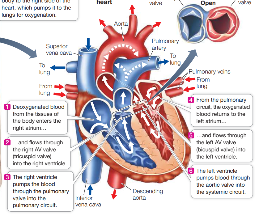
what is the systemic and pulmonary circuit
left pump of heart delivers body throughout the body with the systemic circuit
left side has thicker cardiac muscle wall because it pumps more
right pump delivers blood to the lungs through the pulmonary circuit
what are the major valves in the heart and functions
atrioventricular valve: has a left and right valve, between the atria and ventricles, and opens from atrium towards ventricle so blood goes only in that direction to prevent backflow
right AV valve is tricuspid
left AV valve is bicuspid
pulmonary valve: prevents backflow of blood into right ventricle, directs blood to lungs
aortic valve: prevents backflow of blood into left ventricle, directs blood to aorta
what are the stages of the cardiac cycle
systole - when ventricles contract and blood goes into arteries from atria to muscle
diastole - when ventricles relax
what makes lub dup sound in heart
lub - ventricles contract and atrioventricular valves close, causing pressure to build in ventricles (systole)
dup - the pulmonary and aortic valve open allowing blood in, pressure in ventricles lowers so it relaxes, and now pressure in pulmonary and aortic valves is higher so the valves close which makes the dup sound (end of systole)
what is blood pressure and why does it get high
pressure that blood is pushing onto the vessel walls
*if arteries harden, then they can’t be flexible to accommodate more blood rushing which causes high blood pressure
sinoatrial node
primary pacemaker present on the wall of right atrium at junction between it and superior vena cava
pacemakers are modified cardiac muscle cells that initiate action potentials without stimulation from nervous system
how does action potential/signal travel through the heart during a heartbeat
starts at Sinoatrial node which generates action potential
atria contract
slight delay before Atrioventricular node continues the signal
bundle of His (modified cardiac muscle fibers)
purkinje fibers
ventricles contract
what does blood plasma contain
*the fluid portion of blood, contains:
water
salt (buffer that regulates blood pH)
nutrients like glucose, vitamins, waste products
hormones
proteins, immune response elements
what are the cellular components of blood
red blood cells (erythrocytes) - biconcave and don’t have nucleus when mature; packed with hemoglobin; main function is carrying oxygen
white blood cells (leukocytes) - colorless; involved in inflammation and immune response
platelets - cellular fragments essential for blood clotting; bone marrow produce megakaryocytes which break down into platelets; short lifespan
what is hematocrit
volume of packed blood cells/volume of blood x 100%
measures percentage of red blood cells in your blood
generally higher hematocrit in men
what is the process of forming a blood clot
an injury to blood vessel lining causes collagen fibers to be exposed
platelets are activated and become sticky, adhere to fibers and activate other platelets
prothrombin circulating blood plasma is converted to thrombin
thrombin acts on fibrinogen circulating in plasma to form fibrin
fibrin threads form meshwork that seals wound until vessel wall heals
what are the tissue layers of blood vessels
endothelium - innermost, thin
muscular layer - middle layer of smooth muscle
connective tissue - outer layer with elastic and collagen fibers
elastin layers in between those