PNB Bone Physiology and Articulations
 Call Kai
 Learn
Learn Practice Test
 Spaced Repetition
 Match
Match Flashcards
 Knowt Play
1/98

There's no tags or description
Looks like no tags are added yet.
Name | Mastery | Learn | Test | Matching | Spaced | Call with Kai | Chat |
|---|
No analytics yet
Send a link to your students to track their progress
99 Terms
Functions of bone/skeleton
support, protection, allow movement, storage depot for Ca and P salts, hematopoiesis (making blood cells)
classes of bones
long, short, flat, irregular, sesamoid
long bone example
arms/legs
short bone example
feet/hands
flat bone example
sternum/skull
irregular bone example
vertebrae
sesamoid bone example
patella
parts of long bone
diaphysis, metaphysis, epiphysis, marrow cavity x2
diaphysis
long, skinny shaft
metaphysis
neck - where it gets wider
medullary cavity
center of diaphysis - yellow bone marrow (inactive, fat reservoir)
hollow bubbles
in spongy bone - red bone marrow (active, hematopoietic stem cells)
2 types of bone in long bones
compact and spongy
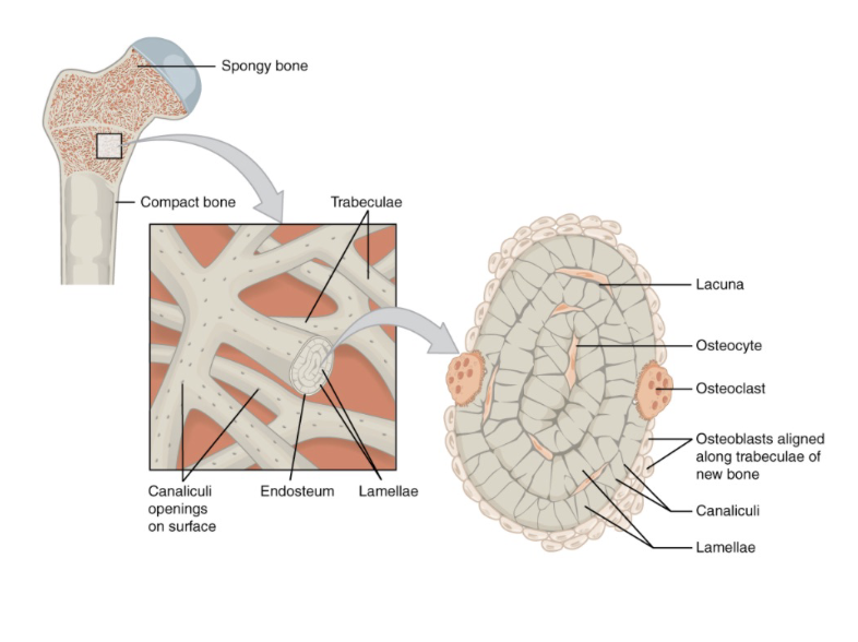
compact bone
osteons w/ concentric layers of lamellae, central canal in middle w/ vessels and nerves
spongy bone
lattice-like network of trabeculae - lighten weight of bone, between is where red bone marrow in epiphysis
layers of long bone
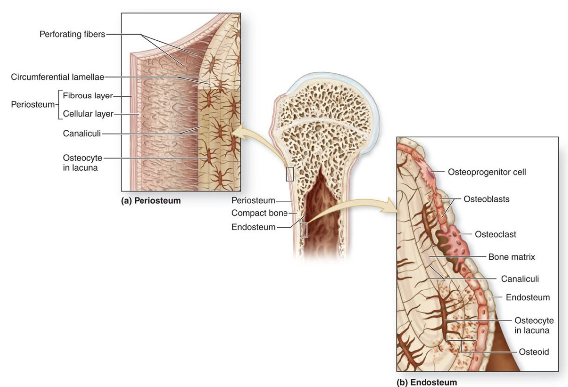
periosteum (fibrous layer and cellular layer) and endosteum
periosteum
wrapping along outside of long bone in two layers - fibrous and cellular
fibrous layer of periosteum
very outside, dense irregular CT - weave around, provide framework, weave bone into tendons/ligaments
cellular layer of periosteum
inside fibrous, contains cells that can deposit calcium salts or take them away — bone remodeling
metaphysis breakdown
contains epiphyseal plate (hyaline cartilage)/epiphyseal line (at 25 yo) — growth plate where bone formation occurs
cartilagenous part of the long bone
articular cartilage - at very end lining of the epiphysis, hyaline cartilage, line the articulations where bones form joint - cushions and reduces frictions
blood supply in bones
very vascularized — blood vessels enter compact bone via Volkmann’s canals (perforating canals) - come in via periosteum, enter perpendicular to central canals

long bone
long bone drawing
compact bone drawing

spongy bone drawing
layers of long bone diagram
two types of bone formation
intramembranous and endochondral
intramembranous ossification
bone forms from mesenchymal cells that differentiate into osteoprogenitor cells that give rise to osteoblasts (forms flat bones)
when skull bones fully ossify
After birth to allow for flexibility through the birth canal — around 2 years
fontanelles
large areas of dense connective tissue on the skull that allow for flexibility during childbirth
endochondral ossification
bone replaces hyaline cartilage (forms most bones in body) — chondrocytes in epiphysis go through hypertrophy, become irregularly shaped, apoptosis, osteoblasts invade empty lacunae made from apoptosis — growth in epiphyseal plate
5 zones of endochondral ossification
1 - resting cartilage, 2 - proliferating cartilage, 3 - hypertrophic cartilage, 4 - calcified cartilage, 5 - ossification
fetal endochondral ossification zones
1 - hyaline cartilage bone model, 2 - periosteal collar, 3 - primary ossification center, 4 - x2 secondary ossification centers, 5 - child bone
periosteal collar
bone added to surface of diaphysis through outside contact with blood vessels — stem cells enter, begin to build matrix around edge = collar)
primary ossification center
source of bone cells — blood vessel invades diaphysis, osteoblasts spreading out with bone matrix (center), diaphysis always transitions into bone first
secondary ossification centers
collar merges with matrix within,blood invading each epiphysis to form 2 more centers
child bone
osteoblasts work to produce matrix until fully enclosed by matrix — lacunae, spongy bone eventually to compact
birth plates
bone maintains ability to grow out length after baby is born because of
2 types of long bone growth
growth in length or growth in width (appositional)
long bone growth in length
cartilage proliferation on epiphyseal side of plate, ossification occurs on diaphyseal side, process continues until chondrocytes stop proliferating and entire growth plate ossifies
appositional growth (width)
adding layers by remodeling marrow cavity, compact bone thickens and strengthens long bone with layers of circumferential lamellae — *can occur in adults if something in lifestyle (ie. weight-training)
bone remodeling in adults
in response to changes in demand (force/load) on bones — weight-training or prolonged bed rest
Wolff’s Law
bones remodel in response to compressive force (higher compressive force=thicker bones and vice versa) — bed rest vs weight-training
calcium reservoir
bones store calcium in excess and break it down in lack of it
two hormones regulating blood calcium homeostasis
parathyroid hormone (PTH) and calcitonin
plasma calcium is low
triggers PTH from thyroid into blood, triggers osteoclasts and reabsorption of calcium, increase Ca to homeostasis
plasma calcium is high
trigger receptors in thyroid, release calcitonin, inhibits osteoclasts, stimulates osteoblasts — create hydroxyapatite, remove Ca from blood, back to homeostasis
osteoporosis
osteoclast activity outpaces osteoblasts as you age— lose bone density, common in older adults and post-menopausal women (lack of estrogen), problematic b/c leads to fractures/decreases calcium reserves/kyphosis
kyphosis
symptom of osteoporosis, excessively curved spine, leads to decrease in lung capacity, decrease in optimal circulation, compress certain nerves
bone repair after break
1 - hematoma (blood vessels throuhgout blood burst and need to contain blood)
2 - cells from endosteum form fibrocartilage around break (internal soft callus), cells from periosteum form hyaline cartilage callus around outside of bone,
3 - osteoclasts reabsorb dead bone fragments and osteoblasts remodel two calli into bony calluses
4 - remodeling (spongy bone back in appropriate places, compact bone in superficial parts)
types of fractures
open/compound (bone breaks through skin - infection), closed (contained w/in skin), and greenstick (bone doesn’t break all way through — children bc not fully ossified)
ALSO includes transverse, spiral, comminuted, impacted, and oblique
articulation
meeting place of 2+ bones
range of motion (ROM)
normal extent of mobility for a specific joint movement — in degrees, differs for everyone (ie. wrist flexion is about 70-90 degrees)
degrees of freedom
number of axes at which movement in a joint occurs, same for everyone (wrist is biaxial joint w/ 2 degrees of freedom and 2 planes of movement)
mobility vs stability
inverse relationship — mobility comes at the risk of stability (ie. shoulder very mobile, more likely to injure)
joint classifications by structure
fibrous, cartilagenous, synovial
joint classifications by function
synarthrosis, amphiarthrosis, diarthrosis
synarthrosis
immoveable (bones meeting in skull)
amphiarthrosis
slightly moveable (public symphesis)
diarthrosis
freely movable (shoulder, hips)
flexion/extension
movements that occur in sagittal plane (front and back)
flexion
bends joint, decreases angle (bend arm)
extension
extends joint, increases angle (extend arm)
ankle movements
dorsiflexion (point up) and plantar flexion (point down)
abduction/adduction
movements that occur in the frontal plane (side to side)
abduction
moving away from midline (raising arm to side)
adduction
moving towards midline (putting arm against body)
circumduction
movement in cone-like shape, both sagittal and frontal plane (moving hand in circle)
rotation
movements that occur around longitudinal axis of moving segment (twisting torso, turning head)
pronation/supination
specialized rotation at the proximal and distal radioulnar joints
supination
palms forward, radius and ulna parallel (anatomical position)
pronation
palms down, radius crosses over ulna
inversion/eversion
special movement of intertarsal joints (ankle)
inversion
bring sole of foot towards midline (in to the body)
eversion
bring sole of foot away from midline (evict from body)
retraction/protraction
forward/backward movement
retraction
pull back (pull in jaw, pull scapula together/back)
protraction
push forward (push out jaw, push scapula forward)
elevation/depression
up and down movement
elevation
move upward (shrug shoulders)
depression
move down (rest shoulders)
opposition
special movement of any finger coming together w/ thumb — pincer grasp, special to few mammals, allows dexterity
joint tidbits
all fibrous and cartilagenous are either gonna be synarthrosis or amphiarthrosis (bc tissue doesn’t allow for a lot of movement)
all synovial joints are diarthrosis (fluid-filled sac that allows for lots of movement)
fibrous joints vocab
sutures of skull, fontanels, syndesmosis
sutures of skull
bound together by short, tight fibers, makes strong articulation — interdigitating (interlock/weave, add strength +stability to joint) — synarthrosis
fontanels
special joints in skulls of fetuses/infants - large areas of dense irregular CT that allow for some movement during birth
syndesmosis joint
connects 2 bones together by tight, longer fibers - interosseous ligament that allows for slight give, very stable (ie. distal tibio-fibular articulation that allows for increased ROM for ankle)
cartilagenous joints vocab
synchodrosis and symphisis
synchondrosis joint
hyaline cartilage connects bones, immovable (ie. long bones, epiphyseal plate during growth or first rib + sternum)
symphisis
fibrocartilage discs unite bones, allows slight movement between articulating bones (between pubic bones, bodies of vertebra)
pubic symphisis during childbirth is special case - opens up a lot more due to estrogen and relaxin that release the ligament itself
synovial joints basic anatomy
joint cavity containing synovial fluid (cushion, lubricate, and nourishes), freely movable joints, contains lots of blood vessels that secrete into fluid, all share common anatomy
other synovial structures
bursa (pillow for synovial fluid, reduces friction and tension) and tendon sheath (synovial fluid-filled structure, forms sleeve around tendons and reduces friction)
types of synovial joints
hinge, pivot, condyloid, saddle, ball-and-socket, and gliding

hinge joint
concave surface articulating with a convex surface, uniaxial, flexion/extension (ie. elbow)

pivot joint
rounder portion of bone rotates w/in a ring of bone/ligament, uniaxial, rotation (ie. atlanto-axial joint)
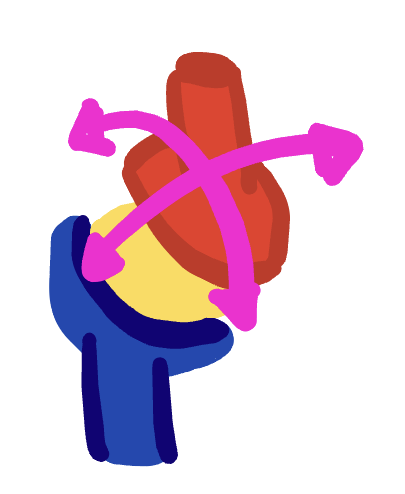
condyloid joint
shallow depression at one end of one bone articulates w/ rounded structure of other bone, biaxial (ie. wrist)
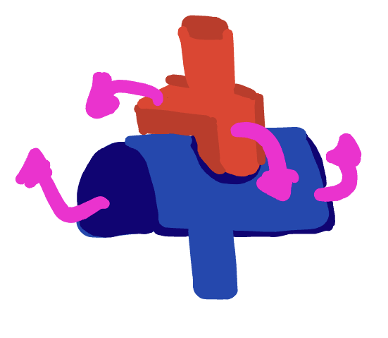
saddle joint
both bones have saddle shape, concave one direction, convex other, rides on saddle, biaxial (ie. first carpo-metacarpal)

ball-and-socket joint
rounded head of one bone fits into concave articulation of adjacent bone, multiaxial (ie. shoulder, hip)

gliding joint
flat/nearly flat articular surfaces that allow gliding in any direction, nonaxial/multiaxial (ie. sternocostal)