Male Reproductive System I
1/32
Earn XP
Description and Tags
Testis and Spermatozoa
Name | Mastery | Learn | Test | Matching | Spaced | Call with Kai |
|---|
No analytics yet
Send a link to your students to track their progress
33 Terms
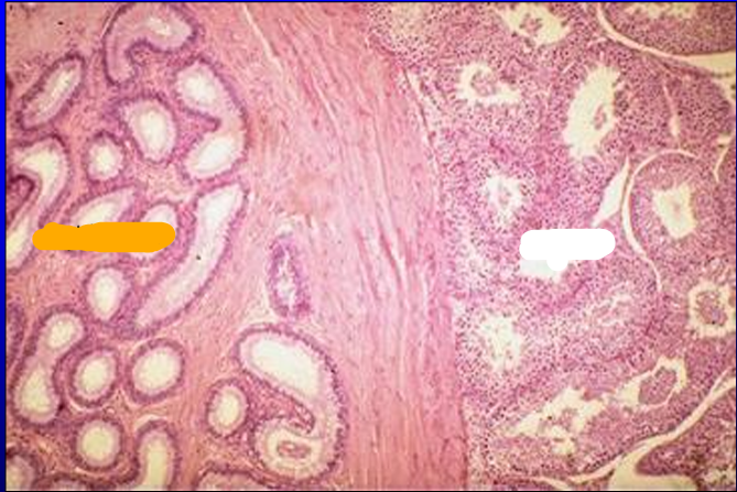
Identify the cut:
Identify the structure:
Specie:
Stain used:
Orange:
White:
Pink:
Magenta:
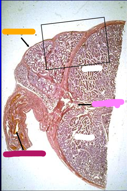
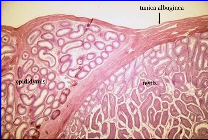
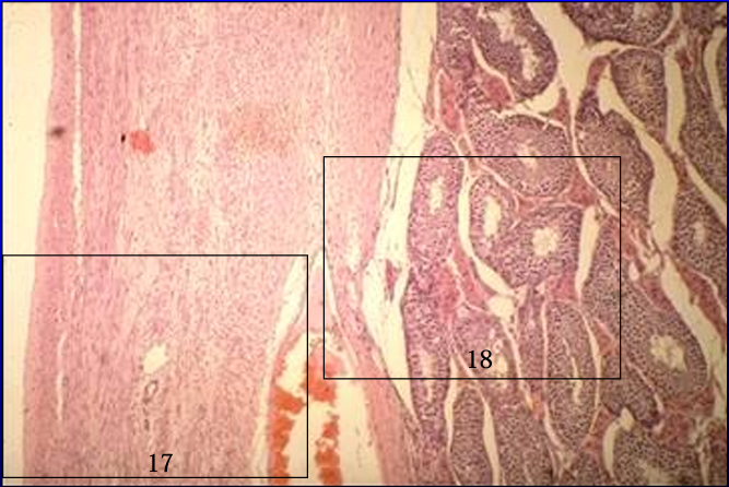
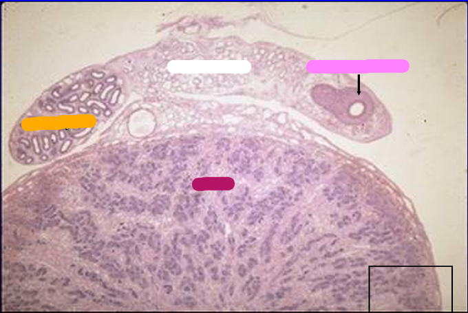
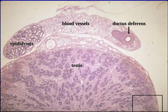
Cross-section of the canine testis (right) and epididymis (top, left), H-E stain.
The section was cut at the level where the head of the epididymis is attached to the testis.
The structure below the epididymis (lower left) contains the blood supply to the testis and epididymis.
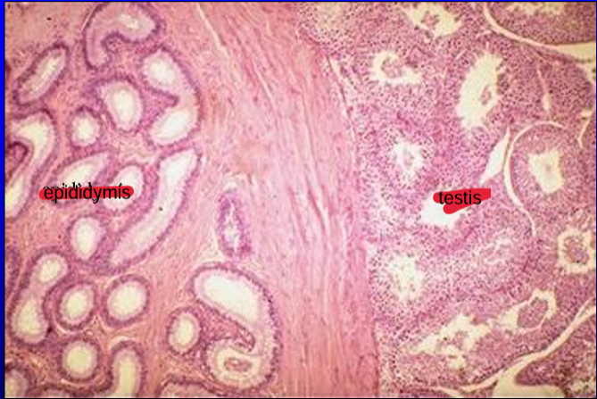
Identify the cut:
Identify the structure:
Specie:
Stain used:
Orange:
White:
Pink:
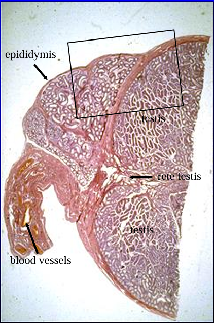
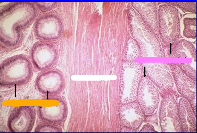
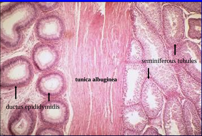
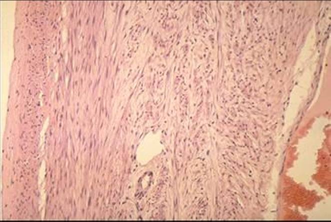
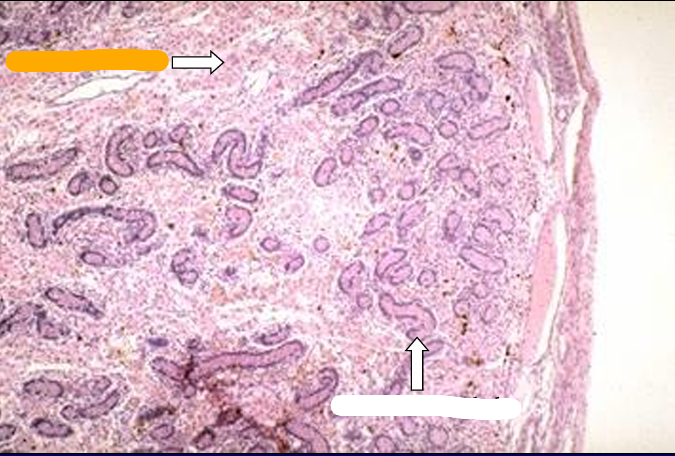
The testis (right) and epididymis (left) are shown at a slightly higher magnification (6X).
The dense connective tissue that surrounds the testis and separates it from the epididymis is the tunica albuginea.
The dense connective tissue that surrounds the testis and separates it from the epididymis is the
tunica albuginea.
Identify the structure:
Specie:
Orange:
White:
Pink:
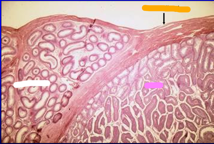
Testis (right) and epididymis (left). 20X. The epididymis contains ducts which are lined by a pseudostratified columnar epithelium; the testis contains tubules lined by a stratified epithelium.
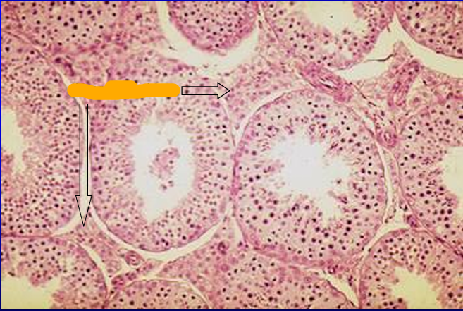
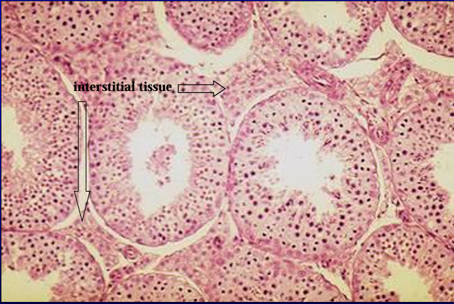
Identify the cut:
Identify the structure:
Specie:
Orange:
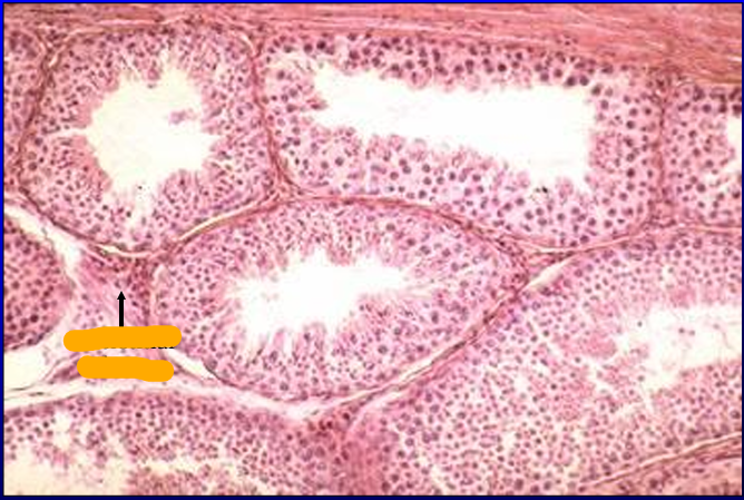
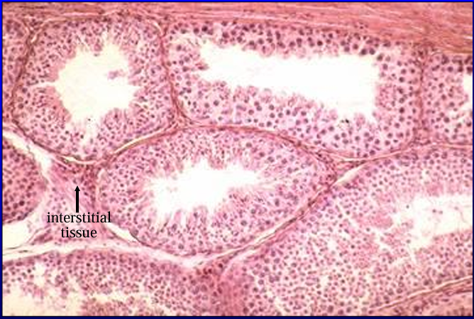
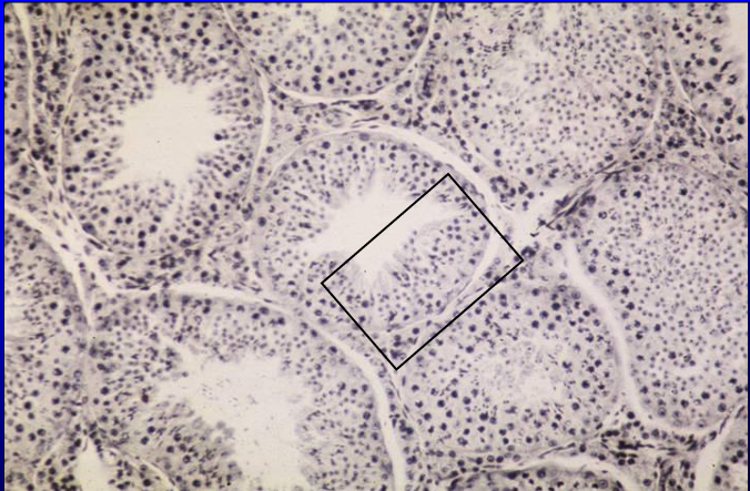
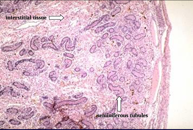
Canine testis. 50X.
The parenchyma of the testis contains convoluted seminiferous tubules; they were cut transversely and obliquely in this slide. Located between the seminiferous is the interstitial tissue
Identify the structure:
Specie:
Orange:
White:
Pink:
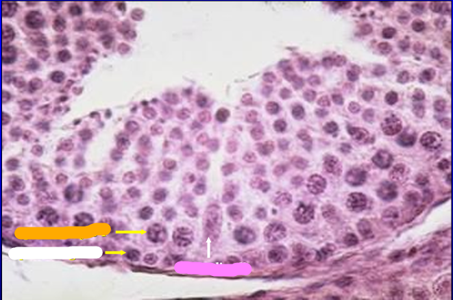
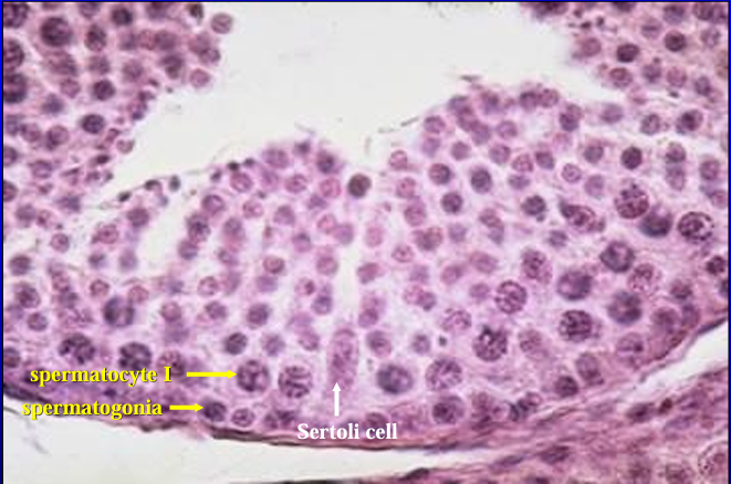
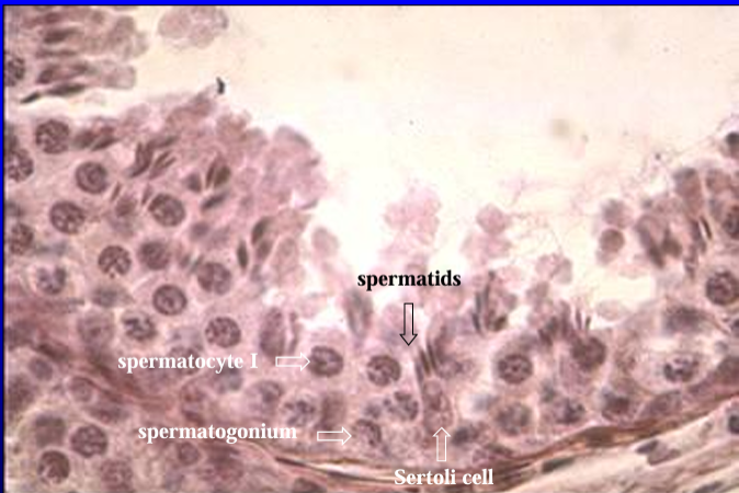
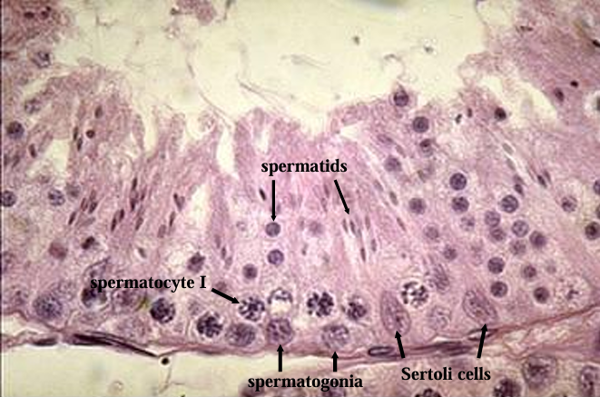
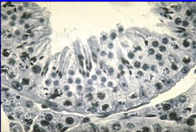
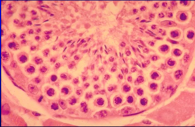
Canine seminiferous tubule.
The seminiferous tubule is lined by a stratified epithelium composed of Sertoli cells and spermatogenic cells.
A Sertoi cell is basically columnar with an oval euchromatic nucleus with prominent nucleolus.
The basal layer of spermatogenic cells is composed of spermatogonia.
The next layer of cells contain large nuclei with thick chromosomes these are the primary spermatocytes.
Toward the luminal side (top) are several layers of cells in more advanced stages of development - the secondary spermatocytes and spermatids
Identify the structure:
Specie:
Orange:
White:
Pink:
Magenta:
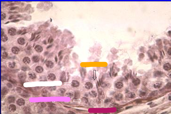
Another view of the seminiferous epithelium.
The spermatogonia, primary spermatocytes, and late spermatids (these have dark elongate nuclei) are seen from the bottom to the top layers.
Identify the structure:
Specie:
Orange:
White:
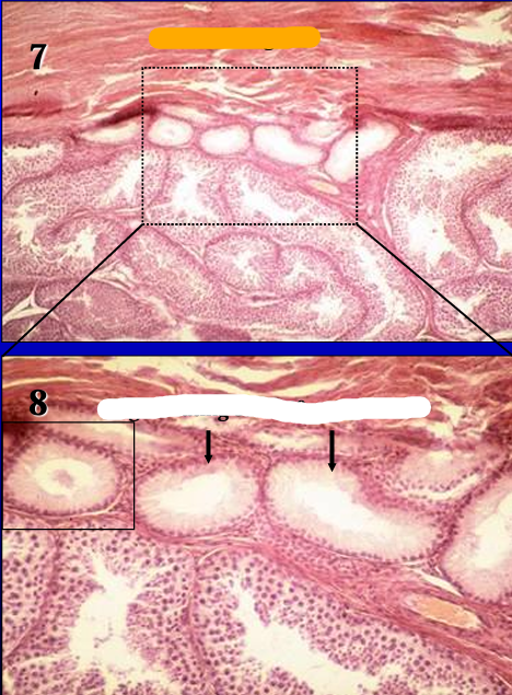
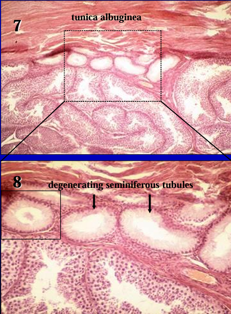
Figure 7 (20X) and Figure 8 (50X) show in increasing magnification the tunica albuginea (dense connective tissue), degenerating seminiferous tubules, and normal seminiferous tubules.
Identify the structure:
Specie:
Lined by:
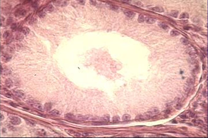
Degenerating seminiferous tubule of the canine testis. 200X.
Note the absence of spermatogenic cells.
The tubule is lined by Sertoli cells only
Identify the structure:
Specie:
Orange:
White:
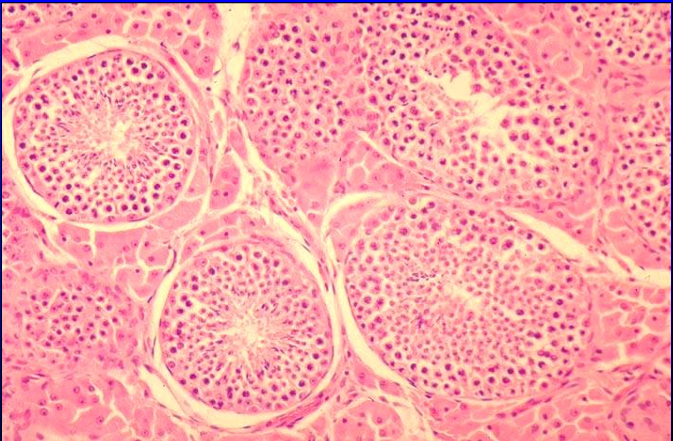
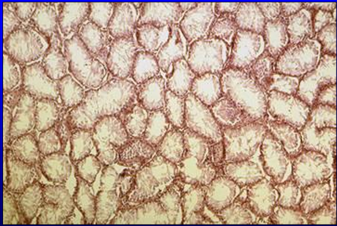
Section of feline testis and epididymis. 20X.
The testis and the epididymis are separated by the tunica albuginea.
Notice the difference in the type of epithelium that lines the ducts of the epididymis and the seminiferous tubules of the testis.
The seminiferous tubules are seen at higher magnifications in the figures that follow
Identify the structure:
Specie:
Orange:
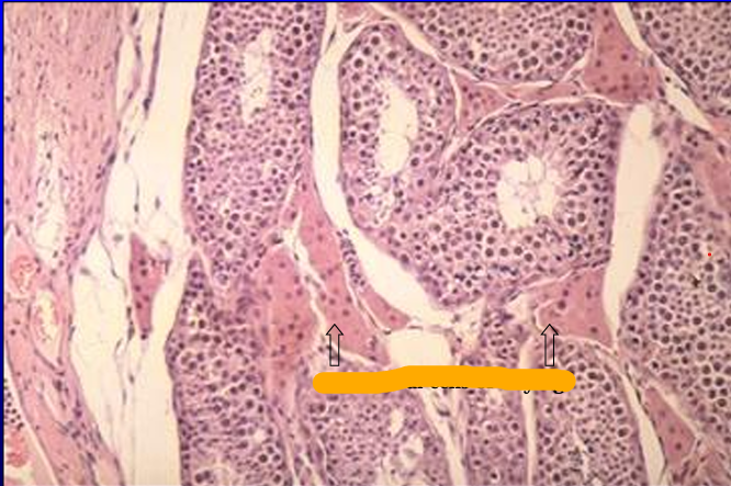
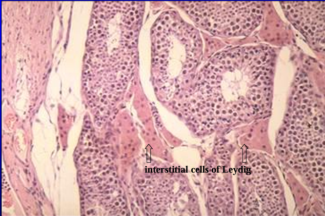
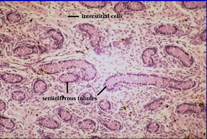
Feline seminiferous tubules.
In this section of the feline testis, note the abundant interstitial tissue between the seminiferous tubules (in comparison to the scanty interstitial tissue seen in the canine testis)
Identify the structure:
Specie:
Orange:
White:
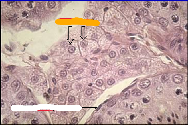
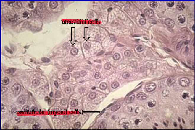
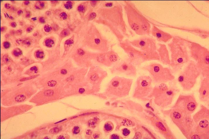
Interstitial cells of Leydig, feline testis.
The interstitial cells are steroid-secreting endocrine cells and they exhibit the characteristic pale vacuolated (foamy) cytoplasm.
What hormone is secreted by these cells? The seminiferous tubule is surrounded by peritubular (myoid) cells; these cells have a flat nucleus and are located just outside the basement membrane (of the seminiferous tubule at the lower right of the figure)
Identify the structure:
Specie:
Orange:
White:
Pink:
Magenta:
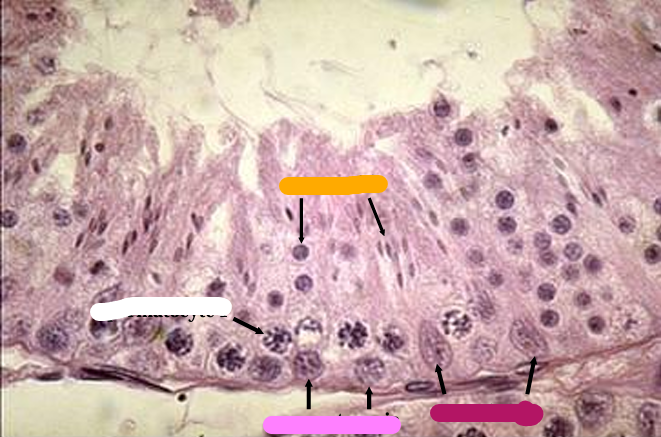
Seminiferous tubule of feline testis.
The seminiferous epithelium shows Sertoli cells (with oval euchromatic nucleus), spermatogonia, primary spermatocytes, and early and late spermatids.
Peritubular (myoid) cells can be seen just outside the seminiferous tubule
Identify the structure:
Specie:
Stain used:
Feline seminiferous tubules, iron-hematoxylin stain.
Iron-hematoxylin stain is used to highlight nuclear details of the spermatogenic cells. Those in the inset are seen at a higher magnification in the next figure.
Identify the structure:
Specie:
Stain used:
Feline seminiferous tubules, iron-hematoxylin stain.
Identify the following structures in this figure: interstitial cells of Leydig and peritubular cells in the interstitial tissue; Sertoli cells, spermatogonia, primary spermatocytes, and early and late spermatids in the seminiferous tubules
Identify the structure:
Specie:
Section of equine testis.
The section shows the tunica albuginea and the seminiferous tubules.
Note the vascular layer in the inner part to the tunica albuginea.
Identify the structure:
Specie:
Equine testis.
This higher magnification of the previous slide shows the presence of abundant smooth muscle fibers in the tunica albuginea of the equine testis
Identify the structure:
Specie:
Orange:
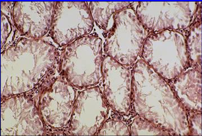
Parenchyma of equine testis.
The convoluted seminiferous tubules and the interstitial tissue are shown.
Identify the structure:
Specie:
Seminiferous tubules and interstitial tissue of equine testis.
Identify the structure:
Specie:
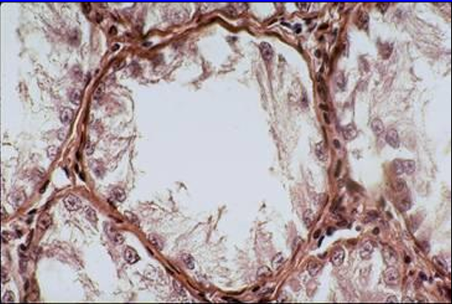
Interstitial cells of Leydig, equine testis.
The interstitial cells are abundant in the equine testis.
The cells exhibit an acidophilic cytoplasm
Identify the structure:
Specie:
Seminiferous tubule, equine testis.
The spaces surrounding the spermatogenic cells are artifacts of preparation.
Identify the interstitial cells of Leydig, peritubular (myoid) cells, spermatogonia, primary spermatocytes, early and late spermatids, and spermatozoa
Identify the structure:
Specie:
Orange:
White:
Pink:
Magenta:
Section of the testis (bottom), epididymis (top left), and ductus deferens (top right) of a 1-day-old pig.
Identify the structure:
Specie:
Orange:
White:
Parenchyma of testis, 1-day-old pig.
The seminiferous tubules and the interstitial tissue are shown
Identify the structure:
Specie:
Orange:
White:
Seminiferous tubules and interstitial tissue of testis of 1-day old pig.
Identify the structure:
Specie:
Orange:
White:
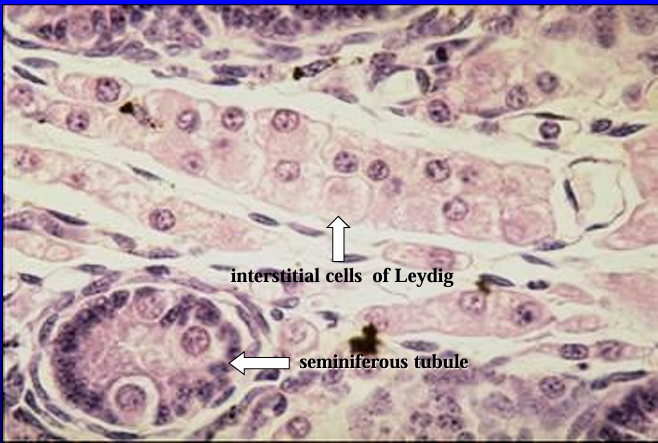
Interstitial tissue of testis of 1-day-old pig.
The interstitial cells are abundant in the porcine testis.
These cells exhibit an acidophilic and vacuolated cytoplasm. A seminiferous tubule is cut transversely in the lower left section of the screen
Identify the structure:
Specie:
Orange:
White:
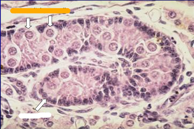
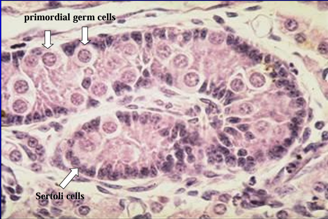
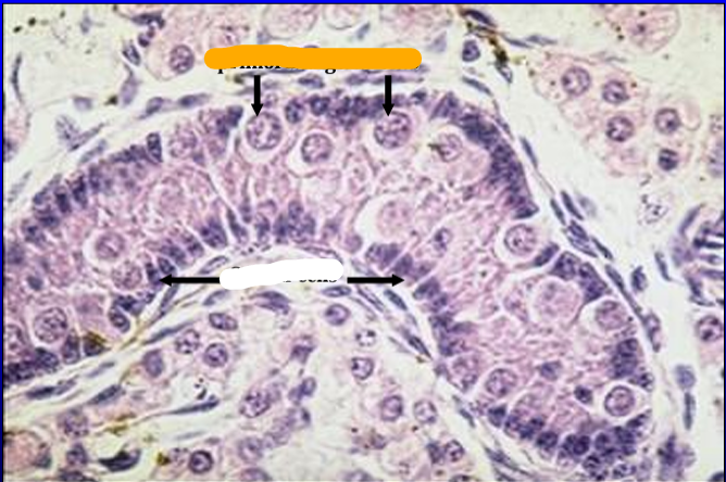
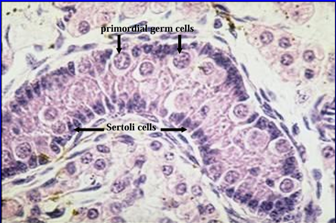
Seminiferous tubules, testis of 1-day-old pig.
The lining epithelium is composed mainly of Sertoli cells (columnar cells with dark nuclei) and primordial germ cells (spermatogonia) - the large round cells.
Spermatogenesis has not yet begun at this early age and, thus, other spermatogenic cells are not seen in the section
Identify the structure:
Specie:
Orange:
White:
Another view of the seminiferous tubule and the interstitial cells of the testis of 1-day-old pig.

Identify the structure:
Specie:
Section of a caprine cryptorchid testis.
Identify the structure:
Specie:
Caprine cryptorchid testis.
Notice the absence of spermatogenic cells in the seminiferous tubules
Identify the structure:
Specie:
Caprine cryptorchid testis.
Notice the absence of spermatogenic cells.
Cells of Leydig are present in the interstitial tissue (they are not, however, abundant in this section)
Identify the structure:
Specie:
Seminiferous tubule, caprine cryptorchid testis.
The principal cell type lining the seminiferous tubules is the Sertoli cell.
Spermatogenic cells are not seen in this section
Identify the structure:
Specie:
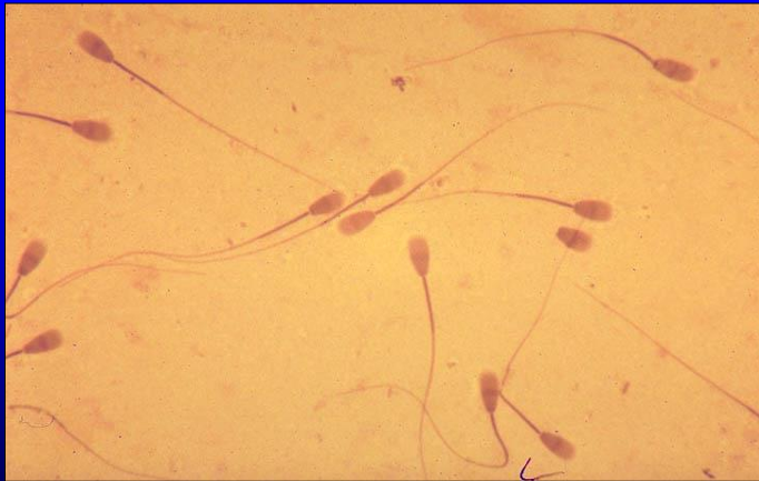
Bovine semen smear.
The head and tail are readily recognizable but the spermatozoa will show better detail when examined under oil immersion.
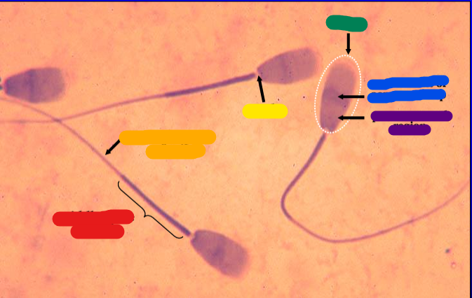
Identify the structure:
Specie:
red:
orange:
yellow:
green:
blue:
violet:
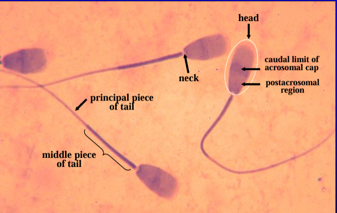
Bovine spermatozoa.
The head contains the nucleus, acrosomal cap, and postacrosomal region.
The neck is the narrow constriction that connects the head and the tail.
The middle piece and principal piece of tail can be identified in all three spermatozoa seen in this figure