Bones and Muscles of Thigh and Knee
 Call Kai
 Learn
Learn Practice Test
 Spaced Repetition
 Match
Match Flashcards
 Knowt Play
1/103

There's no tags or description
Looks like no tags are added yet.
Name | Mastery | Learn | Test | Matching | Spaced | Call with Kai | Chat |
|---|
No analytics yet
Send a link to your students to track their progress
104 Terms
Anterior thigh muscles
Muscles that flex the thigh at the hip and/or extend the leg at the knee
Iliacus
Flexes the thigh at the hip
Iliacus attachment
Hip bone to lesser trochanter
Psoas major
Flexes the thigh at the hip
Psoas major attachment
Vertebrae to lesser trochanter
Sartorius
Flexes the thigh at the hip and also flexes the leg at the knee
Sartorius attachment
Anterior superior iliac spine to tibia
Sartorius fact
Longest muscle in the body
Quadriceps femoris
Extends the leg at the knee joint
Quadriceps femoris attachment
Attaches via quadriceps tendon, patella, and patellar ligament
Four quadriceps muscles
Rectus femoris, vastus lateralis, vastus intermedius, vastus medialis
Rectus femoris
One of the four quadriceps femoris muscles
Vastus lateralis
Lateral quadriceps muscle
Vastus intermedius
Quadriceps muscle located behind rectus femoris
Vastus medialis
Medial quadriceps muscle
Medial thigh muscles
Adduct and flex the thigh
Adductors
Adductor longus, adductor brevis, and adductor magnus
Adductor longus
Medial thigh muscle that adducts and flexes the thigh
Adductor brevis
Medial thigh muscle that adducts and flexes the thigh
Adductor magnus
Medial thigh muscle that adducts and flexes the thigh
Pectineus
Medial thigh muscle that adducts and flexes the thigh
Gracilis
Adducts/flexes the thigh and also flexes the leg at the knee
Gracilis location
Travels along the medial thigh
Lateral thigh
Contains a single muscle that abducts the thigh at the hip
Tensor fasciae latae
Abducts the thigh and connects to the iliotibial tract
Iliotibial tract (IT band)
Band of connective tissue associated with tensor fasciae latae
Gluteal muscles
Posterior hip muscles
Gluteus maximus
Extends the thigh at the hip joint
Gluteus maximus example action
Used when getting out of a chair to stand
Gluteus medius
Abducts the thigh
Gluteus minimus
Abducts the thigh
Gluteus maximus depth
Most superficial of the three gluteal muscles
Gluteus minimus depth
Deepest of the three gluteal muscles
Hamstrings
Posterior thigh muscles that extend the thigh and flex the leg
Hamstring hip action
Extend the thigh at the hip joint
Hamstring knee action
Flex the leg at the knee joint
Three hamstring muscles
Biceps femoris, semitendinosus, semimembranosus
Biceps femoris
Laterally located hamstring muscle
Semitendinosus
Hamstring muscle
Semimembranosus
Hamstring muscle
Hamstring common attachment
Group has similar attachment from ischial tuberosity toward the leg
Knee joint classification
Synovial hinge joint; diarthrosis
Bones associated with knee
Femur, tibia, and patella
Fibula and knee joint
Fibula does not articulate with the femur
Patella location
Embedded in the quadriceps femoris muscle tendon
Quadriceps tendon
Connects quadriceps femoris toward the patella
Patellar ligament
Continuation from patella to tibia
Patellar ligament attachment
Attaches to the tibial tuberosity
Knee collateral ligaments
Provide stabilization and support to the knee
Fibular collateral ligament
Prevents hyperadduction of the leg
Fibular collateral ligament function
Prevents leg from shifting too far medially from the femur
Tibial collateral ligament
Prevents hyperabduction of the leg
Tibial collateral ligament function
Prevents leg from shifting too far laterally from the femur
Cruciate ligaments
Knee ligaments that cross, forming an X
Anterior cruciate ligament (ACL)
Prevents hyperextension of the knee joint
ACL additional function
Prevents tibia from moving too far anteriorly relative to femur
Posterior cruciate ligament (PCL)
Prevents hyperflexion of the knee joint
PCL additional function
Prevents tibia from shifting posteriorly relative to femur
Menisci
Medial and lateral C-shaped fibrocartilage pads within the knee joint
Medial meniscus
C-shaped fibrocartilage pad on medial side of knee
Lateral meniscus
C-shaped fibrocartilage pad on lateral side of knee
Menisci function
Stabilize and cushion the knee joint
Other example of fibrocartilage
Intervertebral discs
ACL injury causes
Hyperextension and twisting movements
ACL rupture
A popping noise may occur when the ligament ruptures
Sports associated with ACL injuries
Sports involving twists, sudden stops, and changes in direction
Unhappy triad
Knee injury commonly caused by a lateral blow to the knee
Three structures of the unhappy triad
ACL, tibial collateral ligament, and medial meniscus
Unhappy triad — ACL
One of the three structures torn
Unhappy triad — tibial collateral ligament
One of the three structures torn
Unhappy triad — medial meniscus
One of the three structures torn
Anterior thigh general action
flex the thigh, extend the leg
Medial thigh general action
flex and adduct the thigh
Posterior thigh (hamstrings) general action
extend the thigh and/or flex the leg
Lateral Thigh general action
abduct the thigh
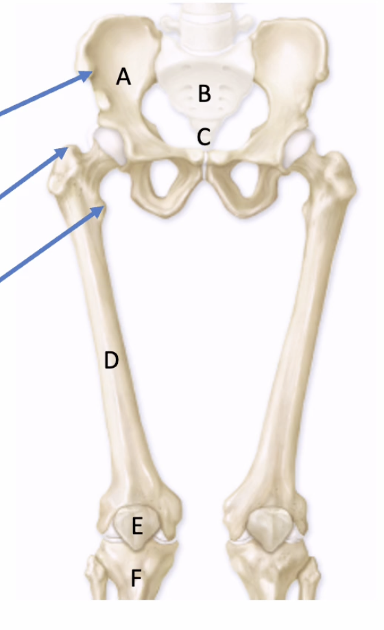
Name letter B
Sacrum

Name letter C
Coccyx
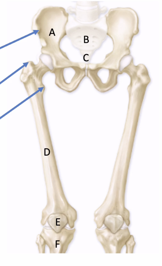
Name what top arrow is pointing to
Anterior superior iliac spine

Name what middle arrow is pointing to
Greater trochanter

Name what bottom arrow is pointing to
Lesser trochanter

Name yellow muscle
Sartorius

Name green muscle
Psoas major

Name blue muscle
Iliacus

Name blue muscle
Recuts Femoris
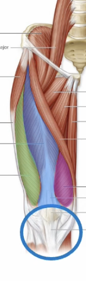
Name green muscle
Vastus Lateralis

Name purple muscle
Vastus medialis

Name purple muscle
Adductors

Name blue muscle
Pectineus
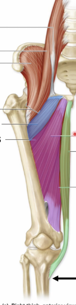
Name green muscle
Gracilis

Name yellow muscle
Tensor fasciae latae

Name green muscle
Gluteus Medius

Name blue muscle
Gluteus maximus

Name purple muscle
Gluteus Minimus

Name blue muscle
Biceps Femoris (laterally placed)

Name green muscle
Semitendinosus
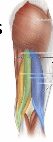
Name yellow muscle
Semimembranosus

name blue outline
Quadriceps tendon

name green outline
Patellar ligament
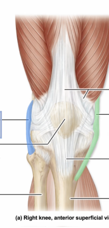
Name blue label
Fibular collateral ligament
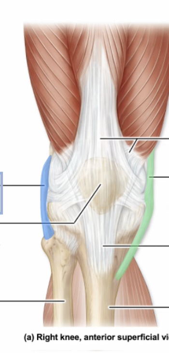
Name green label
Tibial collateral ligament