Immnofluorescence & Autoimmunity
 Call Kai
 Learn
Learn Practice Test
 Spaced Repetition
 Match
Match Flashcards
 Knowt Play
1/33

There's no tags or description
Looks like no tags are added yet.
Name | Mastery | Learn | Test | Matching | Spaced | Call with Kai | Chat |
|---|
No analytics yet
Send a link to your students to track their progress
34 Terms
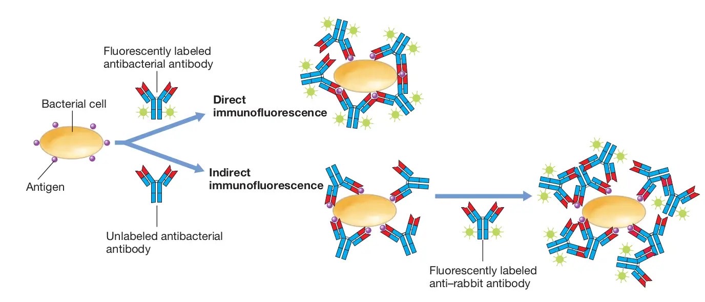
Direct fluorescence
Tagged labeled antibody is added to patient tissue antigen/sera to detect antibody
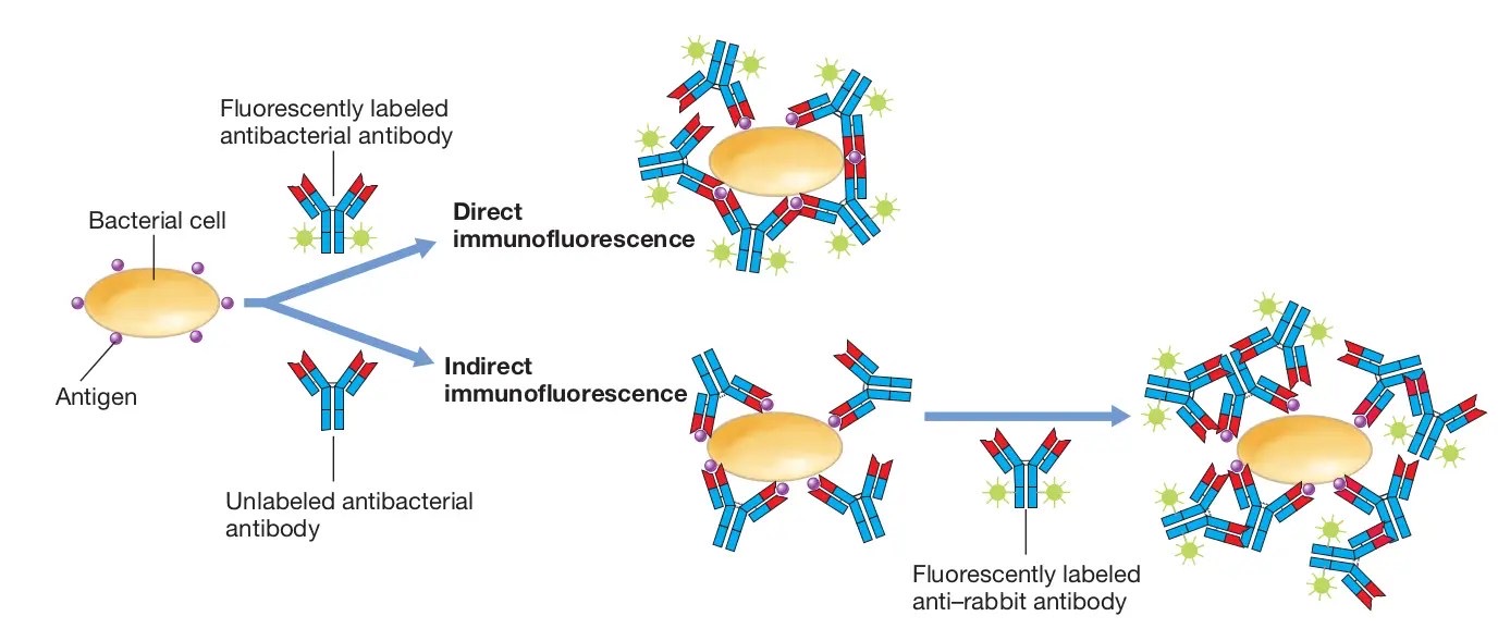
Indirect fluorescence
detects specific antibodies in a patient's serum by using a fluorescently labeled secondary antibody to visualize the binding of the primary antibodies (from the patient's sample) to a known antigen
Use of a conjugate
Autoantibody testing
Antibodies against self cell, circulating immune complex cause auto immune disease
Substrates = animal or human tissue, organs, or cells
Rat kidney/stomach/liver
Treponema, Mycoplasma, Legionella
FITC
an antibody that has been conjugated (labeled) with the fluorescent molecule fluorescein isothiocyanate (FITC), or an antibody that recognizes FITC itself
To visualize and quantify specific molecules or cells
Flow cytometry
ANA antinuclear antibody
Develop into antibody to
DsDNA
SSa
SSb
Sm
RNP
Nucleolar RNA
ANA elevated in
SLE, RA, MCTD, scleroderma, dermatomyositis, malignancies and hypersensitivity
ANA low titers in
Aging, certain viral diseases, chronic hepatitis
Dilute sample to reduce errors
ANA procedure
Hep-2 cells are fixed on slides
Serum added and incubated
Washed then conjugate added
Washed and counterstained = Specific patterns
dsDNA pattern
Homogenous pattern = SLE
RNP pattern
Speckled = MCTD/SLE
Sm (smith)
Speckled = SLE
Scl-70 pattern
Speckled = scleroderma
SSa pattern
Speckled = SLE/ Sjogren’s
SSb pattern
Speckled = Sjogrens
Centromere pattern
Centromere = CREST variant, Scleroderma
Nucleolus pattern
Nucleolar = Scleroderma, Sjogrens or SLE
Chromatin pattern
Homogenous = SLE
Crithidia luciliae
Substrate, Hemoflagellate
Kinetoplast rich in DNA = identify dsDNA antibody
Brighter fluoresence
AMA
Primary biliary cirrhosis
Antibodies against kidney
ASMA
Chronic active hepatitis
Antibodies against rat stomach/mucosa/ smooth muscle
SLE Systemic Lupus Erythematosus
Systemic disease, mainly in fenales
Butterfly rash, renal, CNS, cardiac/respiratory
Reynauds phenomenon
SLE causes
Multiple ANA patterns
Hormonal, genetic, environmental, drug induced
SLE test results
Decreased C4, CH50
Positive DNA antibody
ANA, DNA, ENA (Sm/RNP), Scl-70, SSa/SSb
Sjogrens syndrome
May be seen in conjunction with Rheumatoid arthritis
Systemic
Sicca complex, more common in women
Dry eyes/mouth, arthritis
Antibodies to salivary/tear glands = SSa/SSb
MCTD Mixed connective tissue disease
Symptoms similar to SLE, swelling in hands
Most common in women
95% = +ve RNP
Scleroderma
Fibrotic and degenerative changes in skin, synovium and internal organs
Hardening and tightening of skin and connective tissue
Scl-70, Nucleolar pattern ANA, multiple body systems
CREST variant = skin, hand, face
CREST variant scleroderma
Centromere pattern
Calcinosis = calcium deposits
Raynaud’s phenomenon = spasm of blood vessels
Esophageal dysfunction = acid reflux
Sclerodactyly = thickening and tightening of skin
Telangiectasis = Dilation of capillaries, red marks
Goodpastures syndrome
AMA = Rat kidney
Glomerular basement membrane antibodies
Basement membrane antibodies in respiratory tract
Glomerulonephritis and pulmonary hemorrhage
Pernicious anemia
Gastric parietal cell antibody
Intrinsic factor B12 = Needed for DNA production = Megaloblastic anemia
ASMA test = Smooth muscle/stomach
Drug induced SLE
Isoniazid, nitrofurantoin, procainamide, hydralazine
Homogenous ANA
No renal or CNS involvement
Type 1 diabetes
Insulin dependent diabetes
HLA DR3 and DR4, P-ANCA
Destruction of insulin producing beta cells of islets of langerhans
Autoantibodies to beta cell antigens
ANCA anti neutrophil cytoplasmic antibodies
Antibodies to granules in PMNs
Associated with autoimmune vasculitis
Wegener’s granulomatosis
C-ANCA
P-ANCA
C-ANCA
Cytoplasmic staining pattern
P-ANCA
Perinuclear staining pattern