PATHOLOGY LAB MIDTERMS
 Call Kai
 Learn
Learn Practice Test
 Spaced Repetition
 Match
Match Flashcards
 Knowt Play
1/139

There's no tags or description
Looks like no tags are added yet.
Name | Mastery | Learn | Test | Matching | Spaced | Call with Kai | Chat |
|---|
No analytics yet
Send a link to your students to track their progress
140 Terms
Hypertrophy
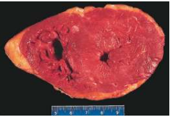
Healthy myocyte
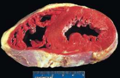
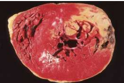
Necrotic myocyte
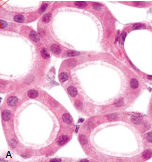
Normal, healthy kidney tubules with viable epithelial cells.
Early (reversible) ischemic injury showing surface blebs, increased eosinophilia of cytoplasm, and swelling of occasional cells.
Kidney
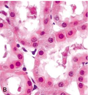
Necrosis (irreversible injury) of epithelial cells, with loss of nuclei, fragmentation of cells, and leakage of contents.
Kidney

Coagulative necrosis.
Kidney
Microscopic view of the edge of the infarct, with a healthy kidney (N) and necrotic cells in the infarct
(I) showing preserved cellular outlines with loss of nuclei and an inflammatory infiltrate (seen as nuclei of inflammatory cells between necrotic tubules).

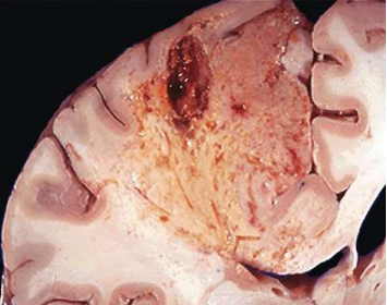
Liquefactive necrosis
Brain
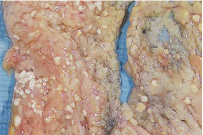
Fat necrosis - white chalky deposits represent foci of fat necrosis with calcium soap formation (saponification) at sites of lipid breakdown in the mesentery.
Caseous necrosis - Tuberculosis of the lung, with a large area of caseous necrosis containing yellow-white and “cheesy” appearing debris.

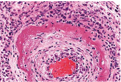
Fibrinoid necrosis in an artery
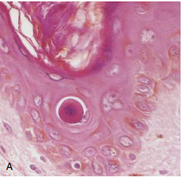
Apoptosis of an epidermal cell. The cell is reduced in size and contains brightly eosinophilic cytoplasm and a condensed nucleus
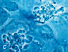
Apoptosis

Blebbing and formation of apoptotic bodies
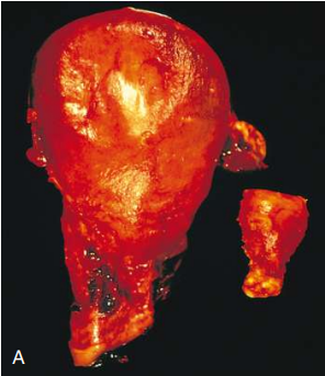
Physiologic hypertrophy of the uterus during pregnancy
Small spindle-shaped uterine smooth muscle cells from a normal uterus

large plump cells from the gravid uterus

Metaplasia of columnar epithelium to squamous epithelium in a bronchus (common with smoking).

Cholesterolosis - Cholesterol-laden macrophages (foam cells, arrow) in a focus of gallbladder cholesterolosis

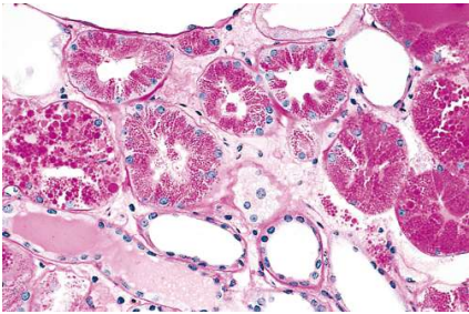
Protein reabsorption droplets in the renal tubular epithelium.
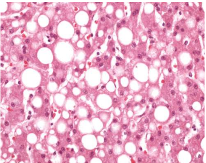
Fatty liver
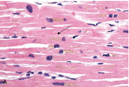
Lipofuscin granules in cardiac myocytes
Dystrophic calcification of the aortic valve - unopened aortic valve in a heart with calcific aortic stenosis.

Early (neutrophilic) infiltrates and congested blood vessels

Later (mononuclear) cellular infiltrates.
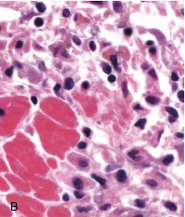
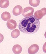
Neutrophils
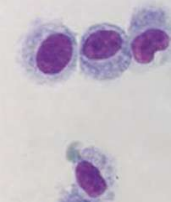
Monocytes
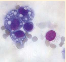
Macrophages
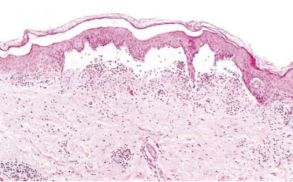
Serous inflammation - skin blister showing the epidermis separated from the dermis by a focal collection of serous effusion.
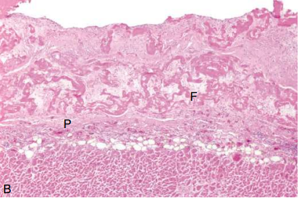
Fibrinous pericarditis
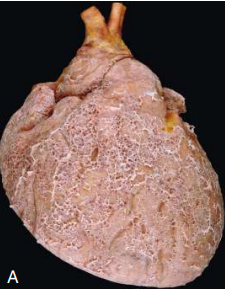
Fibrin exudates
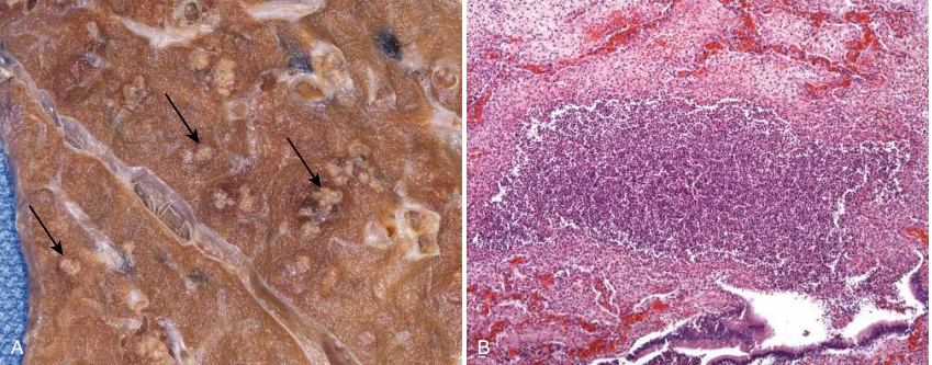
Purulent inflammation - Multiple bacterial abscesses (arrows) in the lung in a case of bronchopneumonia.
The abscess contains neutrophils and cellular debris and is surrounded by congested blood vessels.
Ulcer - chronic duodenal ulcer
(B) Low-power cross-section view of a duodenal ulcer crater with an acute inflammatory exudate in the base.

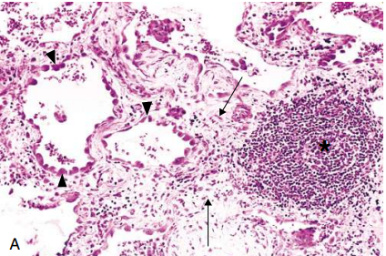
Chronic inflammation in the lung
Asterisk - collection of chronic inflammatory cells
Arrowheads - destruction of parenchyma (normal alveoli are replaced by spaces lined by cuboidal epithelium)
Arrows - Replacement by connective tissue (fibrosis)
Acute inflammation of the lung - neutrophils fill the alveolar spaces, and blood vessels are congested.

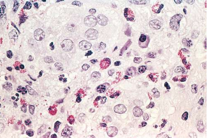
Inflammation containing numerous eosinophils.
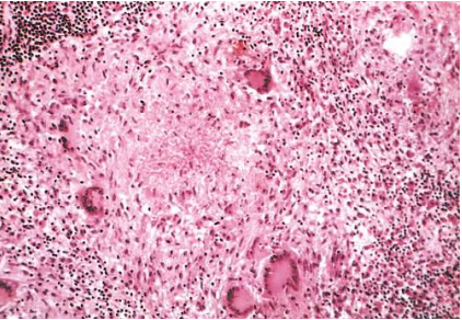
Granulomatous inflammation - Typical tuberculous granuloma showing an area of central necrosis surrounded by multiple giant cells, epithelioid cells, and lymphocytes.
Granulation tissue shows numerous blood vessels, edema, and a loose extracellular matrix with occasional inflammatory cells.
(B) Trichrome stain (collagen) of a mature scar, showing dense collagen with only scattered vascular channels.

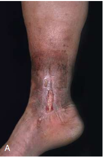
Venous leg ulcer
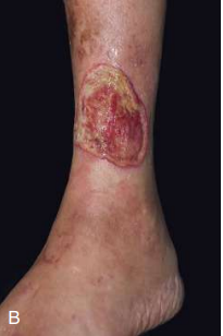
Arterial ulcer, with more extensive tissue necrosis
Diabetic ulcer
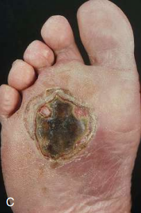
Pressure sore
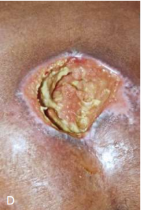
Ulcer crater

chronic inflammation and granulation tissue

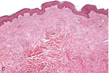
Keloid
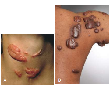
Keloid
Note the thick connective tissue deposited in the dermis.
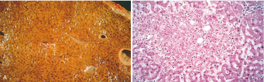
Liver with chronic passive congestion and hemorrhagic necrosis “Nutmeg Liver”
(B) Centrilobular necrosis w/ degenerating hepatocytes and hemorrhage.
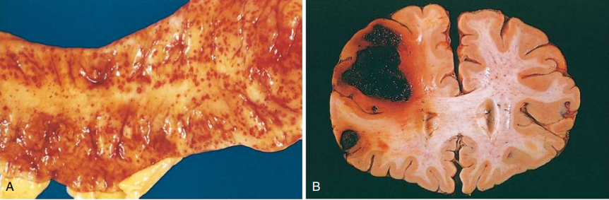
Punctate petechial hemorrhages of the colonic mucosa as a consequence of thrombocytopenia - (B) Fatal intracerebral bleed.
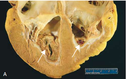
Mural thrombi - Thrombus in the left and right ventricular apices (arrows), overlying a white fibrous scar.
Laminated thrombus in a dilated abdominal aortic aneurysm (asterisks). Numerous friable mural thrombi are also superimposed on advanced atherosclerotic lesions of the more proximal aorta (left side of image).

Thrombosed artery stained for elastic tissue. The original lumen is delineated by the internal elastic lamina (arrows) and is totally filled with organized thrombus.

Embolus from a lower extremity - deep venous thrombosis, lodged at a pulmonary artery branchpoint.

Bone marrow embolus in the pulmonary circulation.
-Cellular elements on the left side of the embolus are hematopoietic cells
-Cleared vacuoles represent marrow fat
-Relatively uniform red area on the right of the embolus is an early organizing thrombus.
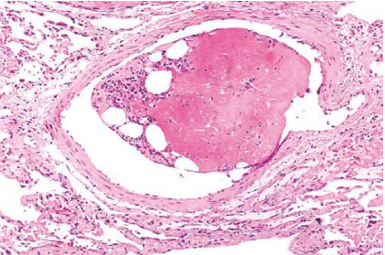
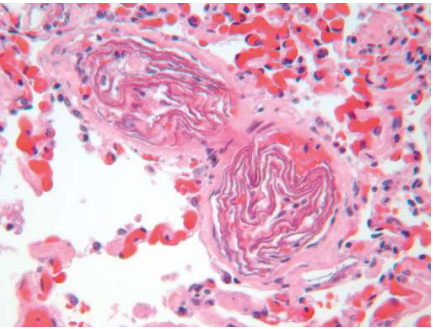
Amniotic fluid embolism - Two small pulmonary arterioles are packed with laminated swirls of fetal squamous cells
-Marked edema and congestion
-Small organizing thrombi consistent with DIC
Kidney infarct replaced by a large fibrotic scar.

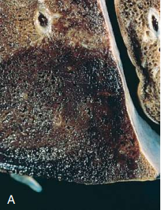
Hemorrhagic, roughly wedge-shaped pulmonary red infarct
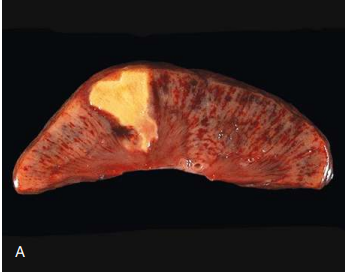
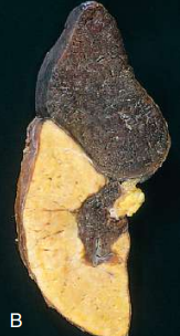
Sharply demarcated white infarct in the spleen.
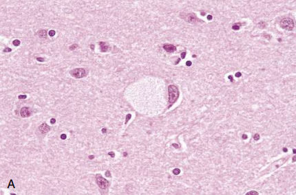
Ganglion cells in Tay-Sachs disease
large neuron has obvious lipid vacuolation
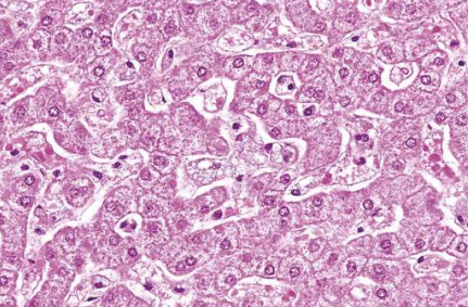
Niemann-Pick disease in liver.
Hepatocytes and Kupffer cells have a foamy, vacuolated appearance due to deposition of lipids.
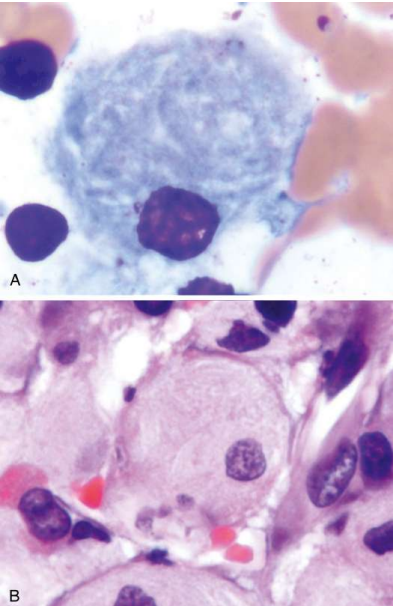
Gaucher disease involving the bone marrow
plump macrophages characteristically have the appearance in the cytoplasm of crumpled tissue paper due to accumulation of glucocerebroside
(A) Wright stain
(B) hematoxylin and eosin.
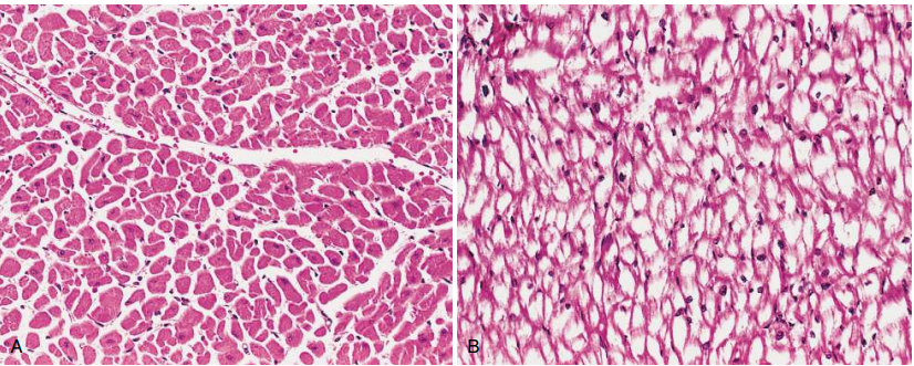
Pompe disease (glycogen storage disease type II)
(A) Normal myocardium with abundant eosinophilic cytoplasm.
(B) Patient with Pompe disease showing the myocardial fibers full of glycogen, seen as clear spaces.
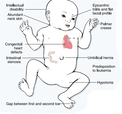
Trisomy 21: Down Syndrome
Trisomy 18: Edwards Syndrome

Trisomy 13: Patau Syndrome
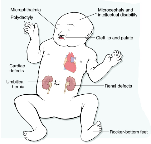
DiGeorge Syndrome - deletion of a probe that maps to chromosome 22q11.2

KLINEFELTER SYNDROME

TURNER SYNDROME

Delayed-type hypersensitivity reaction in the skin
(A) Perivascular accumulation (“cuffing”) of mononuclear inflammatory cells (lymphocytes and macrophages), with associated dermal edema and fibrin deposition.
(B) Immunoperoxidase staining reveals a predominantly perivascular cellular infiltrate that marks positively with anti-CD4 antibody.

(A) Homogeneous staining of nuclei is typical of antibodies reactive with double-stranded DNA (dsDNA) and nucleosomes and is common in systemic lupus erythematosus (SLE).

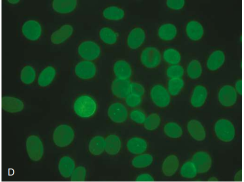
Nucleolar clumpy pattern is typical of antibodies against the nucleolar protein fibrillarin (U3-RNP) and is strongly associated with systemic sclerosis.
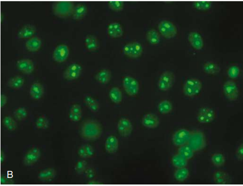
Centromere pattern is seen predominantly in systemic sclerosis, primary biliary cirrhosis, and Sjögren syndrome.

Nuclear envelope pattern is caused by antibodies against nuclear membrane proteins, such as gp210, p68, and lamins, and can occur in patients with autoimmune liver diseases.
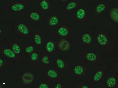
Coarse speckled pattern is seen with antibodies against Sm and U1-RNP, and can be observed in SLE, systemic sclerosis, and mixed connective tissue disease.
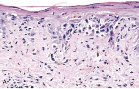
Systemic lupus erythematosus involving the skin - liquefactive degeneration of the basal layer of the epidermis and edema at the dermoepidermal junction.
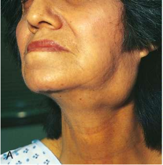
Sjögren syndrome
Enlargement of salivary glands.
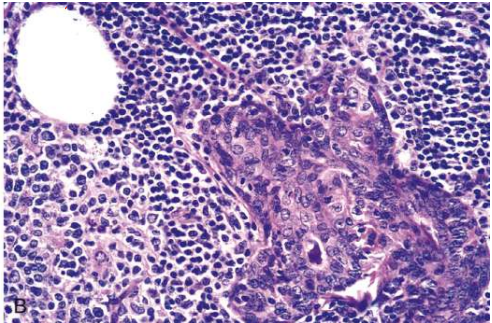
Sjögren syndrome
Intense lymphocytic and plasma cell infiltration with ductal epithelial hyperplasia in a salivary gland.
Normal skin
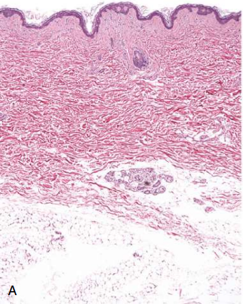
Systemic sclerosis
Note the extensive deposition of dense collagen in the dermis with virtual absence of appendages (e.g., hair follicles) and foci of inflammation (arrow).

Extensive subcutaneous fibrosis has virtually immobilized the fingers, creating a clawlike flexion deformity. Loss of blood supply has led to cutaneous ulcerations.

Bile duct showing grayish-white area of sclerosing cholangitis
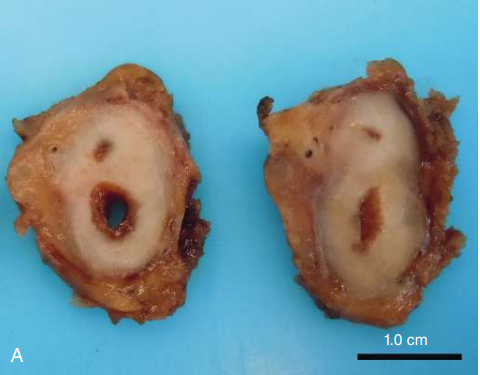
Sclerotic area of the bile duct with storiform fibrosis.
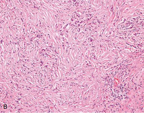
Submandibular gland with infiltrates of lymphocytes and plasma cells and whorls of fibrosis.
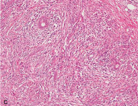
Lacrimal gland stained with an antibody against IgG4, showing large numbers of IgG4-producing plasma cells.

Hyperacute rejection of a kidney allograft showing platelet fibrin thrombi and severe ischemic injury in a glomerulus.

Acute cellular rejection of a kidney graft, manifested by inflammatory cells in the interstitium and between epithelial cells of the tubules (tubulitis). Collapsed tubules are outlined by wavy basement membranes.
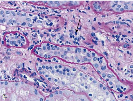
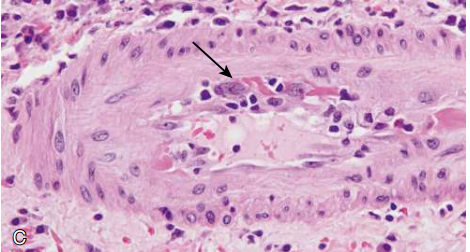
Rejection vasculitis in a kidney graft. An arteriole is shown with inflammatory cells attacking and undermining the endothelium (endotheliitis; arrow).
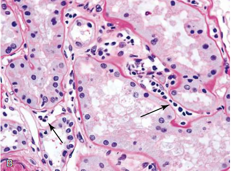
inflammation (capillaritis) in peritubular capillaries (arrows) in a kidney graft.
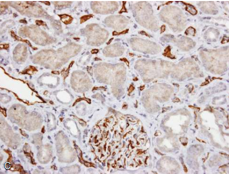
Immunoperoxidase stain shows C4d deposition in peritubular capillaries and a glomerulus.
Graft arteriosclerosis in a cardiac transplant
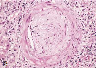
Transplant glomerulopathy - The glomerulus shows inflammatory cells within the capillary loops (glomerulitis), accumulation of mesangial matrix, and duplication of the capillary basement membrane.

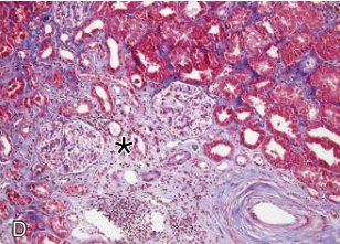
Interstitial fibrosis and tubular atrophy, resulting from arteriosclerosis of arteries and arterioles in a chronically rejecting kidney allograft.
Asterisk - Fibrosis
Bottom right - Arteriosclerosis
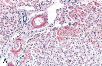
Amyloidosis
Liver stained with Congo red reveals pink-red deposits of amyloid in the walls of blood vessels and along sinusoids.
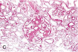
Amyloidosis of the kidney - The glomerular architecture is almost totally obliterated by the massive accumulation of amyloid.
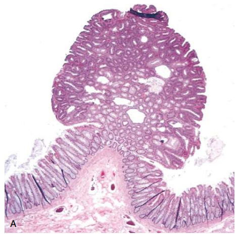
Colonic polyp - adenomatous (glandular) polyp, attached to the mucosa by a stalk, projects into the colonic lumen.
Colonic polyps (arrows)

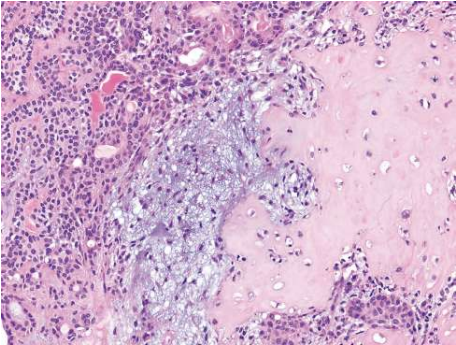
Mixed tumor of the parotid gland - Areas containing nests of epithelial cells (on the left) and myxoid stroma forming cartilage and bone (an unusual feature, on the right)
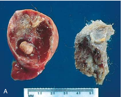
Cystic teratoma of the ovary
presence of hair, sebaceous material, and a tooth.
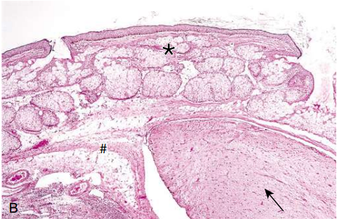
Cystic teratoma of the ovary
sebaceous glands (*)
fat cells (#)
tract of neural tissue (arrow).
Benign tumor (follicular adenoma) of the thyroid - Note the normal looking (well-differentiated), colloid-filled thyroid follicles.
