CHAPTER 22: PRINCIPLES OF DIFFERENTIAL DX AND BIOPSY
 Call Kai
 Learn
Learn Practice Test
 Spaced Repetition
 Match
Match Flashcards
 Knowt Play
1/71

Earn XP
Description and Tags
ppt / book
Name | Mastery | Learn | Test | Matching | Spaced | Call with Kai | Chat |
|---|
No analytics yet
Send a link to your students to track their progress
72 Terms
biopsy
the removal of tissue from a living individual for diagnostic examination
the primary purpose is to determine the diagnosis precisely so that proper treatment can be provided
biopsy = malignancy
clinical examination
anatomic location of the lesion
overall physical characteristics of the lesion
single versus multiple lesions
size, shape, and growth presentation
surface appearance of the lesion
lesion coloration
sharpness of lesion borders and mobility
consistency of the lesion to palpation
presence of pulsation
examination of regional lymph nodes
most fatal locations in the oral cavity
floor of the mouth
posterolateral border of the tongue
dorsum of tongue
this location could originate from epithelial, connective, vascular, neural, muscular, glandular, lymphatic tissues
inner lower lip
this location could consider a minor salivary gland origin, among other possibilities.
herpes zoster
this location of lesions may follow a linear pattern along a nerve pathway.
viral, bacterial, vesiculobullous diseases
commonly produce multiple lesions
small metric ruler
used to measure the lesion.
describe whether the lesion is:
flat → not elevated
slightly elevated
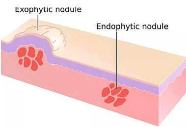
endophytic → grows inward
exophytic → grows outward
sessile → broad-based
pedunculated → attached by a stalk
macule (<10mm)
flat, non-palpable lesion, circumscribed non-elevated area of color change that is distinct from adjacent tissues
examples:
freckle
petechiae
patch (>10mm)
a flat, discolored area on the skin
examples:
café-au-lait spot
vitiligo
port-wine stain
leukoplakia
melasma
papules (<10mm)
circumscribed, small, elevated, solid palpable mass of the skin or mucosa
examples:
mole
lichen planus → most common oral lesion
plaques (>10mm)
flat, slightly elevated superficial lesion
example:
psoriasis
eczema
nodules
dome-shaped lesion → another description
firm lesions that extend into the dermis or subcutaneous tissue
a large, elevated, circumscribed and solid palpable mass of the skin or mucosa
examples:
cysts
lipomas
fibromas
vesicles (<10mm)
clear, fluid-filled blisters, a small blister, circumscribed elevation of skin or mucosa containing serous fluid
example:
herpes simplex
chickenpox
bullae (>10mm)
a blister, an elevated, circumscribed, fluid-containing lesion of skin or mucosa
example:
burns
bullous pemphigoid
pustules
small, cloudy, elevated, circumscribed pus-containing vesicle on skin or mucosa
example:
acne
impetigo
pustular psoriasis
flat lesions
macule
patch
elevated lesions
papule
plaque
nodule
fluid-filled lesions
vesicle
bulla
pustule
clinical discovery of unilateral lesions
herpes zoster (shingles) trigeminal neuralgia (aka: tic douloureu)
managed by tegretol → common brand name carbamazepine
clinical discovery of bilateral lesions
lichen planus
malignant
aggressive, enlarging lesion
cyst with irregular borders might indicate a more aggressive lesion
benign
slower-growing lesion
endophytic
growing inward

exophytic
growing outward
sessile
broad-based

pedunculated
on a stalk
margins of an ulcer
flat
rolled
raised
everted
base of an ulcer
smooth
granulated
fungating appearance
hemorrhagic crust (scab)
covered by fibrin membrane/slough
vascular lesion
dark bluish + blanches on pressure
mucus-retaining cyst
lighter bluish + does not blanch
pigmented lesion
could be a traumatic tattoo or melanotic tumor
white / keratinized lesion
may result from repeated trauma or potentially premalignant changes
red / red-and-white lesion
may indicate more concerning dysplastic changes
has poor prognosis than a white lesion
fluctuant
used to describe the wave-like motion felt during bidigital palpation of a lesion with nonrigid walls that contains fluid
pulsation can be accompanied by:
thrill → palpable vibration
bruit → audible murmur
clinical importance: risk of fatal hemorrhage !
pulsation → rhythmic movement felt during palpation
examination of regional lymph nodes
should be accomplished before any biopsy procedure
bc biopsy can cause reactive lymphadenitis, which may make later diagnosis difficult.
after biopsy, enlarged nodes could be due to:
reaction to the biopsy
infection/inflammation
metastatic tumor
record:
location
size – preferably in centimeters
pain/tenderness
fixation
fixed, matted, movable
texture
soft, firm, hardened
should be systematic:
occipital
preauricular and postauricular
mandibular, submandibular, and submental
deep anterior cervical chain
superficial cervical nodes
along the sternocleidomastoid muscle
deep posterior cervical chain
supraclavicular nodes
buccal lymph nodes may or may not be routinely palpable.
shotty nodes
multiple slightly enlarged
barely palpable lymph nodes
feel like bird shot under the fingers
characteristics of lesions raise suspicion of malignancy
bleeding → bleeds on gentle manipulation
induration → surrounding tissues are firm to touch
growth rate → rapid growth
fixation → feels attached to adjacent structures
erythroplasia → totally red or speckled red-white appearance
ulceration → presents as an ulcer
duration → persisted > 2 weeks
mnemonic: BIGFEUD
indications of biopsy
refractory (poor prognosis)
to confirm a clinical diagnosis
persistent inflammatory signs
unexplained red, white, or pigmented lesions
intrabony lesions that appear to be enlarging
lesions suspected to be malignant or premalignant
lesions that grow rapidly without an obvious reason
persistent lesions that cannot be clinically diagnosed
lesions causing extreme patient concern (cancerphobia)
submucosal swelling beneath apparently normal mucosa
lesions that are firmly attached/fixed to nearby structures
lesions with no identifiable cause that remain for more than 10–14 days despite local treatment
lesions that do not respond to routine treatment or removal of a local irritant within 10–14 days
unknown lesions in high-risk cancer areas, such as:
floor of the mouth, tongue
soft tissue biopsy techniques
nerve block anesthesia
solution be injected at least 1cm away from the lesion → if local infiltration is needed
if within the tissues → artifactual distortion of the specimen
tissue stabilization
tractional sutures & towel clips → can be used to aid immobilization of the tongue or soft palate
hemostasis
the use of a suction device for keeping the surgical field free of blood during the procedure should be minimized as much as possible
use gauze instead
incision
2 football/elliptical-shaped incisions can be made, should converge toward the base
a periphery of normal-appearing tissue should be included in excisional biopsy specimens
this:
produces a good specimen, makes the wound easier to close
if it appears benign → 2-3mm of peripheral tissue
if it appears malignant → 5mm of peripheral tissue
carbon dioxide laser
used when necessary, but can still produce a narrow zone of tissue necrosis.
removal of specimen
a dental curette is used
concave surface → in contact with the bone
convex surface → separates the specimen from surrounding bone
specimen care
be placed in 10% formalin solution (4% formaldehyde)
at least 20 times the volume of the specimen
wound closure
if possible, must be closed through primary closure
use black silk resorbable sutures for deeper layers
polyglycolic acid (Dexon)
polyglactin 910 (Vicryl)
chromic gut
undermined movable soft tissues, distance should be at least the width of the defect
allows tension-free closure of the wound edges.
referral
be placed in 10% formalin solution (4% formaldehyde)
at least 20 times the volume of the specimen
hemostasis techniques

types of biopsy
cytology
aspirational
incisional
excisional
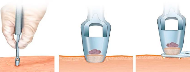
punch biopsy
exfoliative cytologic examination
diagnostic procedure for detection of uterine cervical malignancy or pre-cancer screening method
in the oral cavity, used as an adjunct and not substitute for incisional nor excisional biopsy

oral brush cytology
uses a special brush to collect the epithelial cells
may be a good tool for "monitoring" patients with chronic mucosal changes, such as:
leukoplakia
lichen planus
history of oral cancer

aspiration biopsy
is performed on fluid-filled lesions except a mucocele
the use of a needle and syringe to penetrate a lesion for aspiration of its contents (16-18 gauge needle)
2 main purposes:
determine what is inside the lesion
is it fluid-filled? is it solid?
obtain cells for diagnosis
called Fine-Needle Aspiration (FNA)
used to obtain cells for pathologic examination, especially useful for:
soft tissue masses, neck masses
lesions where surgical biopsy may cause a scar or damage nearby structures
routinely performed on intraosseous radiolucent lesions before entering the bone.
this helps determine:
whether the lesion is vascular or cystic or solid
⚠ This is important because entering a vascular lesion surgically can cause serious bleeding.

clinical discovery of pus
infectious process
clinical discovery of mucus
mucocele, ranula
clinical discovery of air or golden yellow fluid
traumatic bone cyst
clinical discovery of cheese-like, keratin
odontogenic keratocyst
clinical discovery of negative (nothing obtained)
solid lesion
clinical discovery of clear pale, straw-colored
dentigerous cyst
clinical discovery of straw-colored (beer-colored)
cystic (ameloblastoma)
clinical discovery of blood
vascular lesion (aneurysmal bone cyst)
incisional biopsy
a biopsy that samples only a particular or representative part of the lesion
indications:
hazardous location
if there is a suspicion of malignancy
large lesions ( >1cm in diameter)
procedure:
usually a wedge-shaped piece is removed
a narrow and deep specimen is better than a broad and shallow specimen.
the biopsy site should be selected in an area that shows complete tissue changes
the material should be taken from the edge of the lesion to include some normal tissue
[a negative report of a highly suspicious oral lesion suggests that another biopsy specimen is necessary in view of the clinical impressions]
![<p>a biopsy that samples only a particular or representative part of the lesion</p><ul><li><p><strong>indications:</strong></p><ul><li><p><span style="color: red;">hazardous location</span></p></li><li><p><span style="color: red;">if there is a suspicion of malignancy</span></p></li><li><p><span style="color: red;">large lesions ( >1cm in diameter)</span></p></li></ul></li><li><p><strong>procedure:</strong></p><ul><li><p>usually a <span style="color: red;">wedge-shaped piece</span> is removed</p></li><li><p>a narrow and deep specimen is better than a broad and shallow specimen.</p></li><li><p>the biopsy site should be selected in an area that shows complete tissue changes</p></li><li><p>the material should be taken from the <span style="color: red;">edge of the lesion</span> to include some normal tissue</p></li></ul></li></ul><p><em>[a negative report of a highly suspicious oral lesion suggests that another biopsy specimen is necessary in view of the clinical impressions]</em></p>](https://assets.knowt.com/user-attachments/6209c00a-69b8-40e9-b2db-742a17c54755.png)
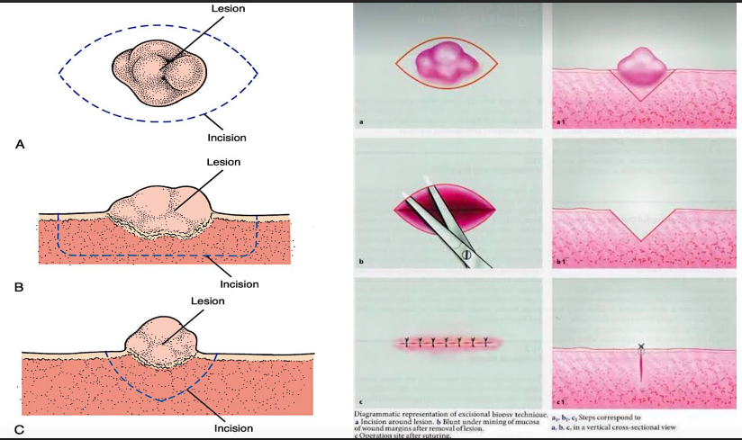
excisional biopsy
removal of the entire lesion along with 2-3mm of normal appearing surrounding tissue is excised
indications:
smaller lesions that on clinical examination appear to be benign (<1 cm)
any lesion that can be removed completely without mutilating the patient
punch biopsy
removal of a small piece of tissue using a punch (a small, hollow cylindrical tube-like bladed instrument)
crusts (crusted)
dried or clotted serum on the surface of the skin or mucosa
dysplasia (dysplastic)
any abnormal development of cellular size, shape, or organization in tissue
erosion
a shallow, superficial ulceration
hyperkeratosis
an overgrowth of the cornified layer of epithelium
hyperplasia (hyperplastic)
an increased number of normal cells
hypertrophy (hypertrophic)
an increase in size caused by an increase in the size of cells, not in the number of cells
keratosis (keratotic)
an overgrowth and thickening of cornified (horned layer) epithelium
leukoplakia
a slowly developing change in mucosa characterized by firmly attached thickened white patches
malignant
anaplastic, a cancer that is potentially invasive and metastatic
scale
a thin, compressed, superficial flake of cornified (keratinized) epithelium
stomatitis
any generalized inflammatory condition of the oral mucosa
ulcer
a crater-like circumscribed surface lesion resulting from necrosis of the epithelium
pathology report and follow-up
patient is generally seen around 1 week after biopsy:
remove sutures, discuss results if available
typical splint duration → 7-10 days
pathology report → 7-14 days
sturge-weber syndrome
the syndrome that would manifest as port wine stain (aka: nevus flammeus)
lucas curette
type: double-ended surgical/bone curette
shape: spoon-shaped, rounded ends
uses: removes granulation tissue, debris, and cystic tissue from extraction sockets
sizes: #84–#88