Scapula and Shoulder Girdle
 Call Kai
 Learn
Learn Practice Test
 Spaced Repetition
 Match
Match Flashcards
 Knowt Play
1/87

There's no tags or description
Looks like no tags are added yet.
Name | Mastery | Learn | Test | Matching | Spaced | Call with Kai | Chat |
|---|
No analytics yet
Send a link to your students to track their progress
88 Terms
shoulder girdle bones
sternum
clavicle
scapula
humerus
sternum - bony land marks
clavicular notch
jugular/suprasternal notch
manubrium
body of sternum
xiphoid process
clavicle - bony landmarks/orientation
superior/anterior:
convex/flat surface
deltoid tubercle
inferior/posterior:
concave
conoid tubercle
groove for subclavius
trapezoid line
impression for costoclavicular ligament
sternal end = bump with vertical flat part
acromial end = flattened out
scapula - bony landmarks
lateral border (side of processes)
superior border
lateral border
inferior angle
anterior:
coracoid process is in front of acromial process
neck
subscapula fossa
posterior:
spine of scapula
supraspinous and infraspinous fossa
lateral:
glenoid cavity
acromion types
modified Bigliani Classification System:
type I-IV
acromion type I
flat - 12%
acromion type II
curved - 56%
considered most common type
acromion type III
hooked - 29%
associated with increased incidence of shoulder impingement → less space for tendons, arteries, etc.
acromion type IV
convex (upturned) - 3%
least common
surgical neck
surgical neck is where humeral head is replaced in surgery
it is an imaginary line
posterior humerus face bony landmarks
anatomical neck
humeral head
greater tuberosity
surgical neck
anterior humerus face bony landmarks
humeral head
anatomical neck
intertubercular groove
lesser tuberosity
greater tuberosity
deltoid tuberosity
shoulder girdle joints
sternoclavicular
acromioclavicular
glenohumeral
scapulothoracic
sternoclvicular joint
synovial saddle joint - convex/concave surfaces (different planes = different congruences)
three planes of motion
articulation between clavicle, manubrium, and 1st rib
poor congruence to mimic planer joint (behaves like ball-and-socket)
ligaments:
anterior/posterior SCL
interclavicular ligament
costoclavicular ligament
articular disk
movements: depression/elevation
interclavicular ligament
holds clavicles together
posterior steronoclavical ligament
on posterior side
holds sternum and clavicle together
anterior sternoclavicular ligament
holds clavicle to anterior/superior portion of manubrium
costoclavicular ligament
connects clavicle to 1st rib
Articular disk of SC joint
forms two compartments
attached to SCLs and interclavicular ligament for stability
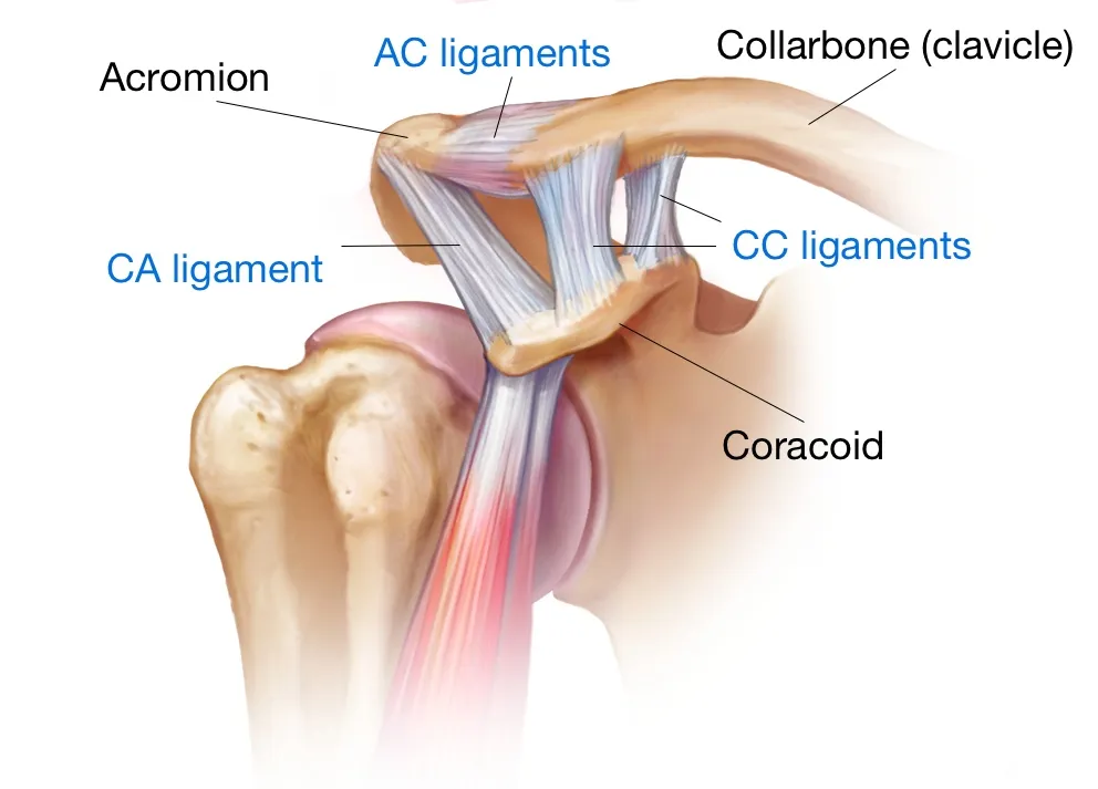
Acromioclavicular joint
planar joint
articulation between acromion process and clavicle
ligaments:
acromioclavicular
coracoacromial
coracoclavicular
tapezoid (anterior and lateral bands0
conoid
little bit more mobility than SC joint
anterior and posterior movement during protraction and retraction
Glenohumeral Joint
ball and socket joint
articulation between glenoid fossa and humeral head
unequal proportion of surface area
about ⅓ of humeral head contacting glenoid fossa (golf ball on a tee)
= decreased stability & increased mobility
glenoid labrum
ligaments:
capsular ligaments
glenoid labrum
fibrocartilage structure
attached to outer margins of glenoid fossa
deepens articulation to increase stability - dynamic and allows some more movement
often tears more due to extending out further and taking on more stress from humeral head
Capsular ligaments - GH joint
merge to encircle entire joint around humeral head
attached:
medially beyond the glenoid labrum (neck)
laterally below anatomical neck
coracohumeral ligament
transverse humeral ligament - holds biceps tendon in place
glenohumeral ligaments - superior, middle, inferior
GH joint posterior capsule
supported by posterior band of IGHL
important in stabilizing from anterior dislocation
contracture results in GH IR deficit (GIRD)
GH movements - sagittal plane
flexion
extension
hyperextension
GH movements - Frontal plane
abduction
adduction
GH movements - transverse plane
IR
ER
horizontal abduction
horizontal adduction
scapulothoracic joint
pseudo-joint/conceptual
lacks bony articulation
slides/glides along posterolateral thoracic wall
scapular motion facilitated by ROM of other joints in the shoulder girdle
does not have ligamentous support - relies on dynamic stabilizers
movements:
elevation/depression
upward/downward rotation
protraction/abduction
retraction/adduction
role of scapula
connects arm to trunk of body
maintains dynamic stability with controlled mobility at the GHJ
proximal to distal transfer of energy
role of scapula: 1. connects arm to trunk of body
provides glenoid fossa for glenohumeral ball and socket joint
allows for movements of the upper extremity (dynamic stability)
role of scapula: 2. maintains dynamic stability with controlled mobility
synergistic co-contractions (force couples)
length/tension relationships for muscles
no isolated movements
role of scapula: 3. proximal to distal transfer of energy
force travels from the legs and trunk to the arm and hand
aids in deceleration in throwing motion (dissipation of energy) - muscles of scapula
scapulothoracic movements - elevation/depression muscles
elevation:
upper trapezius
rhomboids
levator scapula
depression:
lower trapezius
lower serratus anterior
scapulothoracic movements - upwards/downward rotation muscles
upward rotation:
serratus anterior
upper trapezius
lower trapezius
downward rotation:
rhomboids
levator scapulae
scapulothoracic movements - abduction/adduction muscles
abduction:
serratus anterior
adduction:
trapezius
rhomboids
scapulohumeral rhythm - purpose
maximize ROM
static and dynamic stabilizers work to create optimal length-tension relationship for movement to occur
scapulohumeral rhythm - function
the scapula and humerus move at a 1:2 ration (scapula:humerus)
approaches 1:1 ratio within midrange (60o-90o)
ex. arm abducted to 180o will result in 60o of motion via scapula/STJ and 120o via humerus/GHJ
end range is mostly scapular movements
scapulohumeral rhythm: phase 1
30o elevation
humerus: 30o abduction
clavicle: 0o-5o elevation
scapula: minimal movement medially
scapulohumeral rhythm: phase 2
90o elevation
humerus: 40o abduction (up to 90o)
scapula: 20o lateral rotation
clavicle: 15o elevation
scapulohumeral rhythm: phase 3
90o-180o elevation
humerus: 60o abduction, 90o lateral rotation
scapula: 30o lateral rotation
clavicle: 30o-50o posterior rotation, 15o elevation
intramuscular spaces if scapular region
upper triangular region
quadrangular space
lower triangular space
scapulohumeral muscle grouping
attaches humerus to scapula
deltoid
teres major
rotator cuff muscles
coracobrachialis
axioscapular muscle groupings
attaches scapula to the trunk
trapezius
rhomboid minor/major
serratus anterior
levator scapula
axiohumeral muscle grouping
attaches humerus to trunk
pectoralis major/minor
latissimus dorsi
Axilla contents
axillary vein and artery
brachial plexus
lymph nodes
fat
axillary region - apex
upper border of scap, clavicle, and 1st rib
inlet to the cervico axillary canal
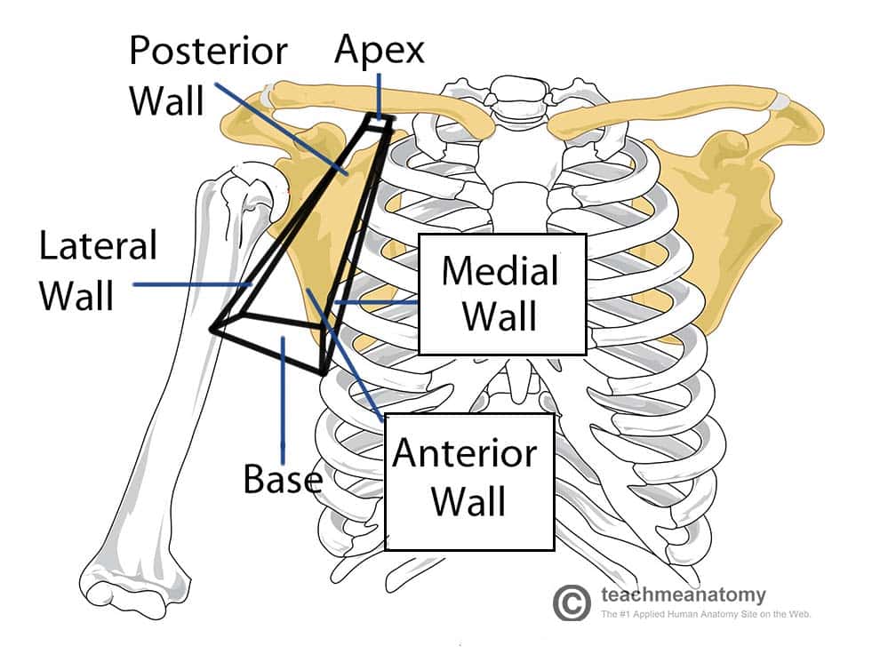
axillary region - base
skin, subcutaneous tissue and fat
outlet to the cervico axillary canal
axillary region anterior border
pectoralis major/minor
axillary fat
axillary region lateral border
intertubercular sulcus
(humerus)
axillary region medial border
serratus anterior
thoracic wall
ribcage
axillary region posterior border
scapularis (?)
teres major
latissimus dorsi
scapula
axillary musculature layers
superficial extrinsic
deep extrinsic
intrinsic
axillary muscles - superfical extrinsic
trapezius
latissimus dorsi
axillary muscles - deep extrinsic
levator scapula
rhomboid major/minor
serratus anterior
axillary muscles - intrinsic
deltoid
teres major/minor
supraspinatus
infraspinatus
subscapularis
deltoid - OINA
O:
lateral ⅓ clavicle
acromion process
spine of scapula
I:
deltoid tuberosity of humerus
N:
axillary nerve
A:
anterior fibers:
shoulder flex
IR
horizontal adduction
posterior fibers:
shoulder extension
ER
horizontal abduction
all fibers - shoulder abduction
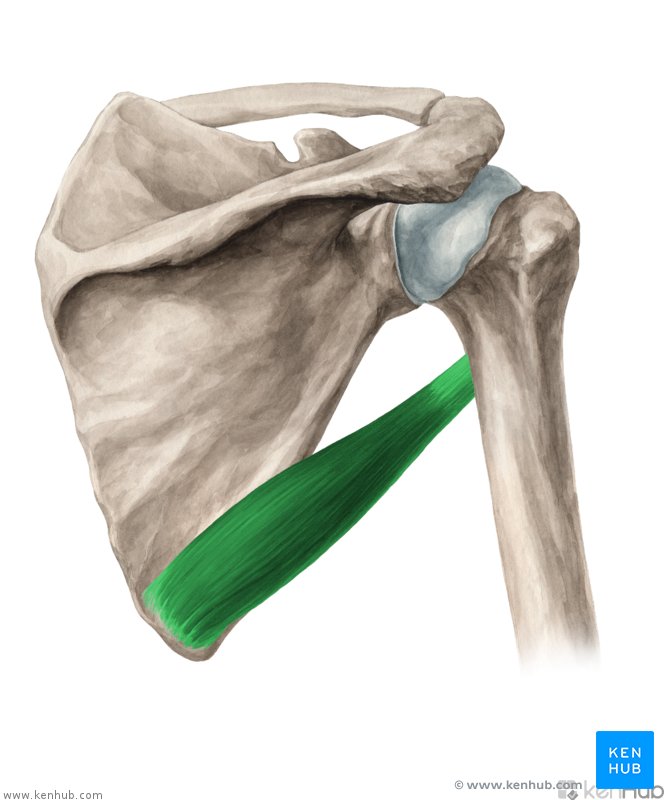
teres major OINA
O:
posterior surface of inferior angle of scapula
I:
medial lip of intertubercular groove of humerus
N:
adducts arm
IR arm
A:
lower subscapular nerve - C5, C6
rotator cuff muscles
SITS
supraspinatus
infraspinatus
teres minor
subscapularis
supraspinatus OINA
O:
supraspinous fossa of scapula
I:
superior facet of greater tubercle of humerus
N:
suprascapular nerve - C4-C6
A:
initiates and assists deltoid in abduction
acts with RC movements
infraspinatus OINA
O:
infraspinous fossa of scapula
I:
middle facet of greater tubercle of humerus
N:
suprascapular nerve - C5, C6
A:
ER arm
acts with RC movements
Teres minor OINA
O:
middle part of lateral border of scapula
I:
inferior facet of greater tubercle of humerus
N:
axillary nerve - C5, C6
A:
ER arm
acts with RC movements
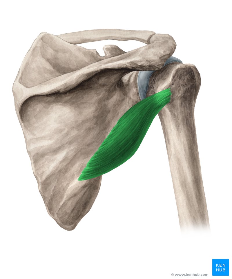
Subscapularis OINA
O:
subscapular fossa
I:
lesser tubercle of humerus
N:
upper and lower subscapular nerve - C5-C7
A:
IR arm
acts with RC movements
brachial plexus
Roots: 5
Trunks: 3
Divisions:
3 anterior
3 posterior
Cords: 3
branches: 5
shoulder pain - causes
global diffuse pain due to close proximity of structures to each other and continual movement
SLAP tear
frozen shoulder
abnormal AC joint
shoulder OA
biceps tendinitis
calcific tendonitis
rotator cuff tear
scapular dyskinesis
loss of motor control of the scapulohemeral motion
normal scapular position is altered
lack of coordinated activation patterns in the scapula stabilizing movements
often found in shoulder impingement pts'
SICK scapular syndrome (causes)
= altered movement of the shoulder girdle
established association to shoulder pathologies
SICK scapular syndrome
S - scapula malpositioning
I - inferior medial roder prominence (off the rib cage and tightness of muscles)
C - coracoid paina nd malposition
K - kinesis abnormalities of the scap
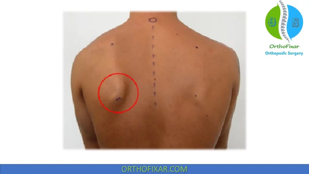
scapular dyskinesis - type I
inferior angle
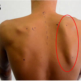
scapular dyskinesis - type II
medial border/winged scapula
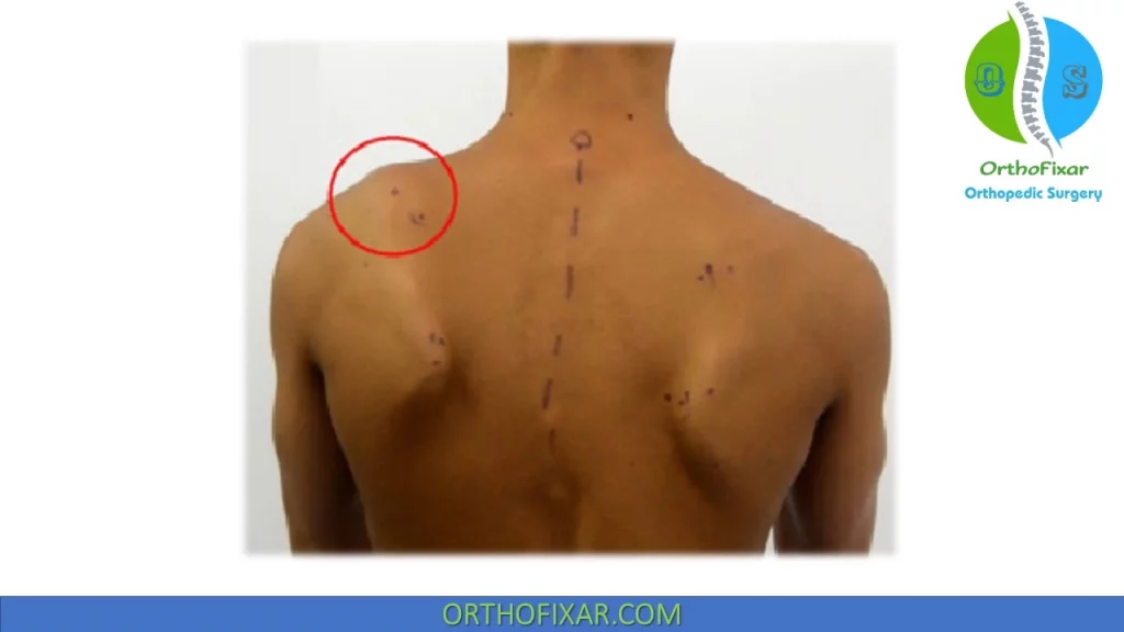
scapular dyskinesis - type III
superomedial border - very dysfunctional
traumatic fx
fx of surgical neck - ORIF
avulsion fx of greater tubercle - ORIF
peripheral nerve injuries
anterior and posterior dislocations
shoulder separation
locations of nerves and injuries
fx of surgical neck may injury axillary nerve (as it wraps around the humeral head)
fx of humeral body in region of radial groove may injure radial nerve
distal humeral fx may injure medial nerve
fx in region of medial epicondyle may injure ulnar nerve
dislocations - general
humeral head can dislocate: inferiorly, anteriorly, & posteriorly
not superior due to acromion process
Direction depends on force application
occurs most frequently in young adults and athletes - traumatically too
anterior dislocation
head of humerus is driven anterior inferiorly where capsule is weakest
fibrous capsule and glenoid labrum may be stripped from anterior aspect of glenoid
flexor and abductor muscle usually pull humeral head into a subcoracoid postion
axillary nerve may be injured
apprehension test
test for glenohumeral instability vs anterior dislocation
go into ER to put stretch on anterior capsule
watch pt facial expression
Hill-sachs lesions
posterolateral humeral head compression fx
typically secondary to recurrent anterior shoulder dislocations as the humeral head comes to rest against the anteroinferior part of the glenoid
shoulder separation: moi/degrees
most common MOI is a fall on the tip of the shoulder
injury to the AC ligament and CC ligament
1st degree - isolated to AC ligament
2nd degree - AC ligament and stretch of CC ligaments
3rd degree - Tear of all three ligaments, AC and both CC heads
(CC= coracoid clavicular)
rotator cuff pathology
repetitive use of the UE, sudden strain/forces, falls
can be degenerative in nature with older pt
why a good hx is needed
involves the musculotendinous rotator cuff and/or long head of biceps tendon
recurrent inflammation of RC tendons can lead to tears
Signs/Sx:
pain
lost of motion most commonly ABD and ER
ABD - supraspinatus involved
ER - teres minor & infraspinatus involved
decreased RC strength
loss of functional mobility
shoulder impingement (syndrome)
reduced sub-acromial space - supraspinatus tendon and bursa squished
more symptoms with abduction because humeral head is moving superiorly
px in middle of ROM
can result in rotator cuff tear
SLAP tear: signs/sx
superior labral tear from anterior to posterior
signs/sx:
locking, popping, catching, grinding, less stable feeling
px with movement of shoulder or holding certain positions
px with lifting objects → especially overhead; still do it becuase other msucles can still move
decrease in shoulder strength
feeling the shoulder will pop out of joint
decreased ROM
pitchers may notice a decrease in their throw velocity or the feeling of having a “dead arm” after pitching
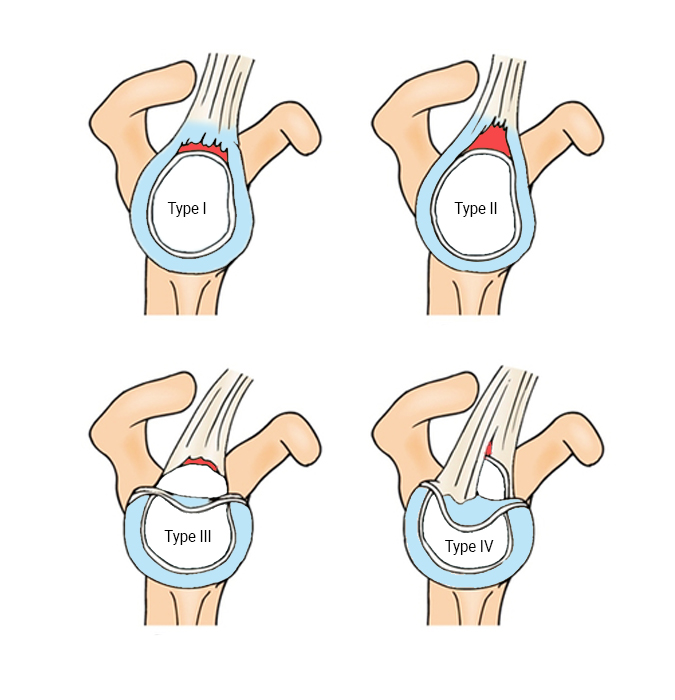
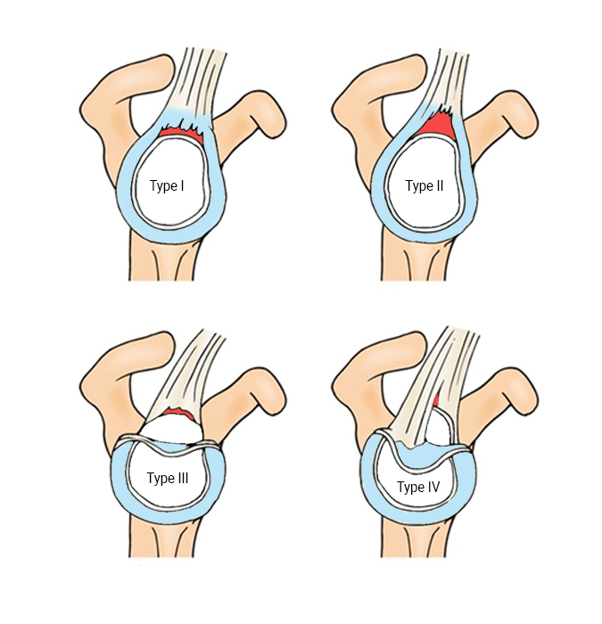
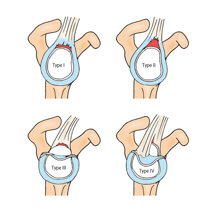
4 types
slap tear type I
frayed cartilag at the edge of labrum
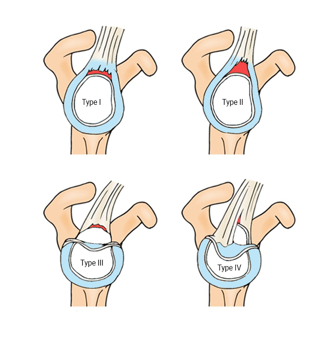
slap tear type II
labrum tears away from glenoid cavity
slap tear type III
bucket handle section tears away from glenoid cavity
slap tear type IV
slap lesion extends and tears the biceps tendon
adhesive capsulitis
aka frozen shoulder
adhesive fibrosis and scarring within the inflamed joint capsule of the GH joint, RC, subacromial bursa and deltoid
common in females 40-60 y/o or with diabetes/metabolic disorders
often insidious onset - can be caused by prior injuries to shoulder (dislocations, fx, tendonitis, RC)
capsular pattern with:
loss of ROM
px
stiffness
loss of functional mobility
3 phases: freezing, frozen, thawing
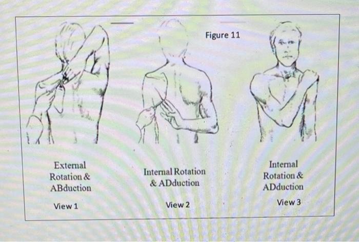
Apley’s scratch test
quick check of AROM
1) horizontal adduction with internal rotation (behind the back)
2) abduction with external rotation (overhead)
3) adduction with internal rotation (across chest)
for frozen shoulder (?)
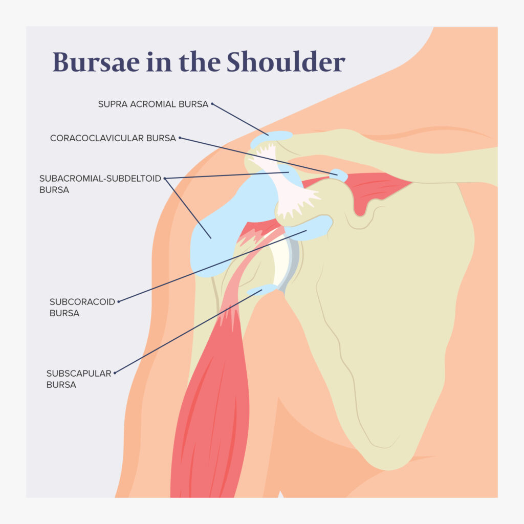
bursitis
inflammation of the bursa(s) in shoulder
infraserratus bursa
supraserratus bursa
trapezoid bursa
infaserratus bursa
subscaular bursa
subdeltoid bursa
subcoracoid bursa
subacromial bursa
shoulder replacement surgery
replacement of shoulder with artificial parts
total shoulder arthroplasty vs reverse shoulder arthroplasty
performed on pt with severe px, loss of UE function from arthritis, fx, RA, exstensive RC/Mm wear and tear
extensive rehab is needed
reverse shoulder arthroplasty
glenoid sphere = convex surfacce
humeral cap = concave surface
flips concave/convex surfaces
increase ROM compared to total SA