Histology of Esophagus

0.0(0)
studied byStudied by 0 people
learnLearn
examPractice Test
spaced repetitionSpaced Repetition
heart puzzleMatch
flashcardsFlashcards
Card Sorting

1/4

encourage image

There's no tags or description

Looks like no tags are added yet.

Study Analytics
Name
Mastery
Learn
Test
Matching
Spaced

No study sessions yet.

5 Terms

1
New cards

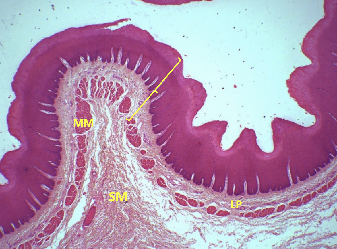
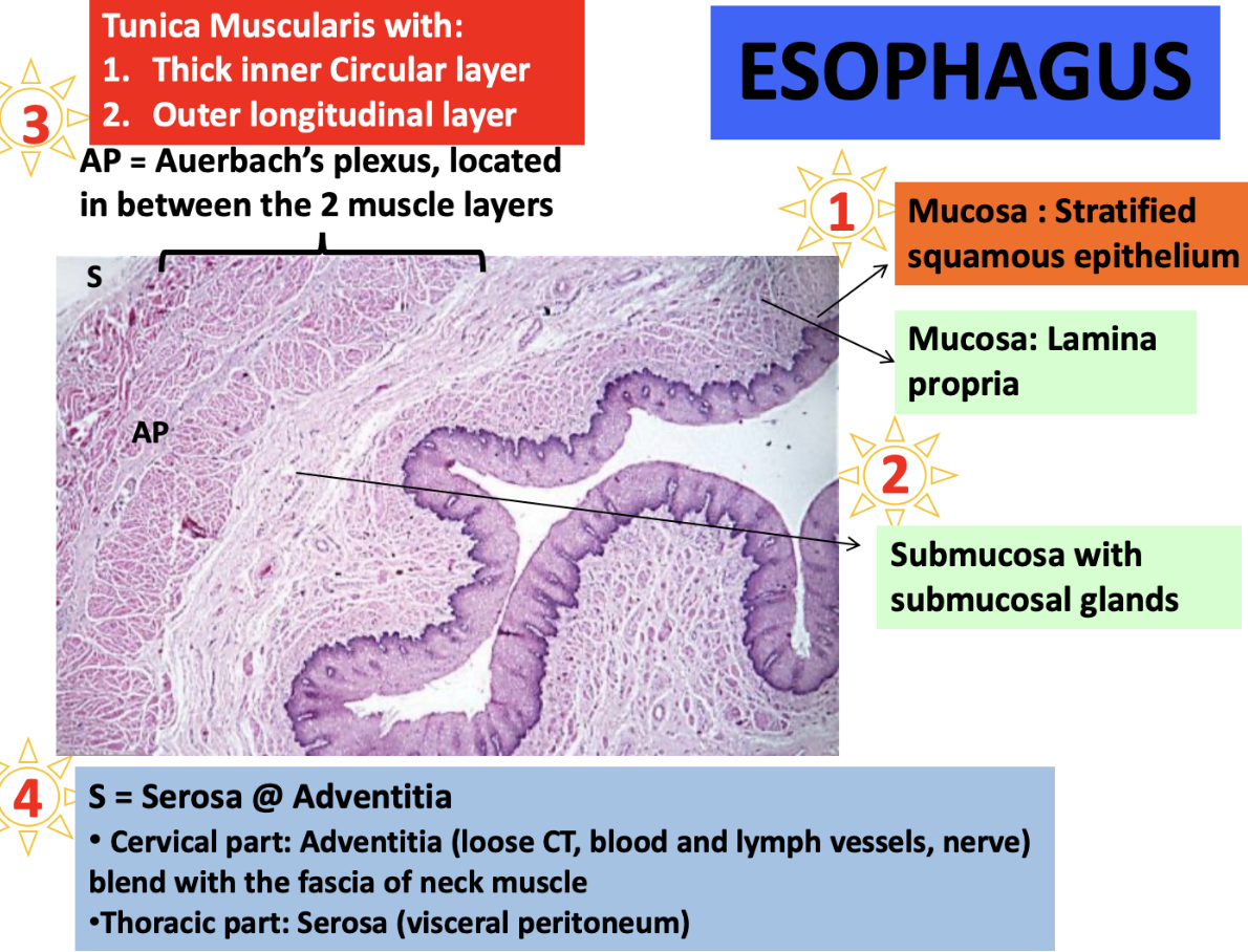
Describe the mucosa layer of esophagus

  • Epithelium

  • Lamina propria

  • Muscularis mucosa

Epithelium: Stratified squamous epithelium

Lamina propria: Loose connective tissue

Muscularis mucosa: Smooth muscle

<p><strong><u>Epithelium:</u> </strong><span style="color: red"><strong>Stratified squamous epithelium</strong></span></p><p><strong><u>Lamina propria:</u> </strong><span style="color: red"><strong>Loose connective tissue </strong></span></p><p><strong><u>Muscularis mucosa:</u> </strong><span style="color: red"><strong>Smooth muscle </strong></span></p><p></p>
2
New cards

Describe the submucosa layer of esophagus

  • Layer made of

  • Consist of

Layer made of: Loose to dense irregular connective tissue

Consist of:

  • Glands that secrete mucus

  • Blood and lymphatic vessels

  • Nerves (Meissner’s plexus)

  • Elastic fibers

<p><strong><u>Layer made of:</u> </strong><span style="color: blue"><strong>Loose</strong></span><strong> </strong>to <span style="color: blue"><strong>dense irregular connective tissue </strong></span></p><p><strong><u>Consist of:</u></strong></p><ul><li><p><span style="color: blue"><strong>Glands</strong></span><strong> </strong>that <span style="color: blue"><strong>secrete mucus </strong></span></p></li><li><p><span style="color: blue"><strong>Blood </strong></span>and<span style="color: blue"> <strong>lymphatic vessels </strong></span></p></li><li><p><span style="color: blue"><strong>Nerves</strong></span><strong> </strong>(<em>Meissner’s plexus</em>) </p></li><li><p><span style="color: blue"><strong>Elastic fibers </strong></span></p></li></ul><p></p>
3
New cards

Describe the muscularis layer of esophagus #fcaa00

  • Arrangement of layers

  • What nerve structure is located between the layers

Arrangement of layers:

  • Thick inner circular layer

  • Outer longitudinal layer

Structure located between the layers: Auerbach plexus

<p><strong><u>Arrangement of layers:</u> </strong></p><ul><li><p><span style="color: #fcaa00"><strong>Thick inner circular layer </strong></span></p></li><li><p><span style="color: #fcaa00"><strong>Outer longitudinal layer </strong></span></p></li></ul><p><strong><u>Structure located between the layers:</u> </strong><span style="color: #fcaa00"><strong>Auerbach plexus</strong></span></p><p></p>
4
New cards

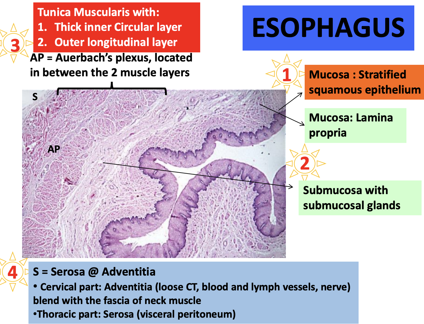
Which part of the esophagus is covered by adventitia and serosa

  • What does each layer consist of?

  • Function

Adventitia: Cervical part of esophagus

  • Not covered by perioneum, surrounded by loose CT instead

  • Blends with fascia of neck muscles

  • Consist of:

    • Loose CT

    • Blood and lymph vessels

    • Nerve

  • Function: Anchors the esophagus in place

Serosa: Thoracic part of esophagus

  • Covered by visceral peritoneum

  • Function: Provides slippery surface to reduce friction

<p><span style="color: purple"><strong><mark data-color="purple" style="background-color: purple; color: inherit"><u>Adventitia:</u></mark> Cervical part</strong></span><strong> </strong>of esophagus </p><ul><li><p><span style="color: purple"><strong>Not covered </strong></span>by <span style="color: purple"><strong>perioneum, surrounded</strong></span><strong> </strong>by <span style="color: purple"><strong>loose CT</strong></span><strong> </strong>instead </p></li><li><p><span style="color: purple"><strong>Blends</strong></span><strong> </strong>with <span style="color: purple"><strong>fascia</strong></span><strong> </strong>of <span style="color: purple"><strong>neck muscles</strong></span></p></li><li><p><strong><u>Consist of:</u> </strong></p><ul><li><p><span style="color: purple"><strong>Loose CT </strong></span></p></li><li><p><span style="color: purple"><strong>Blood </strong></span>and<span style="color: purple"> <strong>lymph vessels </strong></span></p></li><li><p><span style="color: purple"><strong>Nerve </strong></span></p></li></ul></li><li><p><strong><u>Function:</u> </strong><span style="color: purple"><strong>Anchors</strong></span><strong> </strong>the <span style="color: purple"><strong>esophagus</strong></span><strong> </strong>in place </p></li></ul><p><span style="color: #9f00ff"><strong><mark data-color="purple" style="background-color: purple; color: inherit"><u>Serosa:</u></mark></strong></span><strong> </strong><span style="color: #9f00ff"><strong>Thoracic part</strong></span><strong> </strong>of esophagus </p><ul><li><p><span style="color: #9f00ff"><strong>Covered</strong></span><strong> </strong>by <span style="color: #9f00ff"><strong>visceral peritoneum </strong></span></p></li><li><p><strong><u>Function:</u> </strong>Provides<span style="color: #9f00ff"> <strong>slippery surface</strong></span><strong> </strong>to<span style="color: #9f00ff"> <strong>reduce friction </strong></span></p></li></ul><p></p>
5
New cards
<p>Histology of Esophagus </p>

Histology of Esophagus

Histology of Esophagus

<p>Histology of Esophagus </p>