PHAR 555 Exam #1 (Review)
 Call Kai
 Learn
Learn Practice Test
 Spaced Repetition
 Match
Match Flashcards
 Knowt Play
1/161

There's no tags or description
Looks like no tags are added yet.
Name | Mastery | Learn | Test | Matching | Spaced | Call with Kai | Chat |
|---|
No analytics yet
Send a link to your students to track their progress
162 Terms
From least to most stable level of chemical communication in the endocrine system (with examples)…
Ion channels (fastest, taking milliseconds)
Example: Fast-twitch muscles
Modulators (GPCRs) (tweaks things slightly in the positive or negative direction—nothing long-term—at a moderate speed, taking minutes to hours)
Example: Most drugs
Transcription (nuclear) of new proteins and enzymes, altering things like metabolism and the long-term function of a cell
Metabolism (receptor kinases), altering fundamentals of cells
Example: Insulin
glands
secretory organs/tissues that produce and release substances for use in other tissues
exocrine gland
releases substances via ducts (usually epithelial) to specific tissues
endocrine gland
releases substances into the bloodstream, often to distal sites
hormones
longer-acting chemicals that act on distal tissues by transport in the bloodstream
Usually to maintain prolonged homeostasis, aid in bone formation, etc.
Can be either small water-soluble molecules, peptides/proteins, or lipid-soluble steroids
Most then act on specific target tissues (save growth hormone) using receptors, which can elicit further hormone release from those peripheral tissues
How can the hypothalamus send hormones to the pituitary gland?
The hypothalamus…
Sends hormones down axons that lead to the posterior pituitary at which they are released
Receives mostly neural positive feedback
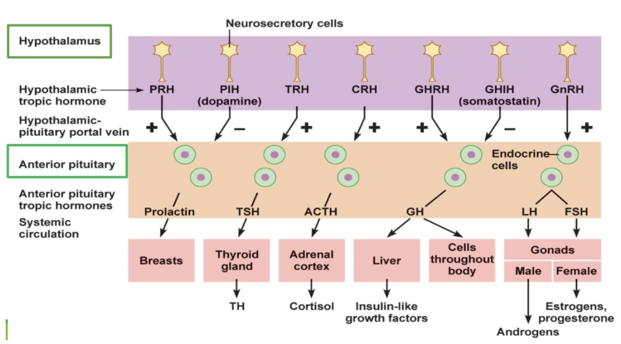
Sends “releasing hormones” through the blood portal system to the anterior pituitary, a hub of different specialized cells that receive the signal and produces/releases “stimulating hormones” to peripheral tissues
Peripheral tissues themselves oftentimes produce their own hormones
Regulated by negative feedback from too high of peripheral hormone levels, as do hypothalamic-releasing hormones
types of anterior pituitary cells and hormones
Gonadotroph
Product: LH, FSH
Target: Gonads
Somatotroph (makes up about half of the cells within the anterior pituitary area)
Product: Growth hormone (GH)
Target: All tissues, liver
Lactotroph
Product: PRL
Target: Breasts, gonads
Thryotroph
Product: TSH
Target: Thyroid gland
Corticotroph
Product: ACTH, β-lipotropin
Target: Adrenal gland, adipocytes, melanocytes
general idea of the HP_ system
The hypothalamus sends out “releasing hormones” as positive stimulation to the cells of the anterior pituitary gland, prompting them to release their own specific “stimulating hormones” into the target peripheral tissues—and then the peripheral tissues themselves can release their own hormones to exert physiological actions
Most releasing hormones are excitatory
supraoptic nucleus
sits directly adjacent to the ventricle receiving blood from external circulation (sensing function) for the pituitary
The pituitary has both arterial and venule circulation to obtain chemical input from the periphery, as well as exit routes to external tissues
hypopituitarism
pituitary gland fails to produce one or more essential hormones
Can be developmental, but is often associated with head trauma or damage to the area
hyperpituitarism
pituitary gland makes too many hormones
Often associated with tumors that lead to excessive production of stimulating hormones
Can cause:
Gigantism, acromegaly → Excess GH (growth hormone)
Goiter and enlarged adrenal cortex → Excess TSH and ACTH, respectively
Precocious sexual development → Excess GnRH
Cushing's syndrome → Excess ACTH and cortisol
Occasionally lactation → Excess prolactin
general treatment approach for “hypo” conditions
Replace and maintain normal peripheral hormone levels
general treatment approach for “hyper-” conditions
Slow or diminish signals—which is often done with either dopamine D2 agonists or somatostatin agonists that broadly inhibit hypothalamic and pituitary activity
How does the posterior pituitary receive signals? What does it then produce?
The posterior pituitary receives direct innervation from the hypothalamus and releases vasopressin (also known as antidiuretic hormone = ADH or arginine vasopressin = AVP) and oxytocin
Vasopressin and oxytocin are relatively stable (lasting minutes to hours) and quite structurally interchangeable
feedback system for posterior pituitary
Feedback is primarily positive neuronal signals for further production and release
What is the trigger(s) for vasopressin release from the posterior pituitary?
Dehydration causes high particle concentration in the blood, sending signals to the hypothalamus to trigger the release of ADH from the posterior pituitary gland until balance is restored
What is the trigger(s) for oxytocin release from the posterior pituitary?
Physical stimulation in the periphery (e.g., contractions in labor, breastfeeding, sexual intercourse) sends positive neuronal signals to the hypothalamus, triggering the posterior pituitary gland to release oxytocin, which amplifies the physical response in a continuous loop until the stimulation ends
physiological actions of oxytocin
Dilation of the birth canal causes a reflex stimulus for the release of oxytocin, leading to rhythmic contractions of the uterus → expulsion of the fetus and placenta
The uterus is 10x more sensitive to oxytocin at term compared to the first trimester. This change in sensitivity dictates that oxytocin is most and only therapeutically useful at term, making it a DOC for inducing labor
Promotes release of milk from mammary glands
What sex hormone antagonizes oxytocin’s actions on the uterus?
High levels of progesterone have wide effects on the body and can help prevent premature labor by antagonizing oxytocin’s actions on the uterus. Progesterone levels then plummet just prior to full term
Note: Effects of oxytocin are reduced when estrogen levels are low, which may suggest a role in co-regulation of libido
drugs for labor and contractions
Oxytocin (Pitocin): primary drug for labor induction
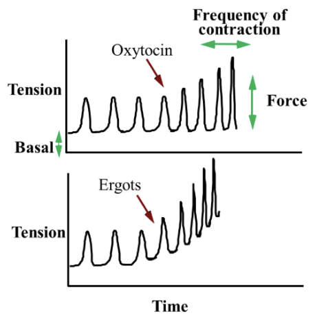
Increases contraction rate and intensity without altering resting tension (i.e., you get full relaxation, unlike ergots)
Ergometrine (ergonovine): ergot derivative with contractile activity, often used to prevent uterine postpartum bleeding
Oxytocin/ergometrine (Syntometrine): given postpartum to reduce bleeding and delivery of placenta
How is the release of vasopressin regulated naturally?
Primary control is volume and osmolarity
Having either a low blood volume or hyperosmolarity (e.g., severe dehydration, infusion with hypertonic solution) causes ADH release to dilute your blood with water content and vasoconstrict your blood vessels
Which drugs affect the release of vasopressin?
Nicotine (and ACh) increases the release of ADH (→ kidneys reabsorb water from the urine and vasoconstrict your blood vessels)
Alcohol decreases the release of ADH (→ kidneys send extra water to the bladder → frequent urination and dehydration)
types of vasopressin receptors
V1A: Located in numerous tissues, particularly smooth muscle of vasculature. Causes IP3-Ca2+ stimulation of blood vessels to promote contraction if your blood volume gets too low
V1B: Cross-regulates anterior pituitary release (mostly ACTH) via similar IP3 intracellular mechanism
V2: primarily acts in the kidney through Gs signaling to promote insertion of aquaporin channels → reabsorption of very clean water from urine if your blood volume gets too low
physiological functions of vasopressin
Renal V2 receptor action: Increases renal tubular reabsorption of water → decreases the volume of urine output (concentrating urine)
Vascular smooth muscle V1A receptor action: Contracts all smooth muscle of the vasculature → causes ↑ in BP and reflexively a ↓ in HR
Dysfunction of vasopressin leads to…
Types
diabetes insipidus (→ excessive urine excretion and constant thirst), which can either be:
Nephrogenic: Failure of the kidney tubules to reabsorb water due to ADH insensitivity
Some drugs (lithium/antivirals) can cause it temporarily
Treatment: Water replacement to prevent dehydration
Neurogenic: ADH deficiency caused by head injury, tumor, cerebral aneurysms, CNS ischemia, etc.
Treatment: Typically desmopressin
vasopressin therapeutics (replacements)
ADH replacement (vasostrict or pitressin)
Disadvantages: Short duration of action; must be injected; both antidiuretic and vasopressor activity are roughly equal (nonselective)
Desmopressin (DDAVP or Nocdurna): Analog of ADH with longer duration of action
In pill/nasal forms
More V2 selectivity to favor antidiuretic activity (hold onto water)
Can be used for nighttime bedwetting
Tolvaptan (Jynarque): V2 selective antagonist (blocker) to excrete water
Can remove water without proportionally getting rid of sodium, which aids in either congestive heart failure or ADH overactivity, conditions that result in hyponatremia
growth hormone-releasing hormone (GHRH)
A 44-amino acid peptide synthesized in the arcuate nucleus
Binds to GHRH receptors on the anterior pituitary → Gs signaling → growth hormone (GH) is released
Can also promote prolactin release
Drug forms of GHRH (1-44; somatorelin) or its truncated analog (1-29; sermorelin) can be used to diagnose pituitary deficiencies, rather than for treatment
growth hormone (GH)
Large peptide of two splice lengths (22kDa = ~90%, 20kDa = ~10%), both of which are equally active
Exogenous is all in the 22kDa form (key for detecting blood doping)
Released in pulsatile fashion from the pituitary gland
While release through the day is irregular, it is generally highest during the initial stage of sleep
Levels generally:
Rise and fall alongside insulin (energy abundance for growing!)
Can be stimulated by both stress and exercise
Increase approaching puberty across the lifespan, then progressively decrease with age
functions of growth hormone (GH)
Directly promotes metabolic switch of…
↓ glucose uptake/use → glucose is spared and fat is burned for energy instead
↑ lipolysis → ↑ fatty acids used for energy
↑ protein synthesis → ↑ tissue/muscle growth
Rather than GH, what hormone is responsible for actual growth and proliferation?
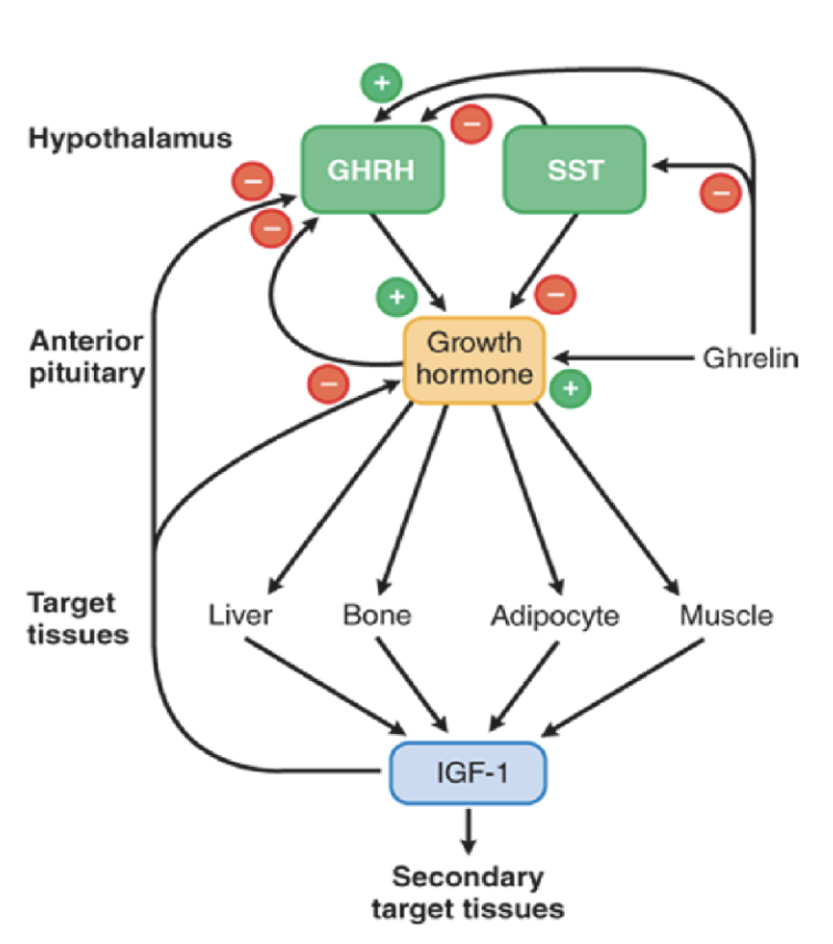
Actual growth and proliferation occurs via the insulin-like growth factor 1 (IGF-1)
IGF-1 promotes growth of bone length, muscle and tissue growth, and burning of fat
High levels of IGF-1 appear to inhibit GHRH and increase somatostatin release, both of which provide negative feedback on GH release
GH receptor
sends signals through the tyrosine kinase JAK/STAT cascade to promote gene expression of IGF-1
Most IGF-1 is produced in the liver
IGF-1 receptor
has extensive overlap in function (and even low-affinity cross-binding) with the insulin receptor
Primarily acts via strong Akt activity to promote cell survival (stop apoptosis)
Secondarily ERK activity to promote cell proliferation (division)
Overproduction of GH is often the result of…
Underproduction of GH is a consequence of…
Overproduction is often the result of a pituitary tumor
Underproduction is often the result of pituitary damage (and, in rare cases, a receptor mutation)
GH disorders
Gigantism: Excess GH signaling during development → Overactivity of IGF-1 → Excessive growth in all directions
Proportional dwarfism: Underproduction of GH → Lack of IGF-1 → Delayed growth, short stature, possible poor muscle development and exercise tolerance
Acromegaly: Post-pubertal GH excess (after epiphyses of bones fuse) → Not all tissues respond equally to growth signals → Abnormal growth of hands/feet, protruding brow and chin, barrel chest, excessive pubertal effects (hair, sweat, voice changes), general swelling of soft tissues
Other common symptoms: Headaches, joint pain, glucose intolerance, and hypertension
treatment for excess GH
Somatostatin analogs (octreotide and lanreotide): formulated in month-long injectable forms to slow GH release and even slow tumor growth in acromegaly
Downsides:
Nasty GI effects and long-term hyperglycemia
Slows or inhibits actions of most anterior pituitary hormones, having widespread potential impacts of energy metabolism, sex hormones, immune function, etc.
Dopamine (D2) agonists (bromocriptine): can lower GH, IGF-1, and prolactin levels in roughly half of acromegaly patients
Neither a first-line option nor as effective as somatostatin (can be combined with somatostatin treatment)
HGH analog antagonist (Pegvisomant (Somavert)): a modified and stable antagonist at the GH receptor that can more selectively slow acromegaly without altering the entire endocrine system
treatment for deficient GH (growth receptor drugs)
HGH (Somatropin): synthetic GH given to deficient children to promote growth through puberty
Can be used for off-label growing and also used controversially in athletic and for anti-aging properties
Due to proliferative effects and the added risk of tumorigenesis, exercise extreme caution
Somatrogon (Ngenla): modified stable hGH analog that can be taken weekly in an autoinjector pen
Mecasermin (Increlex): synthetic IGF-1 given only to children who don’t respond to GH and have extremely low IGF-1 levels
HPG axis
The gonadotropin-releasing hormone (GnRH) couples with Gq/11 proteins to stimulate the release of two stimulatory hormones from the pituitary gland: luteinizing hormone (LH) and follicle-stimulating hormone (FSH), leading to cell development and sex hormone production
FSH and LH receptors (both Gs) are in distinct populations of gonadal cells
Typical levels of sex hormones suppress further GnRH or LH/FSH production and release
Exception: Ovulation and pregnancy violate this notion, as excess during these times leads to more excess

temporal patterns of GnRH in…
Females
Males
Females:
Relatively low and inhibited by estradiol until just prior to puberty, and then exponential increase
Once cycling, pulses become increasingly frequent leading up to just a few days after ovulation, drop dramatically, and then slowly speed up again
Males:
Slow build from youth to puberty (less estradiol suppression), then rapid but shallow pulsatile control
Pulses occur every 2-5 hours
Both sexes show increases of GnRH throughout the lifespan. However, too frequent pulsing or constant stimulation results in the loss of FSH/LH release
Puberty is a result of…
the hypothalamus releasing an increasing number (in both amplitude and frequency) of gonadotropin-releasing hormone (GnRH) → gonadotropic hormones, such as follicle-stimulating hormone (FSH) and luteinizing hormone (LH), are released from the pituitary gland → sexual maturity (sex hormones)
These are found in both males and females but lead to different results depending on sex
LH leads to the release of estrogen from the ovaries and androgen release from the testes
FSH allows for maturation of sperm and eggs
Note: Both males and females produce small amounts of the other gender’s sex hormones (estrogen or androgens). This is largely controlled by aromatase levels
hypogonadotropic hypogonadism
a condition where the brain's hypothalamus or pituitary gland fails to make enough hormones to stimulate the gonads, leading to low sex hormone levels
May be either hormone or receptor functional disruption
Result: Sexual infantilism/delayed puberty, which can be dysfunction at almost all levels of HPG axis
Treatment: Hormone replacement regardless of cause
hypergonadotropic hypogonadism
results from loss of central GnRH inhibition, leading to increased pulsing
Result: Premature/precocious puberty
Diagnosis and treatment: GnRH agonists (e.g. leuprolide depot) to prevent pulsating activity and shut down sex hormone production
prolactin (PRL)
a large peptide structurally related to GH and released by similar stimuli
prolactin (PRL) levels
Levels are primarily controlled by inhibitory signaling through dopamine D2 (Gi) receptor activation on lactotrophs
Thyrotropin-releasing hormone (TRH) plays a minor role in PRL release (only observed in thyroid disorders)
Levels…
Increase dramatically as pregnancy comes to full term and remains with suckling stimulation
Gradually desensitize with time and eventually decline over months
PRL receptor activity
member of the cytokine family, just like GH, binding to a homodimer that activates the JAK/STAT signaling pathway
Primarily initiates and maintains lactation (gene transcription of milk proteins among them)
Estrogen and progesterone block these prolactin actions during pregnancy
Prepares the mammary glands for breastfeeding by promoting the growth of breast tissues (both proliferation and differentiation of mammary ductal and alveolar epithelium)
(GH can also weakly bind to a PRL receptor, so lactation is a possible side effect of acromegaly)
Inhibits LH and FSH release and their effects on ovaries and gonads (If a drug has sexual dysfunction as a side effect… prolactin could be the culprit)
hyperprolactinemia
excessive PRL release due to a number of potential factors:
Dopamine antagonists (reserpine, haloperidol)
Hypothalamic or pituitary disorder
Excess TRH release (simple goiter)
Oral contraceptives
Prolactin-secreting tumors, which come in two clusters
Galactorrhea: excessive or spontaneous lactation
Amenorrhea in women, or even loss of libido/fertility in both sexes
treatment for hyperprolactinemia
any dopamine agonist, with bromocriptine as the DOC
Side effects: N/V, headaches, postural hypotension, CNS activity (in rarer cases and at high doses)
thyroid
crucial for energy balance, temperature regulation, increasing carbohydrate burning and protein synthesis, and fetal limb development
thyrotropin-releasing hormone (TRH)
a small tripeptide (half-life: ~6 minutes) produced in the paraventricular nucleus (PVN) of the hypothalamus and transported to the anterior pituitary
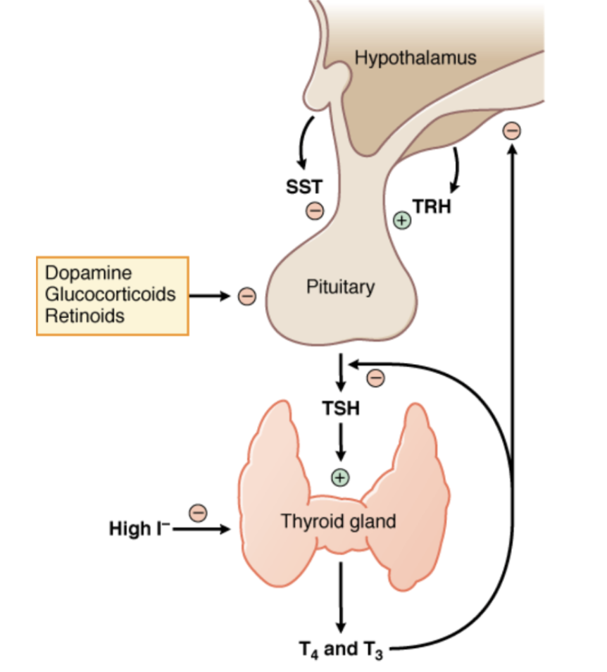
Binds to TRHR (Gq) in thyrotroph cells to stimulate secretion and further synthesis of thyroid-stimulating hormone (TSH) (sometimes referred to as thyrotropin) from the anterior pituitary, which signals the thyroid gland to release thyroid hormones
Besides thyroid hormones, the thyroid also produces calcitonin
How can we inhibit TRH activity?
can be inhibited by dopamine (D2), somatostatin, glucocorticoids, and negative feedback of T3/T4
What synthetic TRH is given IV for differential diagnosis of hypothyroidism?
protirelin (Thyrel TRH)
Measure TSH levels at baseline and 30 minutes post-infusion
Quite outdated now
thyroid-stimulating hormone (TSH)
large glycoprotein structurally related to FSH/LH (same α subunit but different β)
Promotes thyroid hormone production, and binds to TSH receptors in the thyroid to stimulate (Gs) the release of thyroid hormones from stores
Maintains overall health of thyroid
TSH deficiency leads to atrophy
TSH excess leads to goiters
Blood tests for TSH levels is the primary method of determining hypothyroidism, even in asymptomatic cases
What human TSH is given to promote uptake of radioactive iodine (131I) into thyroid tumors?
thyrotropin α (Thyrogen)
Formerly used for diagnostic purposes to examine responsiveness of thyroid to TSH by examining uptake of contrast iodine (123I)
thyroid hormone synthesis
What element on the periodic table do thyroid hormones contain?
What is the major building block?
Heavily involves dietary iodine (i.e., iodized salt) uptake
At any given time, the thyroid has 20-50x the iodine concentration (like a storage house) as plasma
A lot of energy is needed to take up iodine, so the process recycles and scavenges anything in the thyroid that isn’t released as thyroid hormones
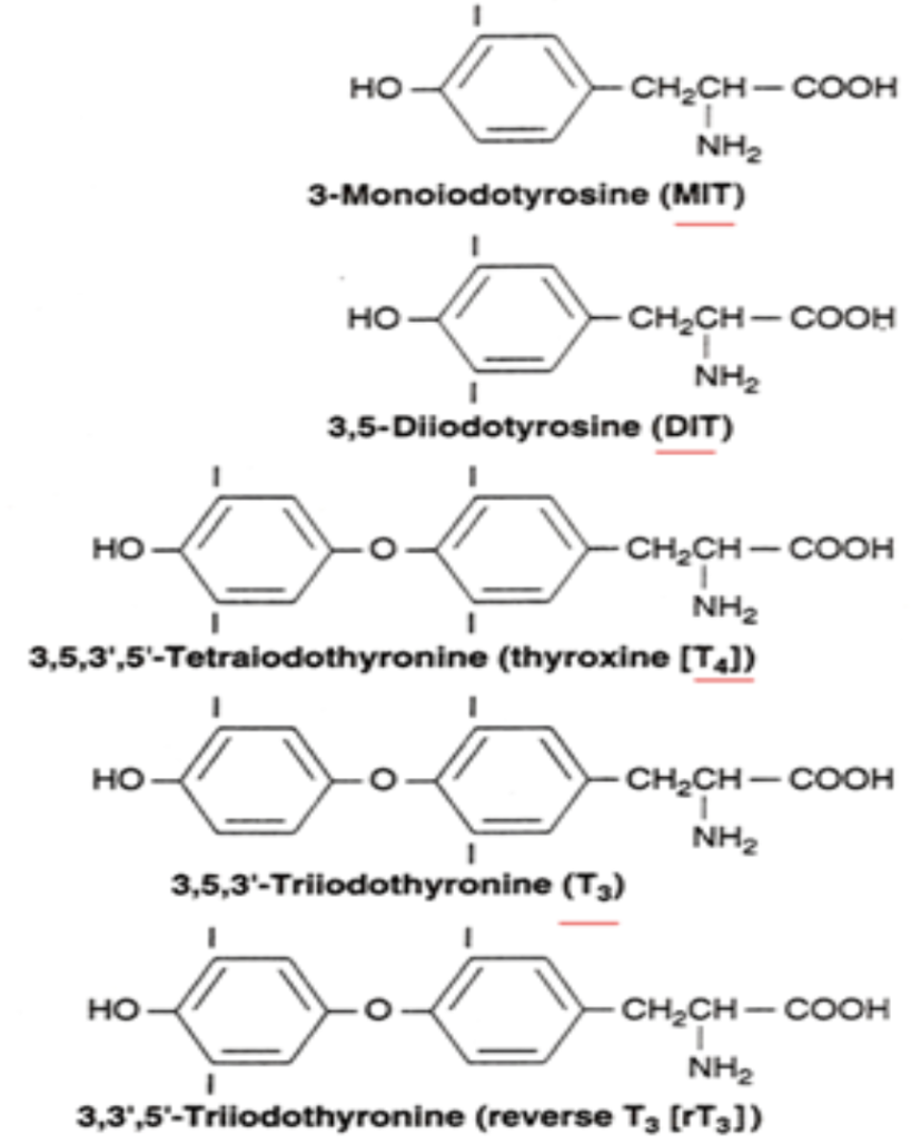
Tyrosine is a major building block that gets iodinated in the process into T4 (thyroxine, 80%) as the primary product, followed by T3 (20%)
thyroid receptors
nuclear receptors that promote gene expression (thyroid hormone response elements)
Includes TRα1, TRβ1, TRβ2
α isoform is primarily expressed in muscle and skeletal tissues
β isoforms primarily expressed in liver, kidney, and brain
TRβ2 can also have a repressive role in gene expression
T3 binding suppresses the transcription of TRH and TSH in the hypothalamus and pituitary, respectively
When unbound, TR acts as…
When bound by T3/T4, TR acts as…
When unbound, TR acts as a gene repressor; when bound by T3/T4, TR recruits co-activator proteins to turn on gene expression
Activation often occurs in heterodimers with other nuclear promoter complexes, most notably the retinoic acid receptors (RXRs)
How is T4 transported into cells?
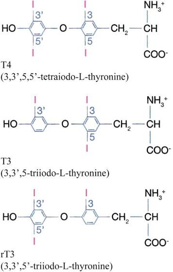
Soluble hormones like T4 are heavily protein-bound and transported into cells primarily via monocarboxylate transporters (MCT8/10) before getting converted into T3
How would you compare the circulating levels of T4 versus T3?
Basal circulating levels of T4 are roughly 30-50x that of T3
Estrogen and analogs can increase protein binding in plasma, while corticosterone and testosterone can lower
Many prescription drugs can alter protein binding and circulating plasma hormone levels
How would you compare the activity of T4 versus T3?
T4 lasts long in the blood and is protein-bound, but has about 10x less affinity for receptors. Thus, most activity is due to T3 conversion in peripheral tissues and binding
How would you compare the stability of T4 versus T3?
Peripheral T4 is more metabolically stable (T1/2 of T4 = 7 days, T1/2 of T3 = 1.5 days), and is converted to either T3 or inactive rT3 based on tissue-specific enzymes
functions of thyroid hormones
Developmental
Cell survival and proliferation, growth of most organs, neurogenesis
T3 mediates metamorphosis of tadpoles into frogs (limb growth and tail resorption)
Thermogenic
T3 can control the efficiency of mitochondrial energy production processes → Burns extra calories to produce heat (it’s how the thyroid determines your thermal setpoint and regulates your body temperature!)
Can increase utilization of carbohydrates and glycogen (although insulin/glucagon usually compensates for actions on glucose levels)
Cardiovascular
Excess thyroid function → Tachycardia and increased stroke volume and tension
Hypofunction → Bradycardia and lowered pulse pressure
baby hypothyroidism disorder
Congenital hypothyroidism (cretinism): deficient thyroid function from birth
The thyroid is critical to brain development, limb differentiation, fertility, and thermal regulation
Treatment: Early detection and supplementing T4
Two ways to have a hypothyroid from birth:
Endemic form: iodine deficiency in pregnancy (mother) and infancy (baby)
Sporadic form: congenital defect of an enzyme or receptor in the process of thyroid synthesis
adult hypothyroidism disorder
Adult hypothyroidism results in edema, thick and rough skin, loss of hair, cold intolerance, lethargy, cognitive impairment, anemia, and infertility (likely mediated by TRH-stimulated prolactin)
Primary: Iodine deficiency → Goiter (still have lots of TSH)
Secondary: TSH deficiency, atrophied thyroid
Tertiary: TRH deficiency, atrophied thyroid
Hashimoto's thyroiditis: autoimmune antibody production against thyroglobulin or other synthetic proteins
Most common cause in the developed world
Falls under primary hypothyroidism
hyperthyroid disorders
Thyrotoxicosis
Graves’ disease
Plummer’s disease (toxic multinodular goiter)
thyrotoxicosis
syndromes from excess T3/T4 hormone release
Symptoms may include:
Goiter with excess hormone release
Increased energy use and alertness/anxiety
Hyperthermia
Symptoms characteristic of SNS overactivity
Graves’ disease
autoimmune production of antibodies that activate TSH receptors directly, bypassing any negative feedback
Recurring, remitting disorder
~5-7x more common in women
Usually manifests in young adults
Characteristic inflammation around eyes (bulging look)
Falls under secondary hyperthyroidism
Plummer’s disease (toxic multinodular goiter)
typical hyperthyroidism in older patients of unknown origin
However, evidence points to mutated TSH receptors that can turn on spontaneously (so secondary hyperthyroidism)
About 20% of cases
Rare ophthalmopathy
“thyroid eye disease (TED)”
a term mostly invented by pharmaceutical companies that refers to a symptom/disorder of Graves’ disease: ophthalmopathy (bulging of eyes and redness/irritation of fatty tissues and tear ducts)
treatment options for thyroid eye disease (TED)
Teprotumumab (Tepezza), veligrotug (Lumvoa)
Monoclonal antibodies that target IGF-1 receptors, the key mediator of causing inflammation in the area of your eyes
Prevents abnormal growth in tissues behind the eye and reduces bulging
Milder cases can be treated with steroids for inflammation and treating the underlying thyroid condition
treatment options for hypothyroidism
Commonly treated via T4 (levothyroxine, Synthroid) for long-acting (lifelong) replacement
Mostly taken orally as tablets
Best absorbed on an empty stomach
Food, drugs that slow acid secretions, and chelating/resorption-blocking drugs can impair uptake
T4 levels will peak within a couple hours, but remain mostly stable for days (half-life: ~7 days)
T3 (liothyronine, Cytomel) is often only given in emergency situations (i.e., coma) (since T3 is the “active form”) for immediate action due to short half-life of few hours
Relatively impractical as replacement therapy
4:1 preparations of T4:T3 (Thyrolar) are available, but the benefits are unclear. Armour Thyroid is ground up pig thyroid gland that provides T4:T3 in similar ratios, but giving synthetic instead of natural has a much lower risk of reactions
main treatment options for hyperthyroidism
Thionamides (MMI and PTU) are antithyroid drugs that interfere with the iodine peroxidase and synthesis of hormones
Does not alter iodide uptake or hormone release
Methimazole is more common for Graves’ and Plummer’s, as it is long-acting enough for QD pills (1-hr versus 6-hr half-life)
Propylthiouracil can also inhibit the conversion of T4 → T3 in the periphery, so it is more commonly given during “thyroid storms”
Lugol’s solution (5% I and 10% KI) or saturated solution potassium iodide (SSKI; 50% KI) consist of excess iodine that increases plasma iodine concentration and slows both uptake and hormone release
Used temporarily to stabilize thyroid activity in a crisis or preoperative (before surgery) to shrink the thyroid
adjuvant treatment options for hyperthyroidism
β-adrenergic antagonists and Ca2+ channel blockers are often used in thyrotoxicosis to counteract sympathetic and cardiac symptoms
How are thyroid tumors treated?
can often be TSH-driven, so high T4 can be given to aid in shrinking the tumor
Typically treated with either surgery or 131I radioactive iodine which concentrates in the overactive thyroid and produces destructive localized radiation (has a low risk of secondary cancers)
PTU can be used with 131I to prevent any integration into circulating hormones in the bloodstream
parathyroid gland
What serum levels does it control?
four small nodular glands located within the thyroid gland
Calcium and phosphate, both of which are typically abundant in diet, central to bone mineralization, and critical to all intracellular function
What four substances specifically regulate levels of circulating calcium and phosphate?
Calcitonin from thyroid
Parathyroid hormone (PTH) from parathyroid
1,25-dihydroxyvitamin D (calcitriol) from kidney
FGF23 from bone cells
parathyroid hormone (PTH)
short-lived peptide (84 AA) with a half-life of minutes once cleaved
Its job is to maintain normal levels of calcium in your blood
Primarily targets 1) kidney to increase calcium reabsorption + phosphate excretion and 2) bone to promote resorption
Activates a kidney enzyme to convert vitamin D into calcitriol, which targets 1) intestines to increase calcium reabsorption + phosphate reabsorption and 2) bone to promote resorption
Describe the feedback loop on the parathyroid hormone (PTH)
The parathyroid glands have a negative feedback loop controlled by parathyroid-secreting cells, which have calcium-sensing receptors (CaSR) that, when occupied by Ca2+, activate:
Gq-mediated suppression of PTH synthesis, AND
Gi-mediated suppression of PTH release
parathyroid hormone 1 receptor (PTH1R)
present in bone and kidney and mediates resorption of calcium-phosphate complexes
Can also be activated by parathyroid hormone-related protein (PTHrP), which is produced in stem cells of bone/cartilage, including excess by tumors
osteoblasts
cells that collect calcium for new mineralization and promote extracellular release
Acted upon by PTH and calcitriol
Note: Although PTH and calcitriol’s effects on phosphate oppose each other, both ultimately increase free Ca2+
vitamin D
family of fat-soluble hormones that bind to nuclear vitamin D receptors (VDR)
Binding to VDRs has rapid, non-genomic actions
Dietary sources provide vitamin D2 (somewhat less stable and potent but a commonly prescribed supplement)
UV interacting with skin produces D3 (cholecalciferol)
What is vitamin D converted into?
Vitamin D is eventually converted into calcitriol, which increases the absorption of Ca2+/phosphate from the intestine (↓ fecal loss), as well as bone resorption
Conversion is activated by PTH, and high levels of calcitriol causes negative feedback onto PTH production itself
PTH also promotes the production of fibroblast growth factor 23 (FGF23) from bone, which increases the metabolism of calcitriol (a miniature negative feedback system to prevent excess resorption and making your bones brittle)
calcitonin
short, 32-AA peptide with really low circulating basal levels and a short half-life (~10 minutes)
Secreted from C cells in the thyroid and has bidirectional (and opposite) control of serum Ca2+ levels as that of PTH
Binds to calcitonin receptors via Gs and Gq to produce opposing actions to PTH that ultimately lower free Ca2+
Aids in maintenance of bone integrity and sustains minimal structure of bone during “calcium stress” events
Inhibits osteoclast activity and differentiation—the cells primarily responsible for bone breakdown
hypoparathyroidism
results from tissue or autoimmune damage, with symptoms commonly related to low blood calcium:
Muscle spasm and tetany, hair/nail/dental demineralization and brittleness
Rare cases of depression and anxiety
hyperparathyroidism
leads to…
Hypercalcemia → Muscle weakness, constipation, flatulence, N/V
Hypercalciuria and hyperphosphaturia → Formation of kidney stones and calcification in kidneys
Hyperparathyroidism can be broken down into…
Primary: tissue damage or lesion (Treatment: Surgical removal)
Secondary: low calcium due to other disorders, such as kidney disease (Treatment: Besides correcting the underlying cause, you may use cinacalcet (sensipar), an allosteric agonist of Ca2+-sensing receptor that lowers PTH)
calcium regulation disorders
Rickets
Osteoporosis
Paget’s disease
rickets
developmental deficiency of vitamin D, resulting in porous and under-mineralized bones and joints
To compensate → Leg curvatures and wide joints, enlarged abdomen, smaller ribs
osteoporosis
Types
lowering of bone mass and risk of fractures, increasing with age
Type I is associated with estrogen loss in women and is responsive to hormone replacement
Type II is natural loss of bone modeling efficiency and PTH activity with age
Paget’s disease
malformations of bone due to bone resorption, followed by disorganized and excessive reformation (i.e., because of excessive osteoclast activity, some parts of bone can end up highly mineralized and parts right next to it can be under-mineralized, which increases the risk of fractures)
Likely a strong genetic component is involved
hormone treatments for parathyroid and calcium regulation disorders
Vitamin D and calcium: treatment for rickets and osteoporosis
Calcitriol: treatment for hypoparathyroidism
Comes in injectable/pill forms
Calcitriol analogs (paricalcitol/Zemplar): treatment for hyperparathyroidism
Designed to reduce PTH activity via a negative feedback loop without affecting bone or calcium
Calcitonin (miacalcin): short-term treatment for Paget’s to reduce remodeling, as well as some forms of osteoporosis to help stave off demineralization
Causes short-term hypercalcemia
Contains synthetic salmon calcitonin, as it’s more active than human form
PEGylated teriparatide (Yorvipath): long-term treatment for hypoparathyroidism
Daily injectable PTH analog, with less need for Vitamin D and calcium supplements
bone formation treatments for calcium regulation disorders
Bisphosphonates
PTH analogs: Teriparatide (Forteo) and abaloparatide (Tymlos)
bisphosphonates
drug class that chelates and binds calcium in bone, and accumulates at active bone remodeling sites → gets taken up by osteoclasts during bone resorption → inhibits osteoclast function and causes apoptosis → can treat Paget’s and osteoporosis (especially postmenopausal)
Integrates into the bone structure, so it can last for a long time
Includes:
Etidronate (Didronel) = daily pill
Risedronate (Actonel), alendronate (Fosamax) = weekly pills
Ibandronate (Boniva) = monthly pill or quarterly injection
Zoledronic Acid (Reclast) = yearly injection
PTH analogs
can treat osteoporosis and reduced fracture in high-risk patients
Uses periodic-burst activation that promotes more osteoblast activity than osteoclasts (Despite it causing a short period of bone demineralization, it is followed by a period of active, aggressive remineralization)
Typically limited to 2-year treatment before it loses its effectiveness
targeted monoclonals for bone formation
Burosumab (Crysvita): targets and inhibits FGF23 = increased phosphate to prevent softening of bones in rare inherited hypophosphatemia and tumor-induced wasting
Denosumab (Prolia): targets receptor activator of the NF-kB ligand RANK-L, a mediator known to cause differentiation and activation of osteoclasts in bone
Blocking can reduce bone loss, though only indicated if bisphosphonates fail or in bone cancers
Romosozumab (Evenity): inhibits sclerostin (known to inhibit bone formation by reducing osteoblast activity) and regulated by PTH/calcitonin
Shown to reduce spinal fracture risk alone or in combination with bisphosphonates
adrenal glands
Receive central blood flow inputs from the aorta and then outputs through the vena cava and renal vein
Produce steroid hormones that regulate carbohydrate metabolism (glucocorticoids) and fluid/electrolyte retention (mineralocorticoids)
Also produce precursor steroids that are building blocks for sex hormones
central regulation of the HPA axis
What provides positive feedback on the axis? What provides negative feedback?
Corticotropin-releasing hormone (CRH/CRF): released from hypothalamus in diurnal fashion (peaks in early morning) to bind to CRH1 receptors in the pituitary gland
In the pituitary gland, CRH1 receptors (Gs) regulate the translation of proopiomelanocortin (POMC), a long peptide that is cut up to create corticotropin (adrenocorticotropic hormone; ACTH) to then be released
Note: Inflammation stimulates the production of CRH and ACTH. Cortisol provides negative feedback to the production of CRH and ACTH (via membrane and nuclear glucocorticoid receptors)
the products of POMC
ACTH and melanocyte-stimulating hormone (MSHs) are released from the pituitary and bind to melanocortin receptors (MC1R and MC2R)
ACTH activates MC2R (Gs) → Synthesis and activation of steroid side-chain cleaving enzyme (CYP11A1) and cholesterol recruitment
ACTH is also key to maintenance and structure of adrenal glands
MSH and lipotropin activate MC1R → Pigmentation
What is the starting material for all steroid hormones (steroids)?
free cholesterol
Note: Cholesterol can either come from LDL/HDL in the blood, or made by the adrenal cell itself
A transport protein shuttles cholesterol into adrenal cells
Once cholesterol is inside an adrenal cell, the steroidogenic acute regulatory protein (StAR) transports it from the outer mitochondrial membrane toward the inner inner mitochondrial membrane, where steroid hormone synthesis begins
types of steroid hormones
Glucocorticoids
Cortisol is the main glucocorticoid in humans; corticosterone is the main glucocorticoid in rodents
Mineralocorticoids
Because they are lipid-soluble, many steroids have to travel through the blood by attaching to binding proteins.
What carries cortisol?
What carries testosterone and estradiol?
What is another protein that some steroid hormones can loosely bind to and travel with?
Corticosteroid-binding globulin (CBG) → carries cortisol
Sex hormone-binding globulin (SHBG) → carries testosterone and estradiol
Some steroid hormones also travel loosely bound to albumin
glucocorticoid receptor (GR) activity
Cortisol is lipid-soluble, so it can cross cell membranes to bind to the nuclear receptor GRα in the cytoplasm
Binding causes dissociation from stabilization proteins
GR is activated and can either:
Increase expression via binding glucocorticoid response elements (GREs), or
Suppress expression of other transcription factors (e.g., POMC promoter, NF-kB production of cytokines)
Note: Cortisol is 3x more potent at the GR than corticosterone. Progesterone is a moderate GR and MR antagonist