2.1 Object and nature of prosthetic Dentistry.
 Call Kai
 Learn
Learn Practice Test
 Spaced Repetition
 Match
Match Flashcards
 Knowt Play
1/84

Earn XP
Description and Tags
Semester two - topic 1
Name | Mastery | Learn | Test | Matching | Spaced | Call with Kai | Chat |
|---|
No analytics yet
Send a link to your students to track their progress
85 Terms
Define Prosthetic Dentistry
It is the dental speciality pertaining to the diagnosis, treatment planning, rehabilitation and maintenance of the oral function, comfort, appearance and health of patients with clinical conditions associated with missing or deficient teeth and/or maxillofacial tissues using biocompatible substitutes.
What is Prosthodontist?
A specialist in Prosthodontics
A dentist who was successfully completed an advanced education program in Prosthodontics that is accredited by the appropriate accrediting body. In the United States, that body is the Commission on Dental Accreditation of American Dental Association / ADA/.
What is a prothesis?
an artificial replacement of an absent part of the human body
a therapeutic device to improve or alter function
a device used to aid in accomplishing a desired surgical result
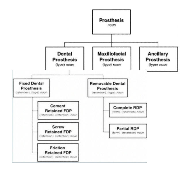
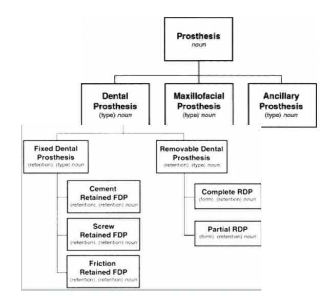
What are the three types of prothesis’?
Dental prosthesis
Maxillofacial prosthesis
Ancillary prosthesis
What are the two types of dental prosthesis?
Fixed dental prosthesis
Removable dental prosthesis
What are the three types of FDP under fixed dental prosthesis?
Cement retained FDP
Screw Retained FDP
Friction Retained FDP
What are the two types of RDP’s under Removable dental prosthesis?
Complete RDP
Partial RDP
Remember!!!
Remember!!!
What is oral facial complex?
in dental prosthetics it refers to the integration and function of prosthetic components within the oral and facial regions with the function of:
Mastication
Swallowing
Breathing
Speech
Special organs : smell, taste and vision
What are the bones of the oral facial complex?
Cranium, maxilla and mandible
What are the joints of the oral facial complex?
TMJ, stylomandibular ligament and sphenomandibular ligament capsular ligament
What are the muscles of mastication?
Temporalis
Masseter
Buccinator
Orbicularis oris
What are the muscles during masticatory movements?
Opening - depressor jaw muscles : mylohyoid/ digastric/ inferior lateral pterygoid
Closing - elevator jaw muscles : medial pterygoid/ superficial masseter / temporalis
What are the extrinsic tounge muscles?
Genioglossus
Styloglossus
Hyoglossus
List the small glands in the oral and facial region
Labials
Buccales
Palatine
List the big glands in the oral and facial region
Parotid
Submandibularis
Sublingualis
How many types of papillae are present on tongue?
4
What are the 4 types of papillae on tongue?
Fungiform papillae
Filiform papillae
Foliate papillae
Cicumvallate papillae
What are the tongue muscles divided into?
Intrinsic and extrinsic
What does the intrinsic muscles of the tongue do?
They change the shape of the tongue
What are the intrinsic muscles of the tongue?
M. longitudinalis linguae
M. transversus linguae
M. verticalis linguae
What does the extrinsic muscle of the tongue do?
Responsible for the tongues protrusion and retrusion
What is an example of an extrinsic muscle of the tongue?
Genioglossus muscle
What is the tongue innervated by?
Innervated by the hypoglossal nerve (cranial nerve XII)
What is the role of the tongue in tongue activity?
Anchoring and moving the tongue
Involved in jaw movements, respiration, speech, taste, mastication, swallowing and sucking
Define a tooth
A tooth is a hard, calcified structure found in the jawbone (either the maxilla or mandible). It’s used for chewing and grinding food during the process of mastication.
What is are dental embrasures?
They are the spaces that you can find around a tooth where the curvature of the tooth surface starts to move away from the adjacent tooth
What are the names for the types of dental embrasures?
Occlusal/Incisal
Facial
Lingual
Gingival
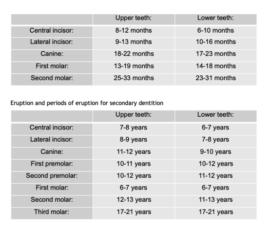
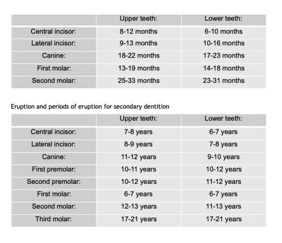
How many teeth make up Primary (Deciduous) Teeth?
consists of 20 teeth
When does Primary (Deciduous) Teeth begin forming?
begin forming during first trimester of pregnancy
when does Primary (Deciduous) Teeth start erupting?
typically start erupting around 6 months of age
At what age would a child have a complete primary dentition of Primary (Deciduous) Teeth?
Most children have a complete primary dentition by 3 years old
Describe the morphology of deciduous teeth
Thinner enamel and whiter than the permanent teeth
Rounded crowns with constricted cervix
More prominent cervical ridge
Roots of anterior teeth are longer and narrower than the permanent
Roots of molars are longer and more slender and flare greatly, comparing to the permanent
How many teeth does Secondary (permanent) teeth consist of?
consists of 32 teeth in most cases
when does Secondary (permanent) teeth start erupting?
being erupting at 6 years of age
when are Secondary (permanent) teeth fully erupted?
most permanent teeth have erupted by age 12
what Secondary (permanent) tooth is an exception in terms of erupting by age of 12?
Third molars (wisdom teeth) are the exceptions, often appearing late teens or early teens
Remember!!!
Remember!!!
What is mixed dentition?
the transition phase in dental development where a person has both primary (deciduous) teeth and secondary (permanent) teeth present in the mouth.
When does mixed dentition usually occur?
Typically occurs between the ages of 6 and 12
What is Hypodontia?
missing one or few teeth due to development issues
What is anodontia?
Rare condition where all primary or permanent teeth are missing
Give two conditions that affect oral function and aesthetics
Hypodontia and Anodontia
Classification: Incisors
function as cutting or shearing instruments for food
Classification: canines
possess the longest roots of all teeth and are located at the corners of the dental arch
Classification: premolars
act like the canines in the tearing of food and are similar to molars in the grinding of food
Classification: molars
located nearest to the temporomandibular joint (TMJ), which serves as the fulcrum during function
What are the tooth surfaces?
Apical, labial, lingual, distal, mesial
Tooth surfaces: apical
pertaining to the apex or root of the tooth
Tooth surfaces: labial
pertaining to the lip; describes the front surface of the anterior teeth
Tooth surfaces: lingual
pertaining to the tongue; describes the back (interior) surface of all teeth
Tooth surfaces: distal
the surfaces of the tooth that is away from the median line
Tooth surfaces: mesial
the surface of the tooth that is toward the median line
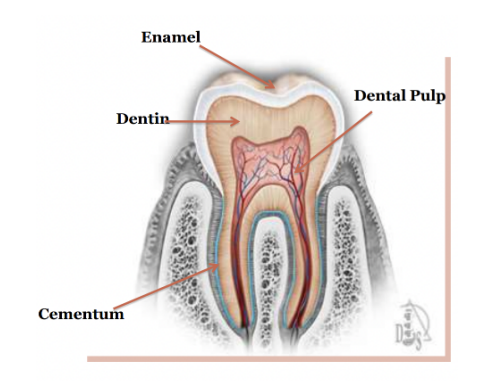
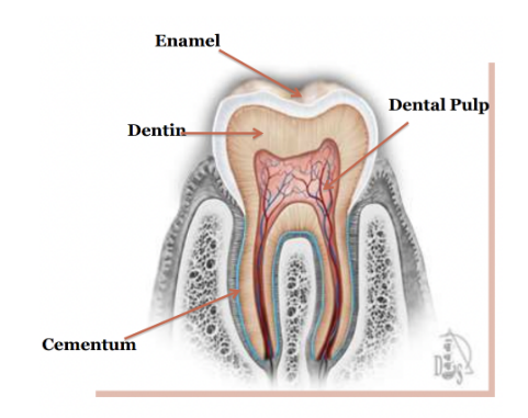
Dental Tissue: Enamel
Outermost layer of tooth, very hard, protects tooth from decay
Dental Tissue: dentin
beneath enamel, also hard, supports enamel and carries some nerve fibers
Dental Tissue: odontoblast layer
these cells create dentin and are located at boundary of the pulp chamber
Dental Tissue: Pulp chamber
Soft tissue in centre of the tooth that contains nerves and blood vessels
Dental Tissue: gingiva
also known as gums, soft tissue that surrounds teeth and covers alveolar bone
Dental Tissue: periodontal ligament
soft tissue that helps anchor tooth to surrounding alveolar bone
Dental Tissue: cementum
hard tissue that covers the tooth root and helps anchor it in place with help of periodontal ligament
Dental Tissue: alveolar bone
part of jawbone that holds the tooth sockets
Dental Tissue: pulp canals
pathways in roots of teeth that carry nerves and blood vessels to the pulp chamber
Dental Tissue: apical foramen
Opening at tip of the tooth’s root where nerves and blood vessels enter tooth
Remember!!
Remember!!
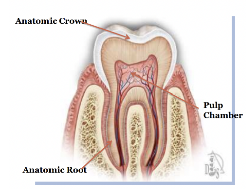
What does the anatomic crown do?
portion of tooth covered by enamel
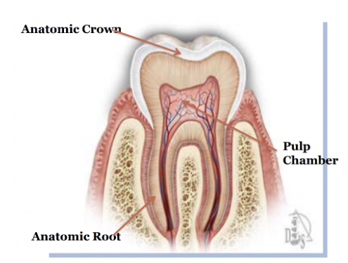
What does the anatomic root do?
It is the lower two thirds of a tooth
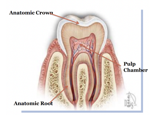
What does the pulp chamber have?
It houses the dental pulp, an organ of myelinated and unmyelinated nerves, arteries, veins, lymph channels, connective tissue cells and various other cells
Tooth sides: incisal
the cutting/tearing edge, in posterior teeth will be occlusal which is used for grinding the food
Tooth sides: mesial
tooth surface towards the median line
Tooth sides: distal
tooth surface away from the median line
List the periodontal tissues
gingiva
alveolar bone
periodontal ligament
cementum
List the hard tissues of the tooth
enamel
dentin
cementum
alveolar bone
What are the soft tissues of the tooth?
pulp chamber
Gingiva
peridontal ligament
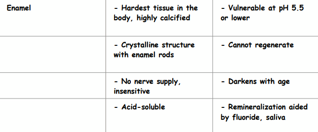
Enamel:
Characteristics:
Hardest tissue in the body, highly……
……structure with enamel….
no nerve supply, ……
…..-soluble
Vulnerability:
Vulnerable at pH ….. or lower
Cannot….
…… with age
Remineralization aided by ….., saliva
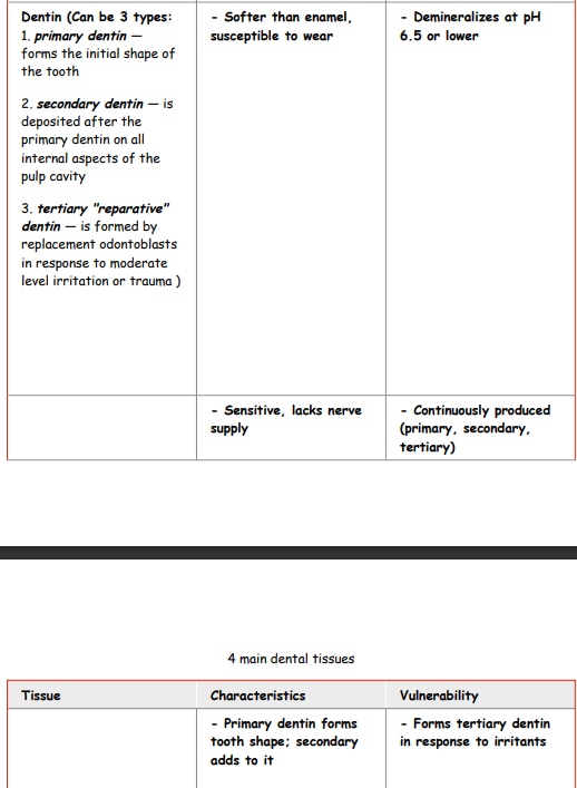
What are the three types of dentin?
primary, secondary and tertiary “reparative” dentin
what does primary dentin do?
forms initial shape of tooth
When is secondary dentin deposited and onto what?
it is deposited after primary dentin on all internal aspects of the pulp cavity
How is tertiary “reparative” dentin formed and in repose to what?
is formed by replacement odontoblasts in response to moderate level irritation or trauma
Dentin
Characteristics:
It is ….. than enamel, susceptible to wear
Sensitive, lacks ……..
Primary dentin forms…….; secondary adds to it
Vulnerability:
Demineralizes at pH …. or lower
……produced (primary, secondary, tertiary)
Forms ……dentin in response to irritants
softer
nerve supply
initial
6.5
continuously
tertiary
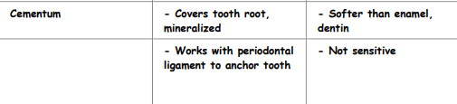
Cementum
Characteristics:
covers….., mineralized
works with periodontal ligament to…..
Vulnerability:
…..than enamel, dentin
not…..
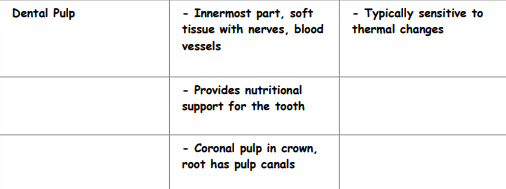
Dental Pulp
Characteristics:
…..part, soft tissue with nerves, blood vessels
provides…….for the tooth
……..in crowns, root has pulp canals
Vulnerability:
Typically sensitive to …….
Tooth sides: Lingual
pertaining to the tongue; describes the back surface of the teeth
Tooth sides: Labial
pertaining to the lips; describes front surface of the teeth
Tooth sides: Apical
pertaining to the apex or root of the tooth
The dental tissue: Pulp chamber
The soft tissue in the centre of the tooth that contains nerves and blood vessels