Antebrachialis Muscles, Cubital Fossa and Its content
 Call Kai
 Learn
Learn Practice Test
 Spaced Repetition
 Match
Match Flashcards
 Knowt Play
1/72

There's no tags or description
Looks like no tags are added yet.
Name | Mastery | Learn | Test | Matching | Spaced | Call with Kai | Chat |
|---|
No analytics yet
Send a link to your students to track their progress
73 Terms
depression on the anterior aspect of the elbow
Cubital Fossa
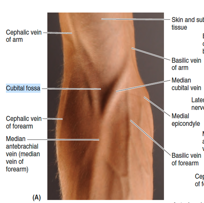
Margins of cubital fossa
Superior: Imaginary horizontal line connecting the humeral epicondyles
Lateral: brachioradialis
Medial: Pronator teres
Floor:
Lateral: Supinator
Medial: brachialis
Roof:
Bicipital aponeurosis
Fascia
Skin

Contents of the cubital fossa from lateral to medial
Radial nerve (passes between the brachioradialis and brachialis)
Biceps brachii tendon
Brachial artery
Median nerve

Contents of the cubital fossa (superficial layer to deep)
Skin
Subcutaneous
Median cubital vein
Medial and lateral cutaneous nerves of the forearm
Deep fascia
Bicipital aponeurosis
Deep cubital fossa
Biceps tendon
brachial artery
median nerve
radial nerve
Floor
Brachialis
Supinator
Blood supplies of the cubital fossa
Brachial artery
Ulnar artery
Radial artery
Median Cubital vein
Cephalic vein
Basilic vein
common site for sampling and transfusion of blood, and IV injections
Median cubital vein
lies directly on deep fascia
Runs diagonally from cephalic vein to basilic vein
Median cubital vein
Ulnar neuropathy (Cubital tunnel syndrome)
happens when the ulnar nerve (the "funny bone" nerve) gets squeezed or stretched as it passes through the inside of your elbow, particularly at the cubital tunnel retinaculum/arcuate ligament of Osbourne
•Distal humeral fracture
•Common in children
•70% is due to fall on outstretched hand
•Up to 10% are associated with damage to neurovascular structures
Supracondylar fracture
•During fracture and reduction
•MC with posterolateral displacement
•Superior to brachial muscle
Brachial artery injury
•MC: anterior interosseous nerve injury
•Runs with brachial artery
Median Nerve injury
branch of median nerve most commonly injured
Anterior interosseous nerve (AIN nerve)
injured with the same frequency as median nerve
Radial nerve
much less common, may be injured with flexion type fractures
Ulnar nerve
the result of an untreated ischemic injury that causes muscle and nerve damage in the involved compartment of the extremity.
Volkmann’s Ischemic Contracture
Bicipital myotatic reflex confirms the integrity of what nerve?
Musculocutaneous nerve
dense connective tissue sheet between the radius and ulna
Interosseous Membrane of the Forearm
puts forearm in its most stable mid-prone position/ position of function
Taut fibers
deep fascia enclosing the arm like a sleeve
-deep to the skin and subcutaneous tissue
-continuous with the antebrachial fascia
Brachial fascia
deep fascia of the forearm
Antebrachial fascia
extend from the deep surface of the brachial fascia to the central shaft and medial and lateral supra-epicondylar ridges of the humerus
Medial and lateral intermuscular septa

Fascial compartments of the forearm
ANTERIOR/ FLEXOR/ PRONATOR
POSTERIOR/EXTENSOR/ SUPINATOR
The fascial compartments of the limbs generally end at the joints; therefore, fluids and infections in compartments are usually contained and cannot readily spread to other compartments. The ______ compartment is exceptional in this regard because it communicates with the central compartment of the palm through the ______.
anterior compartment; carpal tunnel
long flexor tendons that form its roof pass directly beneath the flexor retinaculum to enter the palm
carpal tunnel
TRUE OR FALSE:
•ANTERIOR/FLEXOR/PRONATOR - MEDIAN N
•POSTERIOR/EXTENSOR/SUPINATOR - RADIAL N
True
Common flexor attachment of the first layer muscles of the forearm
Medial epicondyle of the humerus
All muscles of the anterior forearm cross the elbow joint except ___
•the 3 deep muscles.
All of the muscles in the anterior forearm are supplied by the median nerve except for the ff:
•FCU- ulnar nerve
•Medial part of FDP- ulnar nerve
•The tendons of most of the flexor muscles are locate on the anterior surface of the wrist and are held by:
palmar carpal ligament
flexor retinaculum
ALL anterior superficial muscles of the forearm cross the wrist joint except:
Pronator teres
•Fusiform
•Most lateral of the superficial forearm flexors
•Forms medial boundary of the cubital fossa
Pronator teres
•Long fusiform
•Medial to pronator teres
•Belly replaced by long flattened tendon in the middle of forearm
•Passes thru canal in lateral part of flexor retinaculum thru a vertical groove in the trapezium
•Has own synovial tendinous sheath of FCR – guide to radial artery
Flexor carpi radialis
•Fusiform
•Absent in one or both sides, usually left, in 14% of people
•Guide to the median nerve at the wrist : tendon lies deep and slightly medial to the nerve
Palmaris longus
•Most medial of the superficial flexors
•Simultaneously flexes and adducts the hand at the wrist if acting alone
•With FCR: flexes wrist
•With ECU: adducts wrist
•The only superficial muscle innervated by ulnar nerve
Flexor carpi ulnaris
•Forms intermediate layer between the superficial and deep muscles
•2 heads: humero-ulnar and radial heads
•Median N and ulnar A enter the forearm by passing between the 2 heads
Flexor digitorum superficialis
•Muscle belly splits into 4 distinct tendons near the wrist
•Pass deep to the flexor retinaculum thru carpal tunnel to the fingers
•At level of proximal phalanx, splits to create small openings called Chiasma of Camper, allowing FDP tendons to pass
Flexor digitorum superficialis
•Tendons are enclosed in a synovial common flexor sheath
•Action: flexes the middle phalanges of the medial 4 fingers at the PIP joints
•Capable of flexing each finger it serves independently
Flexor digitorum superficialis
•the only muscle that can flex the Distal interphalangeal joints
•“clothes” the anterior aspect of the ulna
•Crucial element for grip strength and pincer grip
Flexor digitorum profundus
•Long flat flexor of the thumb
•Lies lateral to the FDP
•Clothes the anterior aspect of the radius distal to the attachment of the supinator
Flexor pollicis longus
•Quadrangular muscle
•Prime mover for pronation
•Deepest muscle in the anterior aspect of the forearm
•The only muscle that attaches only to the ulna at one end and only to the radius at the other end
Pronator quadratus
Initiates pronation and is assisted by the Pronator Teres when more speed and power are needed
Pronator quadratus
Supination muscles
•Biceps brachii
•Supinator
Pronator muscles
•Pronator teres
•Pronator quadratus
puts the forearm in mid-prone position
Brachioradialis
True or false:
•Muscle of the posterior forearm are Extensor-supinator muscles
True
True or false:
Muscles of the posterior forearm are All are innervated by radial N
True
True or false:
Muscles of the posterior forearm are held in place at the wrist by the extensor retinaculum to prevent bowstringing of tendons
True
Posterior Forearm Muscles that extend and abduct/ adduct the hand at the wrist joint:
ECRL
ECRB
ECU
Posterior Forearm Muscles that extend the medial 4 fingers
Extensor digitorum
Extensor digiti minimi
Extensor indices
Posterior forearm muscles that extends the thumb
Abductor Pollicis Longus
Extensor Pollicis Longus
Extensor Pollicis Brevis
The only muscles that do not cross and are incapable of acting at the wrist
Brachioradialis
Supinator
•Fusiform muscle
•Lies superficially on the anterolateral surface of the forearm
•Forms lateral border of the cubital fossa
•Rotated to the anterior aspect of the humerus and thus flexes forearm at the elbow
Brachioradialis
•Powerful elbow fixator
•Radial N bifurcates before diving deep to this muscle
•Insertion: proximal to radial styloid process
Brachioradialis
•Relatively short muscle belly and longer tendon
•Indispensable when clenching the fist
•Wrist extension and abduction
ECRL
•Larger muscle belly, shorter tendon
•Last superficial muscle of the posterior forearm supplied by the radial N before it passes thru the supinator
Extensor Carpi radialis brevis
•Principal extensor of the medial 4 digits
•Occupies much of the posterior surface of the forearm
Extensor digitorum
•Accessory extensor of the little finger
•Functions to extend the 5th digit
Extensor digiti minimi
•Extensor and adductor of the wrist
•Short muscle belly with a long flat tendon that converges distally into a round tendon
•Inserts at the medial base of metacarpal V
Extensor carpi ulnaris
•Triangular in shape
•Small muscle that provides accessory support to other posterior forearm muscles
Anconeus
•Has superficial and deep heads that help supinate the forearm
•Passing between the 2 heads is the deep branch of the radial N
•Prime mover for slow, unopposed supination
Supinator
•Thumb abduction bet metacarpal I and the trapezium
•Assists in wrist abduction
•Inferior margin of the anatomical snuffbox
Abductor pollicis longus
•Inferior margin of the anatomical snuffbox
Extensor pollicis longus
•Larger, tendon longer than EPB
•Adducts the extended thumb and rotates it laterally
Extensor pollicis longus
•Triangular depression on the lateral aspect of dorsum of hand
Anatomical snuffbox
The tendons of the APL and EPB bound the anatomical
snuff box _____, and the tendon of the EPL bounds it
______
Anteriorly; posteriorly
lies in the floor of the snuffbox
Radial artery
•palpated proximally in snuffbox
Radial styloid process
Palpated distally in snuff box
Base of the first metacarpal
Floor of the snuffbox
Scaphoid
Trapezium
most frequently fractured carpal bone
Scaphoid
•Initial X-rays may not reveal fracture
•Repeat X-rays taken 10-14 days reveal bone resorption
•Union of fractured parts may take up to 3 months because of poor blood supply
•Avascular necrosis of the proximal fragment of scaphoid
SKL