Human Bio WACE Exam
1/168
There's no tags or description
Looks like no tags are added yet.
Name | Mastery | Learn | Test | Matching | Spaced |
|---|
No study sessions yet.
169 Terms
Validity
- Testing what is meant to be tested
Data represents the phenomenon you are measuring
Reliability
An experiment gives the exact same result each time it is performed
Exocrine
- secretes into a duct that exits the body
- Eg, sweat and salivary glands
Endocrine
secretes hormones into the extracellular fluid that surrounds the cells that make up the gland.
Cellular effects of hormones
- Activates a gene/s
- Changes shape of enzymes
- Changes the rate of transcription and translation
Protein and amine hormones
Water soluble, Attaches to receptor proteins on the membrane of the target cell, The combination of the hormone with the receptor causes a secondary messenger to be released and diffuse through the cell and activate particular enzymes
Steroid hormones
Lipid soluble, Combines with a receptor inside the cell. Activates the genes to synthesise a particular protein
Functions of the hypothalamus
- Controls secretions to the pituitary gland via releasing and inhibiting factors
- Regulates body temperature, water balance and heart rate (homeostasis)
- Produces hormones that are carried to the pituitary gland
- Hormones produced here either inhibit or stimulate hormone production in the anterior pituitary
- Other hormones pass through the nerve fibres to the posterior where they are secreted
- Makes hormones for posterior pituitary
Structure of the Pituitary Gland
- Lies under the hypothalamus and is joined to it by the infundibulum
- Consists of an anterior and posterior lobe
- Anterior lobe is at the front and is connected to the hypothalamus by a complex network of blood vessels
- Posterior lobe is to the rear and is joined to the hypothalamus by nerve fibres, does not make hormones
- Secretes hormones that control other endocrine glands
What hormone does the thyroid produce and what effects does it have
Thyroxine,
targets most cells,
increases metabolic rate and therefore oxygen consumption and heat
What hormone does the parathyroid produce and what effects does it have
Parathyroid hormone (PTH)
targets bones, kidneys
bones release calcium, reabsorption of calcium
What hormone does the thymus produce and what effects does it have
Thymosins,
targets t lumphocytes
stimulates development and maturation of t lymphocytes
What hormone does the pineal gland produce and what effects does it have
melatonin,
controls sleep patterns and circadian rhythms, stimulated by darkness and inhibited by light
Adrenal cortex
aldosterone- kidney, reduces amounts of sodium and increase amount of potassium in the urine
cortisol- most cells, promotes normal metabolism, helps the body deal with stress, promotes repair of damaged tissue
What hormone does the Adrenal Medulla produce and what effects does it have
Adrenaline and Noradrenaline- most tissues, prepares the body for fight or flight responses. increases rate and force
Pancreas
Insulin- most cells, stimulates uptake of glucose. lowers blood glucose levels.
Glucagon- liver and fat storage, stimulates the breakdown of glycogen and fat to increase blood glucose levels
What hormones do the testes produce and what effects do they have
Androgens- many tissues, stimulates sperm production, growth of skeleton and muscles and secondary sexual characteristics
What hormones do the ovaries produce and what effects do they have
Estrogen- many tissues, stimulates the development of the female characteristics and regulates the menstrual cycle
Progesterone- uterus and mammary gland, regulates the menstrual cycle, pregnancy and prepare the mammary glands for milk secretion
Enzyme amplification
- Series of chemical reactions
- 1 hormone activates 1000’s of enzyme molecules
- 1 hormone causes a hormonal cascade which could trigger the production of over a billion enzyme molecules
Hormone clearance
- Hormone is turned off
- The breaking down of a hormone once it’s finished its job
- Broken down at kidney or liver
- Excreted in bile or as urine
How are hormone secretions regulated
by negative feedback
Negative Feedback Loop
- Negative feedback is when the response produced by the secretion of the hormone is the opposite of the stimulus that caused the secretion
- Releasing factors stimulate the release of a hormone
- Inhibiting factors slow down the secretion of a hormone
What is a neuron
A nerve cell
What is a nerve
A bundle of nerve fibres (Any long extension of cytoplasm of a nerve cell body, although the term usually refers to an axon) held together by connective tissue
CNS vs. PNS
Central Nervous System: Consists of the spinal cord and the brain.
Peripheral Nervous System: Nerves that connect the central nervous system with the receptors, muscles and glands of the body
Myelinated vs. Unmyelinated fibres
Myelinated fibres: An axon with the myelin sheath covering
Unmyelinated fibres: An axon without the myelin sheath covering
Grey matter vs. White matter
Grey matter: Consists of nerve cell bodies and unmyelinated fibres
White matter: Composed of myelinated fibres (fatty tissue)
Sensory/receptor neurons (unipolar) structure and function
Carry messages from receptors in the sense organs or skin to the central nervous system (brain and spinal cord)
Carry messages from receptors in the sense organs or skin to the central nervous system (brain and spinal cord)
Motor/effector neurons (multipolar) structure and function
Carry messages from the central nervous system (brain and spinal cord) to the muscles and glands
Has 1 axon and multiple dendrites extending from the cell body
Interneurons (bipolar) structure and function
Connects sensory and motor neurons together, found in the spinal cord and for reflexes in the grey matter of the spinal cord
Cell body is to one side of the axon
Synapse-
- The junction between the branches of adjacent neurons, - Occurs between a branch at the end of an axon and a dendrite or the cell body of another neuron
Transmission across a synapse-
1) An action potential arrives at the pre-synaptic axon terminal
2) Local depolarisation causes voltage gated calcium ion channels to open
3) Calcium ions from the extracellular fluid diffuses through the presynaptic membrane of the axon terminal and enters the cytoplasm of the axon terminal
4) The calcium ions cause neurotransmitter vesicles to migrate to the pre-synaptic membrane of the axon terminal
5) The neurotransmitter leaves the vesicles and enters the synaptic cleft through exocytosis
6) The neurotransmitter diffuses across the synapse to the post-synaptic membrane of the dendrite of the adjacent neuron
7) Sodium ions flood in, causing depolarisation in the postsynaptic dendrite
8) An action potential will be generated
Nerve impulse-
1) A resting neuron has a positive charge on the outside of the membrane and a negative charge on the inside (resting membrane potential -70mV)
2) There is a high concentration of positive sodium ions on the outside and a high concentration of positive potassium ions on the inside
3) There is a greater concentration of negatively charged ions on the inside of the membrane than positive potassium ions making the inside negatively charged
4) A stimulus causes voltage gated sodium ion channels to open and sodium ions rush into the intracellular fluid
5) -55mV is threshold for voltage gated sodium channels
6) The inward movement of positively charged sodium ions reverses the charges either side of the membrane
7) The cell becomes depolarised, the charge on the inside is positive and the charge on the outside is negative
8) After the inside of the membrane becomes flooded with sodium ions, gated potassium channels open and allow the potassium ions to move to the outside
9) As soon as the potassium ions are released, the sodium ion channels close (membrane is repolarised)
10) The sodium potassium pump restores the concentration of sodium and potassium ions when the membrane is a resting state
Nerve impulses in myelinated vs. unmyelinated fibres
Unmyelinated | Myelinated |
Depolarisation of one area of the cell membrane causes an action potential to flow onto the membrane immediately adjacent to the stimulus. (1)
| Depolarisation of one area of the cell membrane causes an action potential to jump from one node of Ranvier to another. (1) |
The nerve impulse/exchange of ions (NOT action potential) moves along the entire length of the neuron/axon. (1) | The nerve impulse/exchange of ions (NOT action potential) only occurs at the nodes of Ranvier or cannot occur where the axon is myelinated. (1)
|
Lower concentration gradient of ions either side of the membrane. (1) | Higher concentration gradient of ions either side of the membrane at the nodes of Ranvier. (1)
|
The nerve impulse / message (NOT action potential) travels along the whole length of the fibre, reducing its speed. (1) | The action potential jumps from one node of Ranvier to the next on the myelinated fibre (saltatory conduction), the impulse can travel faster. (1)
|
Saltatory conduction-
when the nerve impulse jumps from one node of Ranvier to the next
Divisions of the PNS
Afferent (sensory)
Somatic: Carries impulses from receptors around the muscle and skin to the CNS
Visceral: Carries impulses from the internal organs to the CNS
Efferent (motor)
Autonomic: Carries impulses from skeletal muscles to the CNS
Somatic: Carries impulses to the heart and other involuntary muscles
Autonomic
Sympathetic: Fight or flight response, prepares body for strenuous activity
Parasympathetic: Rest and digestion, maintains body during quiet and restful conditions
Autonomic vs. Somatic
Characteristic | Autonomic | Somatic |
Effectors | Involuntary muscles/organs | Skeletal muscles (voluntary) |
General function | Adjustment of the internal environment | Response to the external environment |
Efferent (motor) pathway | 2 nerve fibres from the CNS to the motor neuron with a synapse in a ganglion. | One nerve fibre from the CNS to the motor neuron, no synapse and no ganglion |
Neurotransmitter at effector | Noradrenaline = sympathetic Acetylcholine = parasympathetic | Acetylcholine at neuromuscular junction |
Control | Involuntary | Voluntary |
Nerves to target | 2 sets, sympathetic and parasympathetic | One set, excitation of skeletal muscles |
Effect target organ | Excitation or inhibition | Always excitation |
Effects of the Sympathetic vs. Parasympathetic NS
Neurotransmitter | Noradrenaline | Acetylcholine |
Heart | Increases rate and strength of contraction | Decreases rate and strength of contraction |
Lungs | Dilates bronchioles | Constricts bronchioles |
Stomach | Decreases movement | Increases movement |
Liver | Increases breakdown of glycogen and released as glucose | Increases uptake of glucose and synthesis of glycogen |
Iris of the eye | Dilates pupils | Constricts pupils |
Sweat glands | Increase sweat secretion | No effect |
Salivary glands | Decreases saliva secretion | Increases saliva secretion |
Blood vessels of:
Skin
Skeletal muscles
Internal organs |
Vasoconstriction
Vasodilation
Vasoconstriction (except heart and lungs)
|
Little effect
No effect
Little effect |
Urinary bladder | Relaxes muscles of wall | Constricts muscles of wall |
Adrenal medulla | Stimulates hormone secretion | No effect |
Compare the Nervous System and the Endocrine System
|
Nervous System |
Endocrine System
|
Speed
|
Rapid |
Slow |
Duration
|
Quick |
Long |
Transmission
|
Neurons |
Hormones |
Nature
|
Electro-chemical |
Chemical |
Cells affected
|
Muscles and glands |
All body cells |
Type of response
|
Specific/local |
Widespread |
What things are responsible for the protection of the NS
Bone (outer) | Cranium and vertebral canal = tough outermost protective layer |
Meninges (connective tissue)
- Outer layer (dura matter) - Middle layer (arachnoid matter) - Inner layer (pia matter) |
Tough and fibrous Loose mesh of fibres Contains blood vessels that stick closely to the brain and spinal cord
|
Cerebrospinal fluid (between pia and arachnoid matter) | Shock absorber, cushions the brain and spinal cord |
Function of the cerebral cortex
Higher order functions such as thinking, reasoning, memory, learning and conscious awareness of surroundings. Comprised of 4 lobes
Function of the corpus callosum
Nerves fibres that allow communication between the 2 cerebral hemispheres
Function of the cerebellum
Coordination of fine contraction of voluntary muscle for smooth movements, balance and posture
Function of the hypothalamus
Homeostasis; appetite, thirst, metabolism, body temperature, response to fear, water levels, sleeping
Function of the medulla oblongata
Regulates heart, breathing and diameter of blood vessels
Cardiac centre- Regulates rate and force of heart beat
Respiratory centre- Regulates rate and depth of breathing
Vasomotor centre- Vasoconstriction and vasodilation of blood vessels
Function of the spinal cord
Made up of grey matter and surrounded by white matter
contains sensory axons carrying impulses towards the brain (ascending tract)
contains bundles of motor axons carrying impulses from the brain to the muscles (descending tract)
Responsible for spinal reflexes, which are reflexes by nerve cells in the spinal cord without input from the brain
What is homeostasis
the ability of an organism to maintain a constant internal environment/steady state despite changes in either internal or external conditions to maintain optimal functioning of cells/cell processes/metabolic processes
Types of Receptors-
Receptor | Location | Detects |
Thermoreceptors | Hypothalamus (central) Skin (peripheral) | Core body temperature External temperature |
Osmoreceptors | Hypothalamus | Osmotic pressure |
Chemoreceptors | Nose Mouth Blood vessels Medulla oblongata | Regulation of chemicals in the body. pH levels |
Touch receptors | Skin (lips, fingertips, eyelids and genitals) | Responds to touch |
Pain receptors | Skin Mucous membranes Most organs (not brain) | Damaged tissues |
Pressoreceptors | Aorta Right carotid artery Right atrium | Blood pressure |
Define a reflex and describe its properties
A rapid autonomic response to a change in the internal or external environment
Properties
1) Requires a stimulus
2) Involuntary and occurs without conscious thought
3) Response is rapid, only a small number of neurons involved
4) Response is stereotyped, the impulse travels the same way each time it happens
What occurs in a spinal reflex arc?
Pathway of a nerve impulse travels from a receptor to an effector
1) A receptor which is on a sensory neuron reacts to a change in the internal or external environment by initiating a nerve impulse along the sensory neuron
2) A sensory neuron carries the impulse from the receptor to the CNS (brain or spinal cord)
3) The nerve impulse is passed to an interneuron via a synapse which is in the grey matter of the spinal cord
4) The impulse is then passed to the motor neuron via a synapse in the grey matter of the spinal cord.
5) The motor neuron carries the nerve impulse to an effector
6) The effectors carry out the appropriate response, normally a skeletal muscle cell or secretory cells
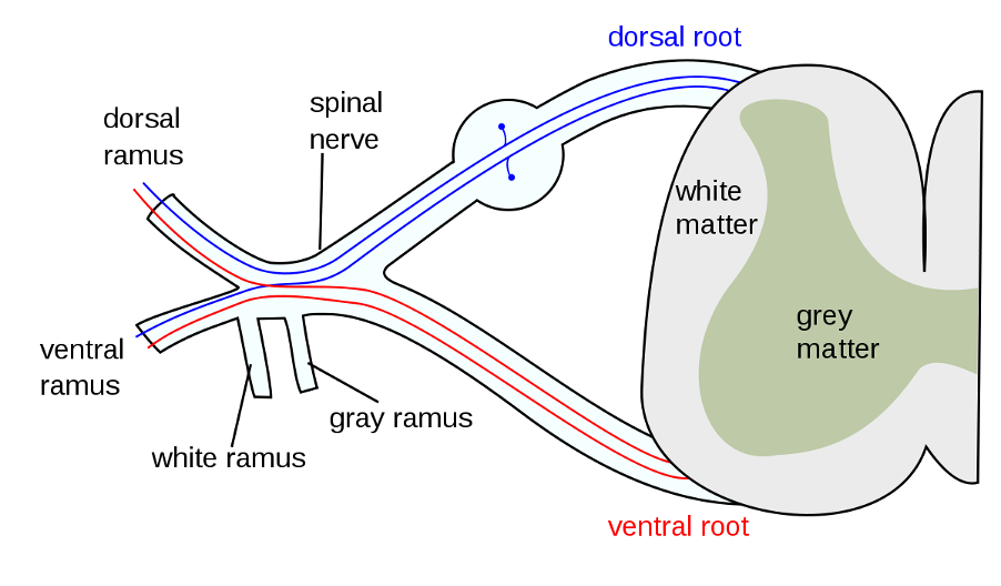
Label the parts of a spinal reflex
Parts of the negative feedback loop (sister ruby munches eggs really fast)
Stimulus: the change in environment causing the system to operate, Eg core temp above 37
Receptor: detects the change, Eg thermoreceptors
Modulator: the control centre, process the information from the receptor and sends the information to the effector to act. Hypothalamus
Effector: carries out appropriate response. Eg, sweat glands
Response: counteracts the effect of the stimulus, Eg sweat secretion
Feedback: the original stimulus has been changed by the response, Eg core temp lowered to 37
Positive Feedback loop-
- No role in homeostasis
- Response to stimulus is intensified
- For body processes that must be completed quickly
- Release of oxytocin from posterior pituitary during child birth
Feedback loop- heat
Feedback loop- osmoregulation
Feedback loop- blood sugar
Feedback loop- blood oxygen
Compare insulin and glucagon and their role in blood glucose regulation
Insulin (beta cells) | Glucagon (alpha cells) |
Decrease blood sugar levels | Increase blood sugar levels |
1) Accelerates transport of glucose from the blood inti the cells, especially skeletal muscles | Converts glycogen into glucose |
2) Accelerates the conversion of glucose into glycogen | Glucose is released into blood, increasing blood sugar levels |
3) Also stimulates conversion of glucose into fat and adipose tissues | Produce new sugar molecules from fats and amino acids (gluconeogenesis) |
Chemical sensors in beta cells stimulate secretion of insulin |
|
Glycogenolysis | Glycogenesis |
What is Insulin’s role in BG regulation?
Effector: Beta cells |
Processes · Glycogenesis – conversion of glucose to glycogen in liver/muscles. · Lipogenesis – conversion of glucose to lipids in adipose tissue |
What is glucagon role in BG regulation?
Effector: Alpha cells |
Processes · Glycogenolysis – conversion of glycogen to glucose in liver/muscles. · Gluconeogenesis – conversion of lipids and amino acids to glucose in the liver |
What is adrenaline/noradrenaline’s role in BG regulation?
Effector: Adrenal medulla |
Processes · Glycogenolysis – conversion of glycogen to glucose in liver/muscles. · Glycogenolysis (Cori cycle) – lactic acid produced can be transported to liver and converted into glucose |
What is thyroxine’s role in BG regulation?
Effector: Thyroid gland |
Processes · Increases absorption of glucose from the small intestine into the bloodstream · Increases rate of cellular respiration, leading to increased rate of glucose absorption into all cells of the body. |
Define glycogenesis
Conversion of glucose into glycogen from other carbohydrates, especially glucose
Define glycogenolysis-
Breakdown of glycogen to glucose
Define gluconeogenesis
Conversion of fats and amino acids into glucose
How is breathing stimulated?
- As carbon dioxide is dissolved in water, it forms carbonic acid which then dissociates to form hydrogen and bicarbonate ions
- Increase in hydrogen = decrease in pH
- Hydrogen/carbon dioxide stimulate peripheral chemoreceptors (aortic and coarotid bodies) which then send impulses to the respiratory centre of the medulla oblongata
- The impulses from the medulla stimulates the diaphragm and intercostal muscles via the phrenic and intercostal nerves respectively
Describe the receptors responsible for stimulating breathing
Central chemoreceptors | Peripheral chemoreceptors |
In medulla oblongata | In aortic and carotid bodies |
Detect CO2 concentration, pH of blood plasma and cerebrospinal fluid | Measures changes in pH (hydrogen ion concentration), CO2 and O2 of blood plasma |
Takes a few minutes to respond and communicate | Responds quicker due to their location |
Responsible for majority control of breathing | Small control over O2 breathing rate |
hyperventilation-
- Rapid and deep breathing to provide more oxygen and remove more carbon dioxide than necessary
- More O2 in, more CO2 out- Can occur due to severe physical and emotional stress
- It will correct itself as the chemoreceptors will not be stimulated since they are more receptive to co2 and breathing will not be required until it returns to normal
- Can be done intentionally to hold breath
- However, as the breathing reflex responds to CO2, the person may require O2 before CO2 levels rise to the point where the body stimulates the breathing reflex.
This could result in a lack of oxygen to the brain, causing the person to fall unconscious
Disruptions to homestasis: Diabetes-
high blood glucose levels where insulin is not being produced or not enough of it is being produced
Type 1 vs. Type 2
| Type 1 | Type 2 |
Causes | Caused by autoimmune response on beta cells in the pancreas | Caused by lifestyle factors, eg overweight, lack of exercise |
Insulin | Does not produce insulin but cells do respond to it | Does produce insulin but cells don’t respond to it |
Treatment | Requires daily injections of insulin to manage conditions | Require management of diet, eg increase activity |
When | Normally begins in childhood/early life | Normally begins onwards of 40 |
effects of high blood glucose levels-
- Damage to blood vessels
- Blindness
- Kidney failure
- Cardiovascular disease
- Loss of sensation
- Ulcers and gangrene
- Amputation of toes or foot
Causes of hyperthyroidism
- Thyroid gland produces too much thyroxine
- Overactive thyroid
Diseases | Graves’ disease = immune system attacks thyroid |
symptoms of hyperthyroidism
- Increased metabolism
- Rapid heart beat
- Unexplained weight loss
- Increased appetite
- Fatigue
- Sweating
- Anxiety
Protruding eyeballs
Treatment for hyperthyroidism
- Surgery to remove part of the thyroid gland
Drugs that block the thyroids’ use of iodine
hypothyroidism- causes, symptoms, treatment, diseases
Causes | - Lack of iodine available to the thyroid gland - Thyroid not producing enough thyroxine |
Symptoms | - Slow heart rate - Unexplained weight gain - Fatigue - Lack of energy - Intolerance to cold |
Treatment | - Synthetic hormones containing iodine |
Diseases | Hashimoto’s disease |
Infectious disease
a disease passed from one person to another by infection with micro-organisms
Pathogen
a disease-causing organism
Vector
an agent capable of transferring a disease-causing organism from one person to another
Non-specific defences-
defences of the body that act against all pathogens
Specific defences-
defences of the body that are directed against specific pathogens
Structural features of bacteria and their functions
Structure | Description |
Slime layer | For protection of the bacteria from antibiotics |
Cell wall | Made of peptidoglycan, a combined carbohydrate protein |
Cell membrane | Controls movement of substances in and out of the cell |
DNA | No nuclear membrane, so DNA forms a tangle inside the cells. Some DNA is in the form of loops called plasmids |
Plasmids | DNA in loops inside the bacteria |
Flagella | Allow for movement |
Cytoplasm | Where the organelles hang out in |
Capsule | Formed of complex carbohydrates for protection. |
Methods of Transmission for pathogens
Transmission | Description | Example |
Transmission by contact | Direct: physically touching an infected person Indirect: touching an object which has been touched by an infected person | STI’s |
Transmission of body fluids | From an infected person which comes into contact with the mucous membrane or bloodstream of the uninfected person | HIV Hepatitis B and C |
Droplets | Contains the pathogen which is breathed in or ingested. Emitted through coughing, sneezing or breathing | Measles Colds Influenza
|
Ingestion | Eating or drinking food which had been contaminated with pathogens | Typhoid fever Salmonella Dysentery |
Airborne | Similar to transmission by droplets. If virus and bacteria survive it can cause infection once inhaled | Chickenpox Measles |
Vectors | An agent which is capable of transferring a pathogen from one person to another. Vectors can spread directly or indirectly. Vectors are specific to a disease | Malaria = mosquitos Dengue fever |
Bacteria vs. Virus
Feature | Bacteria | Virus |
Size | 0.5-5 micrometres | 20-400 nanometres |
Protein coat | No | Yes |
Cell wall | Yes | No |
Plasma membrane | Yes | No |
Cytoplasm | Yes | No |
Nucleus | No | No |
Membrane-bound organelles | Yes | No |
DNA/RNA | DNA and RNA | DNA or RNA |
Diseases caused | Salmonella Pneumonia Norovirus Listeria E. coli. | HPV HIV AIDS Warts |
External non-specific pathogens-
External non-specific defences | Description | Location |
Skin | Physical barrier. Secretes sebum which is an oily fluid which kills pathogens. Fatty acids and salts in sweat also prevent micro-organisms from growing | Outside of the body |
Sebum | An oily secretion produced by sebaceous glands which kill some pathogenic bacteria | Secretes into the blood |
Sweat | Secreted by sweat glands and contains salts and fatty acids which prevent the growth of many micro-organisms | Skin Mucus membranes |
Mucous membrane | Lines the body cavities that open to the exterior. Secrete mucus which prevents the entry of micro-organisms to the body | Lines the body cavities |
Internal non-specific responses- phagocytes
Cells which ingulf and digest micro-organisms and cell debris
Internal non-specific responses- leucocyte
Have the ability to leave the blood capillaries and migrate through tissues to the place of infection
Macrophage
macrophages are involved in specific and non-specific defences. Non-specific = engulfing pathogens. they Secrete substances that kill bacteria Specific = alerts the immune system to the presence of foreign material. They are phagocytic
Internal non-specific responses- inflammation
Shows signs of redness, swelling, heat and pain. Occur as a result of the process which shows in response to an infection
Internal non-specific responses- fever
Elevation of body temperature caused by an increase in the body’s thermostat (controlled by hypothalamus). Inhibits growth of bacteria/viruses and speeds up the rate of chemical reactions which allow cells the repair at a faster rate
Internal non-specific responses- pyrogens
Chemicals released by white blood cells during an inflammatory response which acts directly on the hypothalamus, causing it to increase body temperature
what happens during an inflammatory response
1) When stimulated by mechanical or chemical damage, mast cells release histamine, heparin and other substances. Mast cells stimulate and co-ordinate inflammation by releasing chemicals
2) Histamine increases blood flow through the area and causes the walls of the blood capillaries to become more permeable so that fluid is filtered from the blood. This increase in blood blow causes the heat and redness associated with inflammation, and the escape of fluid from the blood causes swelling
3) Heparin prevents clotting, so the release of heparin from the mast cells prevents clotting in the immediate area of the injury. A clot of the fluid around the damaged area does form and this slows the spread of the pathogen into healthy tissues
4) The chemicals released by the mast cells attract phagocytes. Macrophages and leucocytes actively consume micro-organisms and debris by phagocytosis
5) The abnormal conditions in the tissue stimulate pain receptors, and so the person feels pain in the inflamed area
6) The phagocytes, filled with bacteria, debris and dead cells, begin to die. The dead phagocytes and tissue fluid form a yellow liquid called pus
7) New cells are produced by mitosis and repair of the damaged tissues takes place
Specific Resistance: Humoral vs. cell mediated responses
Characteristics | Humoral (antibody-mediated response) | Cell mediated | |
Type of cell | B-cells | T-cells | |
Where are they produced | Bone marrow | Bone marrow | |
Location of maturation | Bone marrow | Thymus gland | |
Where are they found | Lymphoid tissue Lymph node Spleen Thymus gland Tonsils | ||
Location of resistance to infection | Extracellular fluid (blood, lymph) | Intracellular fluid | |
How antibodies act on pathogens
1) Combine with foreign enzymes or bacterial toxins to inactivate them
2) Bind to the surface of viruses and prevent them from entering cells
3) Coat bacteria to make them easier to be consumed by phagocytes
4) Agglutination, making phagocytosis easier.
5) Dissolve organism/antigen/pathogen
6) Combining with soluble antigens to make them insoluble
Response to a Foreign antigen in body
1) B-cells are sensitised by non-self-antigens in the extracellular fluid
2) B-cells enlarge and divide into groups called clones
3) Most of clones become antibody-secreting plasma cells, the rest become memory cells
4) Antibodies circulate in the blood, lymph and extracellular fluid
5) Antibodies combine with antigens to form antigen-antibody complexes
6) Memory cells spread to all body tissues, so that a rapid response can occur should the antigen enter the body again
Antigen
any substance capable of causing a specific immune response. Proteins on surface of cell
Antibody
substance produced in response to a specific antigen. Combines with the antigen to neutralise it or destroy it
First response vs. Secondary response
Primary response | Secondary response |
First exposure to non-self-antigen | Second exposure to non-self-antigen |
Slow response | Fast response |
B-cells to multiply and differentiate into plasma cells | There’s already heaps of plasma cells and memory cells from the primary response |
Takes several days to build up large amounts of antibodies | Antibodies are released straight away preventing severe symptoms |
Killer T-cells
migrate to site of infection and attach to invading cells and secrete a substance that will destroy the antigen
Helper T-cells
Intensifies the immune response by attracting more macrophages, intensifies the phagocytic activity of macrophages
Suppressor T-cells
slows down immune response after infection is dealt with successfully. Releases substances that inhibit T and B cell activity