neuro 4 Autonomic Nervous System & Blood Supply of the CNS
 Call Kai
 Learn
Learn Practice Test
 Spaced Repetition
 Match
Match Flashcards
 Knowt Play
1/66

There's no tags or description
Looks like no tags are added yet.
Name | Mastery | Learn | Test | Matching | Spaced | Call with Kai | Chat |
|---|
No analytics yet
Send a link to your students to track their progress
67 Terms
what is the Somatic nervous system
functional division of the PNS “Somato” (Greek) means “body
Somatic nervous system is characterized by conscious
sensation and conscious control of the body.
Everything that we have talked about up to now has
been somatic – sensory and motor
what is the Autonomic Nervous System?
Another functional division of the PNS
Think “autonomous” or “automatic”
Autonomic nervous system is characterized by unconscious/involuntary control
Innervates the organs and is therefore sometimes referred to as the visceral nervous system.
Why is the ANS important?
It is critical for regulating organ function and maintaining homeostasis:
1. Cardiovascular System
2. Respiratory System
3. Gastrointestinal System (bowel)
4. Urinary System (bladder)
5. Reproductive System
are discorders involving the cns common?
yes
what 2 components does the autonomic ns have
sensory component
motor componenet
what does sensory component do?
sometimes referred to as visceral afferents or visceral sensory fibres.
◦ Responds to stimuli such as stretch, pressure, ischemia, and inflammation.
◦ Involved in reflex arcs that are important for regulating ANS activity
what does motor component do?
innervates the following target tissues:
◦ Cardiac muscle
◦ Smooth muscle (within the walls of blood vessels, GI tract, urogenital tract, and respiratory tract)
◦ Glands (salivary, sweat, adrenal medulla)
structure of sensory component
◦ 1 neuron projecting from a target tissue to the CNS
◦ Cell body in dorsal root ganglion
what about structure of Motor component (efferent)
◦ A 2-neuron chain connects the CNS to the target tissue.
◦ The synaptic connection between the 2 neurons occurs within an autonomic ganglion
what are the two neurons and what do they do
1. Pre-ganglionic neuron: cell body of the 1st neuron in the CNS; it projects to a ganglion
via a myelinated axon.
2. Post-ganglionic neuron: cell body of the 2nd neuron is found in the ganglion; it projects
to the target tissue via a non-myelinated axon
The efferent/motor component of the ANS is divided into the?
Sympathetic Nervous System → “Fight or Flight
Parasympathetic Nervous System → “Rest & Digest
Where does the sympathetic pathway start
The cell bodies of sympathetic preganglionic neurons are located in the spinal cord from: t1 to l2
Sympathetic NS is therefore referred to as having thoracolumbar output/origin.
function of symp ns
Activated during the “Fight or Flight” response:
◦ Increases heart rate and blood pressure.
◦ Increases diameter of airways (bronchodilation).
◦ Inhibits salivary glands and decreases GI motility (directs
blood away from GI tract).
◦ Stimulates adrenal gland.
◦ Activates sweat glands .
◦ Dilates pupil
Where does the parasympathetic system originate
Parasympathetic preganglionic neurons originate from two locations:
Brainstem → associated with cranial nerves III, VII, IX, and X
Sacral spinal cord → S2–S4
Where are post parasympathetic ganglia?
Very close to or actually within the target organ
That means the preganglionic axon has to travel a long distance from the CNS before reaching the ganglion.
pathway for para sym ganglia
CNS ───────── LONG preganglionic ─────────→ ganglion → SHORT postganglionic → 🫀 target
What does S2–S4 supply?
innervate the pelvic viscera (distal colon, bladder, and reproductive organs).
Functions of the Parasympathetic NS
Active when at rest – “Rest and Digest”.
◦ Decreases heart rate and blood pressure.
◦ Decreases diameter of airways (bronchoconstriction).
◦ Stimulates salivary glands and increases GI motility.
◦ Constricts pupil
What does "functionally antagonistic" mean
Antagonistic = having opposing effects.
Many organs receive innervation from both sympathetic and parasympathetic divisions. Often, one division increases an activity while the other decreases it.
For example, the heart:
Sympathetic → increases heart rate
Parasympathetic → decreases heart rate
example of this effect in smooth muscle
If both divisions innervate a particular smooth muscle, they can have opposing effects:
One division → contraction
Other division → relaxation
Sympathetic + parasympathetic activity work together to regulate the overall activity of an organ.
what is an exception to this rule?
Sweat glands → sympathetic only
Most smooth muscle of blood vessels → sympathetic only
So these generally do NOT have the typical dual sympathetic + parasympathetic innervation.
Instead, the sympathetic nervous system can regulate them by changing its level of activity.
what is acitivty of ans controlled by?
controlled by the hypothalamus (diencephalon)
how does hypothalamus do it?
◦ Sends projections to the brainstem and spinal cord and thereby regulates the activity of pre-ganglionic
sympathetic and parasympathetic neurons
◦ Also regulates the strength of autonomic reflexes and coordinates activity of the sympathetic and parasympathetic NS (bowel and bladder function)
break down peripheral nervous system each subdivision and function

how much oxygen does the brain consume?
The brain accounts for only 2% of body weight, but it consumes 20% of the oxygen.
where does brain get its blood from?
Internal carotid arteries (anterior circulation)
Vertebral arteries (posterior circulation)
what does the Internal carotid arteries (anterior circulation) supply
supply the telencephalon (anterior, medial, & lateral surfaces) and diencephalon
what does the Vertebral arteries (posterior circulation) supply?
supply the posterior and inferior parts of telencephalon (occipital lobe and inferior temporal lobe), brainstem, cerebellum, and cervical spinal cord
what do neurons depend on?
The CNS also has a very limited ability to store energy, so neurons depend heavily on continuous delivery of: Oxygen + glucose through the blood
what are the 3 Major branches of internal carotids
Posterior communicating arteries
Anterior cerebral arteries
Middle cerebral arteries
what does Posterior communicating arteries do?
connect the anterior circulation to the posterior circulation
what does Anterior cerebral arteries do?
supply the medial and superior surfaces of the frontal and parietal lobes
what are The two anterior cerebral arteries are connected by?
anterior communicating artery (very small)
what does the Middle cerebral arteries
do?
largest branches of the internal carotids. They supply most of the
lateral surface of the cerebral hemispheres, as well as subcortical areas.
◦ Supply a large portion of the motor areas in the frontal lobe.
◦ Supply a large area in the parietal lobe.
◦ Supply the superior surface and anterior pole of the temporal lobe
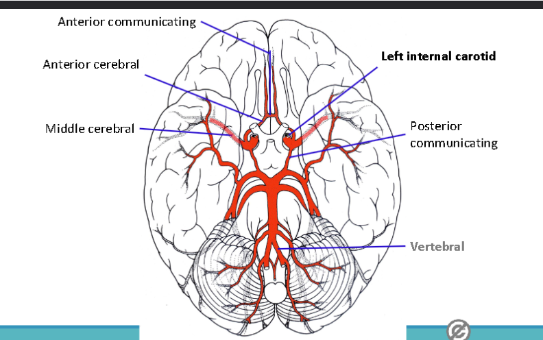
Which artery is formed when the two vertebral arteries merge
basilar artery
at the junction of the pons and medulla
what does the basilar artery do?
The basilar artery travels upward along the anterior surface of the pons.
Along the way, branches of the vertebral and basilar arteries help supply structures including the:
Spinal cord + cerebellum + pons
Then, near the upper end of the pons, the basilar artery splits again.
what does the Basilar artery split into?
The single basilar artery divides into:
The basilar artery travels on the anterior surface of the pons before splitting
Left posterior cerebral artery (PCA)
Right posterior cerebral artery (PCA)
PCAs supply areas including?
Occipital lobe
Inferior temporal lobe
Midbrain
Connected to the anterior circulation via the posterior communicating arteries.
what is the Circle of Willis?
Acts as a backup system for cerebral blood flow
interconnected arterial circle that provides an alternate route for cerebral blood flow if an artery becomes blocked.
what happens if their is a blockage in circle of willis?
If there is a blockage somewhere in the circle, blood can flow in the opposite direction to compensate (not perfect, but it helps
what arteries make up the circle (5)
1. Anterior cerebral arteries
2. Anterior communicating artery
3. Internal carotid arteries
4. Posterior communicating arteries
5. Posterior cerebral arteries


what do superficial veins do?
The superficial cerebral veins drain blood from the more external portions of the cerebral cortex.
drain the lateral surfaces of the cerebral cortex
what do deep veins do?
Deep veins (internal cerebral veins and basal vein) – drain the deep structures of the brain (basal nuclei and diencephalon)
what do all veins drain into?
dural venous sinuses
what are dural venous sinuses?
Sinus:
◦ Channel within the dura that carries venous blood.
◦ Functionally similar to a vein but has a different structure.
A dural venous sinus is a channel within the dura mater that carries venous blood away from the brain
pathway of venous blood
Brain → cerebral veins → dural venous sinuses → internal jugular veins → heart
what 3 sinuses carry the most of brains blood?
Superior sagittal sinus
Inferior sagittal sinus
Straight sinus
these sinuses are unpaired
where is superior sagittal sinus
found at the superior margin of the falx cerebri
where is inferior sagittal sinus found?
found at the inferior margin of the falx cerebri. It flows into the
where is Straight sinus found?
found at the junction of the falx cerebri and tentorium cerebelli
where do the The superior sagittal and straight sinuses meet?
at the confluence of sinuses
A confluence of sinuses is basically a meeting point where multiple dural venous sinuses come together.
what are paired sinuses?
Paired sinuses simply means there are two of that dural venous sinus: one on the left side and one on the right side of the skull.
For example, the transverse sinus is paired:
Left transverse sinus ← 🧠 → Right transverse sinus
what are the major paired sinuses (4)
transverse sinus
superior petrosal sinus
inferior petrosal sinus
sigmoid sinus
where can the sinuses be seen?
seen as grooves on the inner surface of the skul
blood flow in dural venous sinuses
superficial veins —> superior sagittal sinus —->inferior sagittal and deep veins —-> straight sinuses —-> confluence —->transverse —-> sigmoid —->internal jugular
The superior and inferior petrosal sinuses drain smaller veins coming from the brainstem and cerebellum (and the orbit)
types of strokes
Ischemic
Hemorrhagic
Stroke is the 3rd most common cause of death in Canada
what is ischemic stroke?
blood supply to an area becomes blocked
(blood clot or plaque).
◦ >80% of strokes are ischemic and the MCA is most commonly
affected
what is Hemorrhagic stroke?
an artery ruptures, resulting in blood loss
(hypertension adds to the risk)
In the case of superficial vessels (ie. aneurysms of the
circle of Willis) blood leaks into the subarachnoid space
what are common causes of ischemic strokes?
thrombosis
embolism
what is thrombosis
a clot that forms within an artery, often coincident with atherosclerosis
what is embolism
a ‘travelling mass’, most commonly a blood clot (thromboembolus), or a piece of
atherosclerotic plaque that becomes detached, travels through the vasculature and becomes
lodged within an arteryBoth result in interruption of blood flow to the area supplied by the artery.
what happens to Anterior cerebral artery during stroke
paralysis and impaired sensation affecting the contralateral lower limb
what happens to middle cerebral artery at stroke?
paralysis and impaired sensation affecting the contralateral upper limb and the face
what happens to posterior cerebral artery
visual deficits affecting the contralateral half of the visual field and memory deficits
what is the signs of stroke occuring.
FAST
Face - is it drooping
Arms - can you raise both
Speech - is it slurred
Time - to call 911 right awauy