Skeletal System: Anatomy, Diseases, and Medical Procedures
1/105
There's no tags or description
Looks like no tags are added yet.
Name | Mastery | Learn | Test | Matching | Spaced | Call with Kai |
|---|
No analytics yet
Send a link to your students to track their progress
106 Terms
Ossification
Begins after 3-months gestation and continues through adolescence.
Osteoclasts
Carry off old or damaged bone.
Osteoblasts
Build the bones back up.
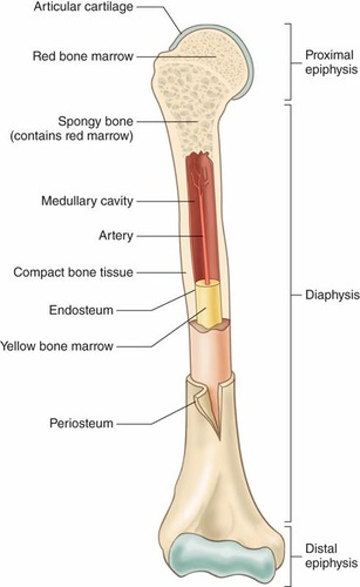
Periosteum
Tough, fibrous tissue forming outermost covering of bone.
Compact bone
Dense, hard strong bone forming protective outer layers of bone.
Spongy bone
Porous, thus more susceptible to fractures.
Red Bone Marrow
Located inside the ends of long bones and certain short bones.
Medullary cavity
Center of long bones; storage site of red and yellow bone marrow.
Endosteum
Lines medullary cavity.
Yellow bone marrow
Fat storage in long bone medullary cavity.
Cartilage
Smooth, rubbery connective tissue acting as shock absorber.
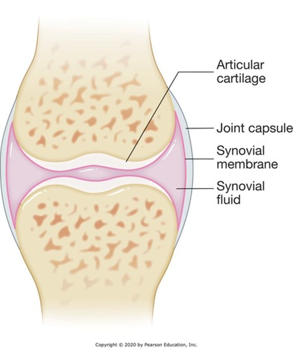
Articular cartilage
Covers surface of articulations (joints) allowing smooth joint movement.
Meniscus
Curved fibrous cartilage in some joints.
Diaphysis
Shaft of long bones.
Epiphysis
Wider ends of long bones.
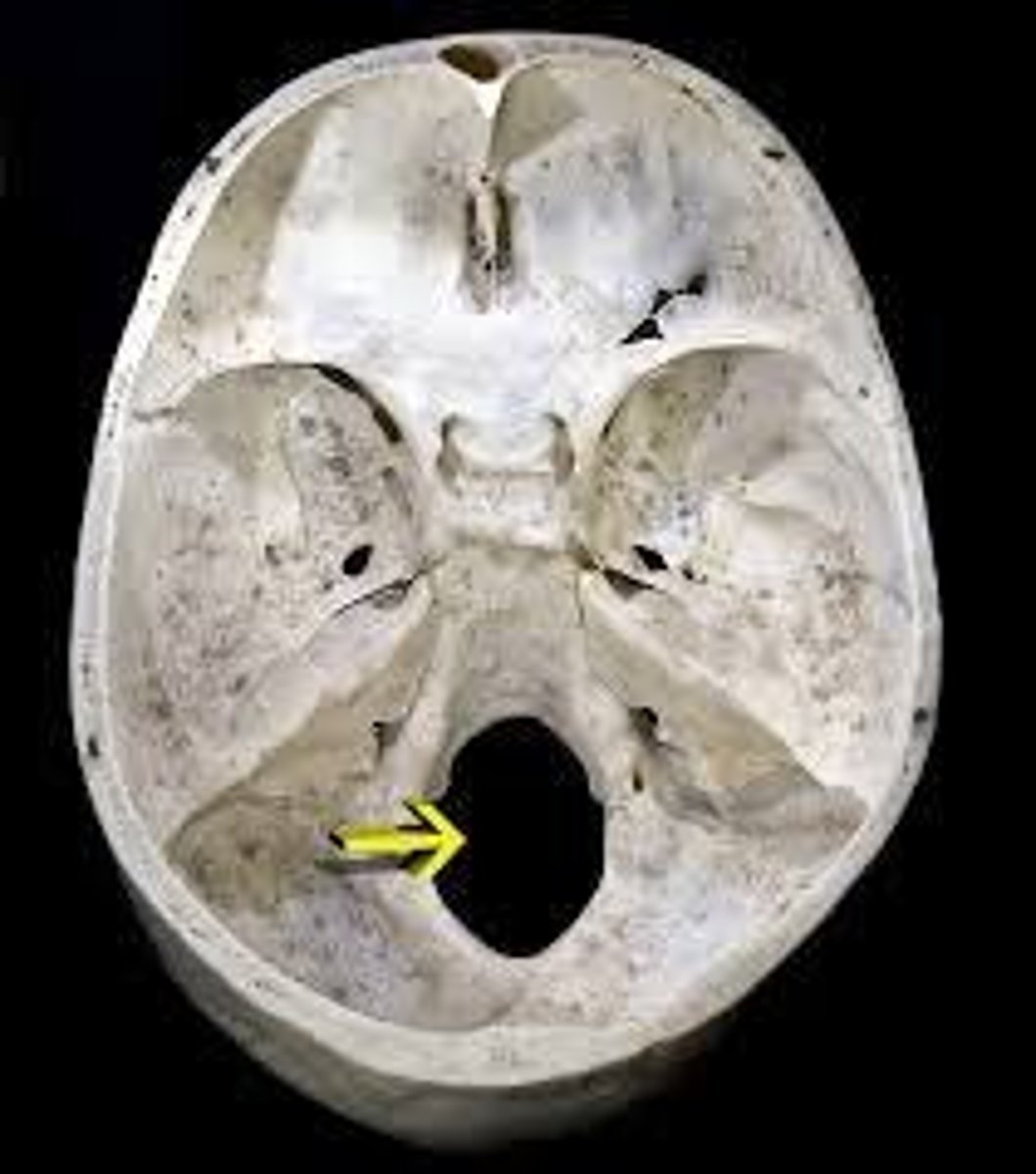
Foramen
Opening in a bone allowing passage of blood vessels, nerves, and ligaments.
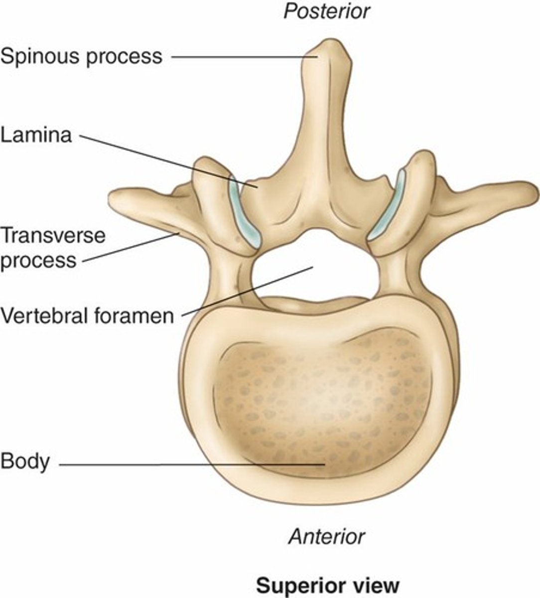
Process
Projection allowing for the attachment of muscles or tendons.
Fibrous Joints
Inflexible layers of dense connective tissue that hold bones tightly together.
Cartilaginous Joints
Consist of bones connected by cartilage allowing slight movement.
Synovial Joints
Articulation of joints permitting variety of movements.
Synovial capsule
Outermost layer, resembling a sleeve.
Synovial membrane
Lines capsule and secretes synovial fluid.
Ligaments
Fibrous tissues connecting one bone to another bone.
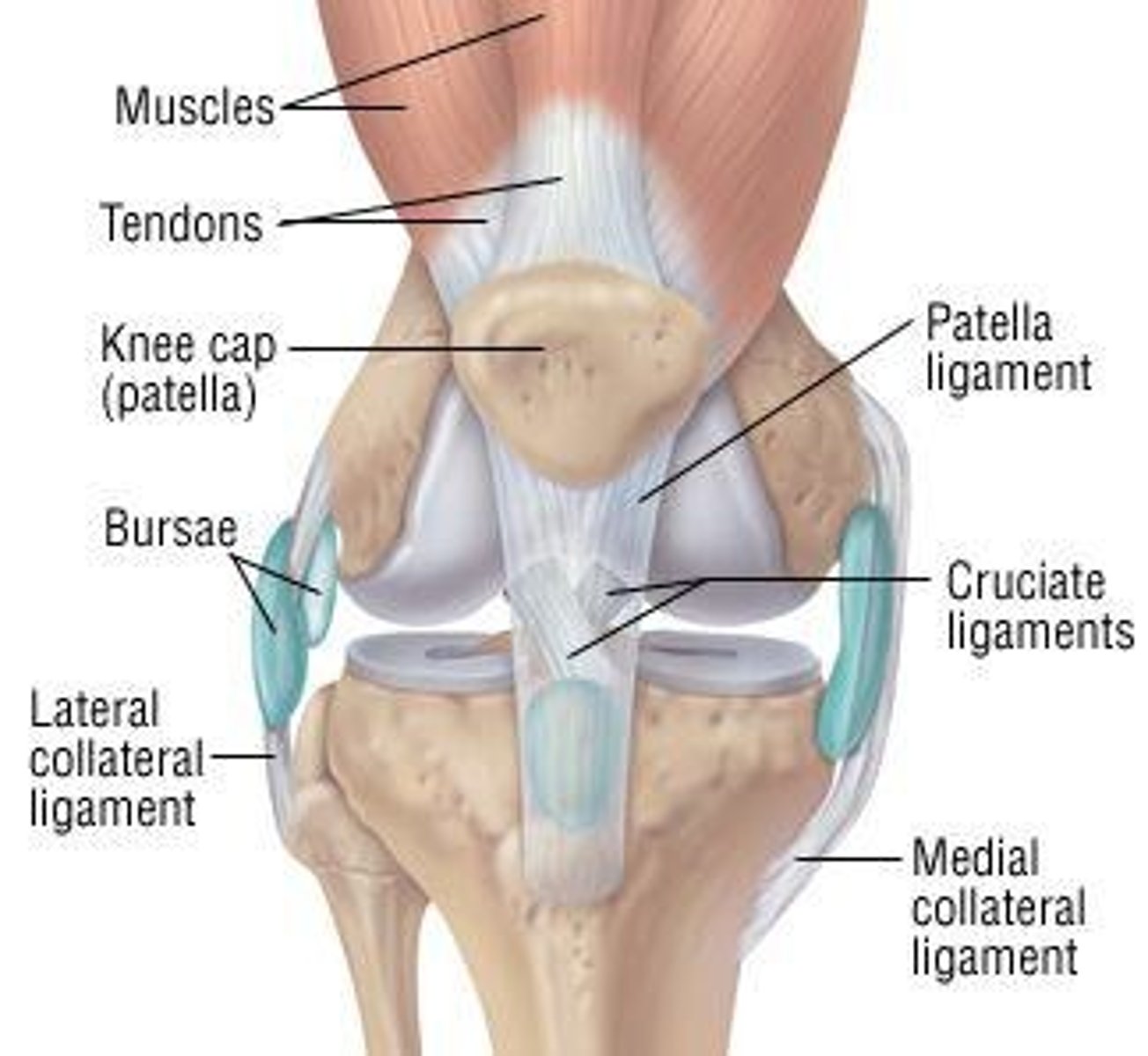
Bursae
Fibrous sac acting as cushion, easing movement in areas subject to friction.
Axial Skeleton
Consists of the bones of the head, chest, and back.
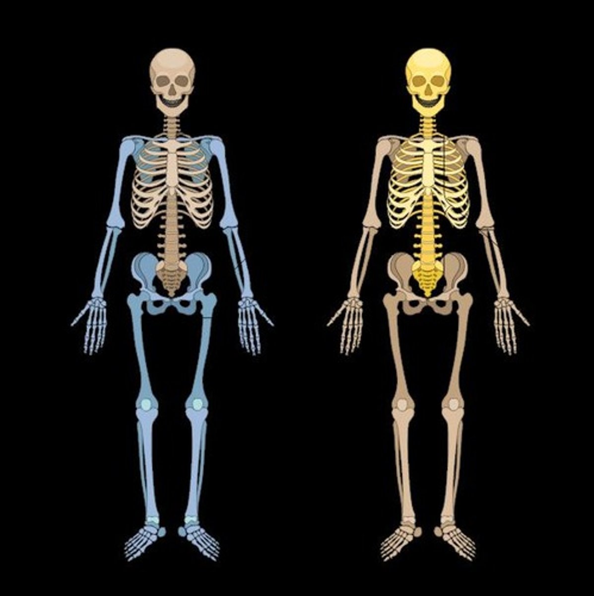
Appendicular Skeleton
Consists of the bones of the shoulders, arms, hips, and legs.
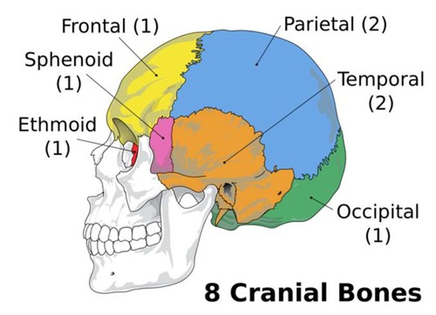
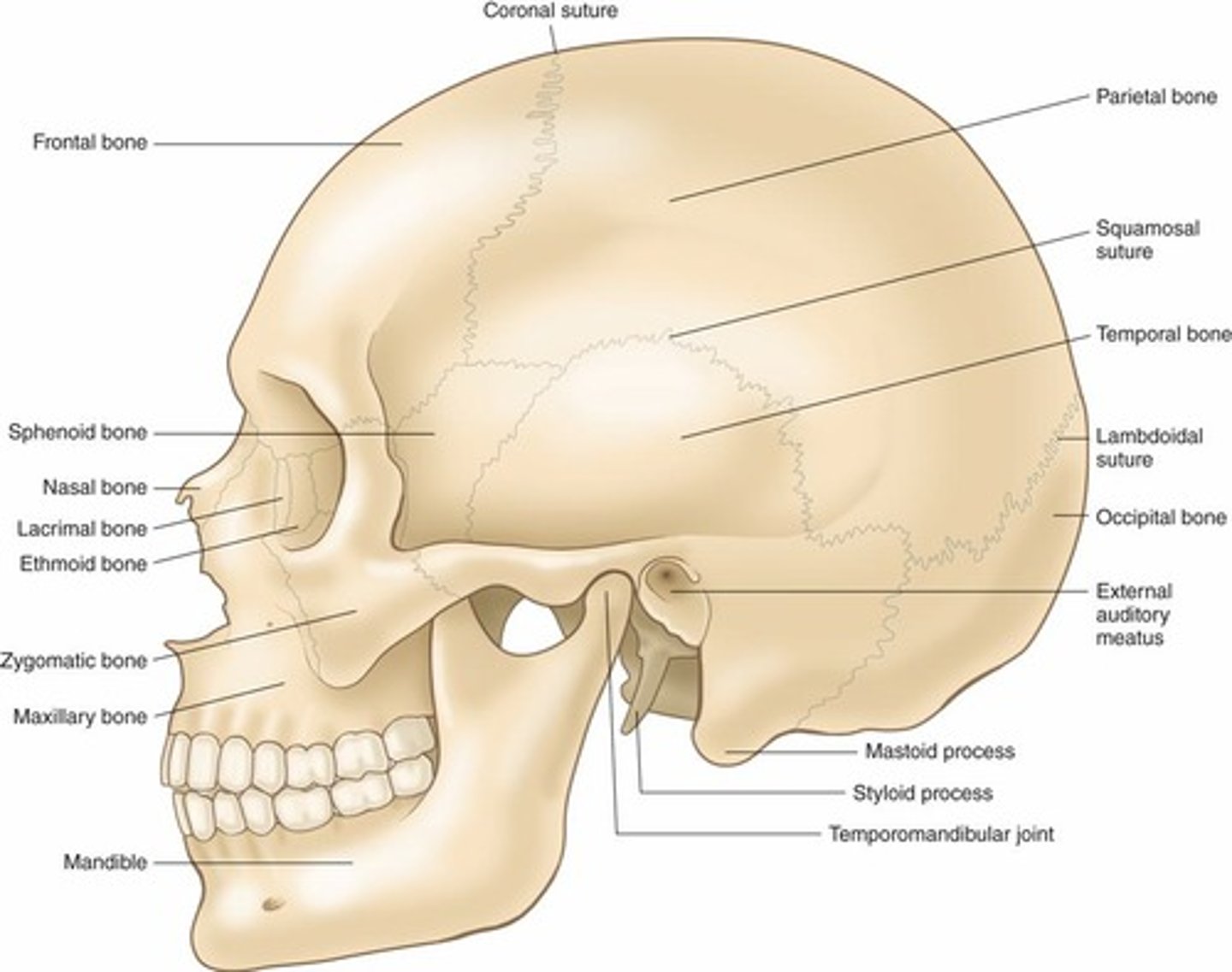
Cranium
8 bones.
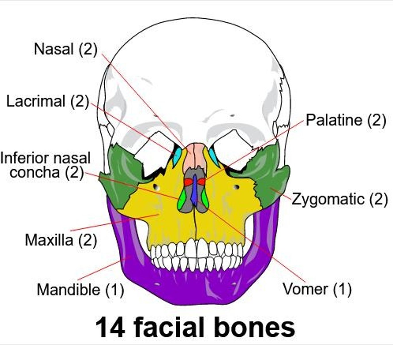
Face
14 bones.
Middle ear
6 bones.
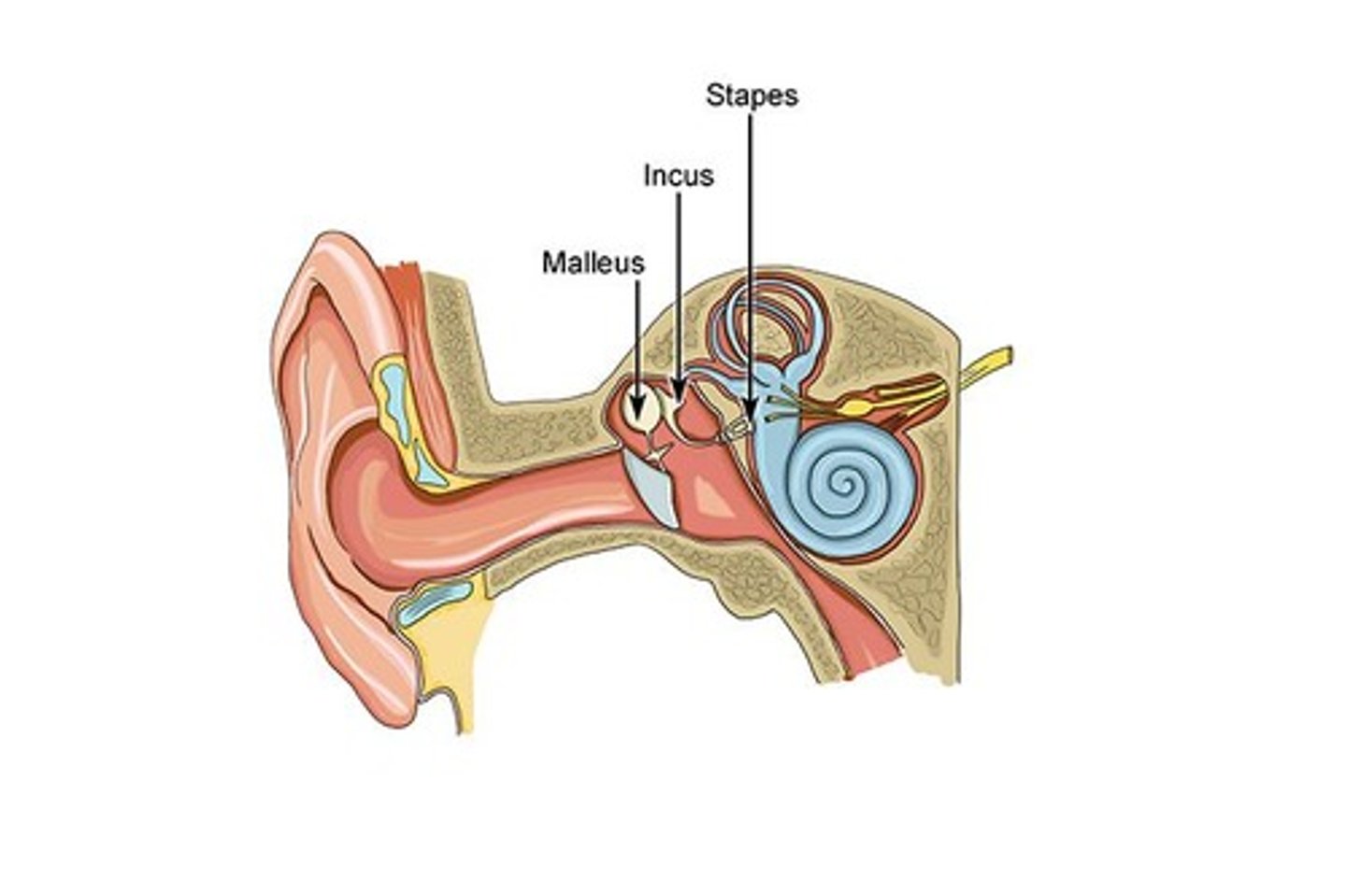
Auditory Ossicles
Three tiny bones located in each middle ear: Malleus, Incus, Stapes.
Fontanelles
Also known as 'Soft Spots', allow cranial bones to move together during birth and grow apart as the brain grows.
Thoracic Cavity
The chest contains the Rib Cage
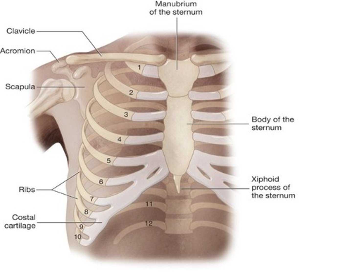
Rib Cage
12 pairs of ribs
True ribs
Ribs 1-7
False ribs
Ribs 8-10
Floating ribs
Ribs 11-12
Combing Form
cost/o = rib
Sternum
Flat bone located in middle of the chest
Manubrium
Forms upper portion of the sternum
Body (sternum)
Forms middle portion of the sternum
Xiphoid process
Made of cartilage; forms lower portion of the sternum
Clavicle
Collar bone; connects manubrium to scapula
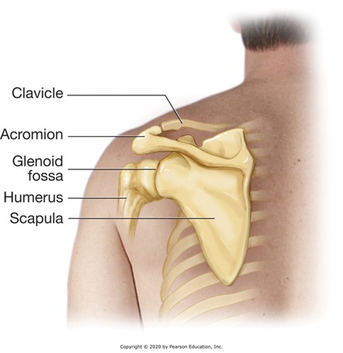
Scapula
Shoulder bone
Acromion
Extension of scapula; forms high point of the shoulder
Humerus
Upper arm
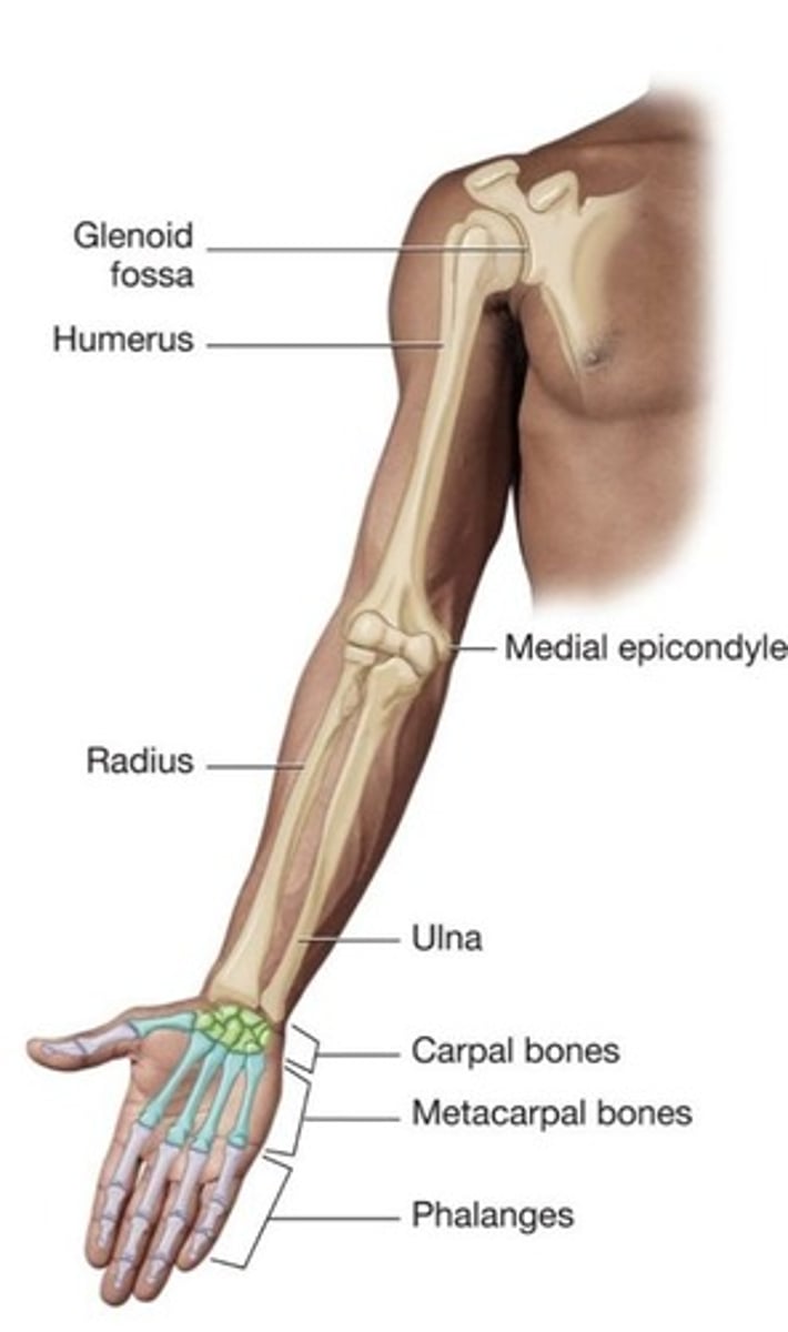
Radius
Smaller, shorter bone in forearm
Ulna
Larger, longer bone of forearm
Carpal bones
Eight bones that form the wrist
Metacarpal bones
Five bones that form the palms of the hand
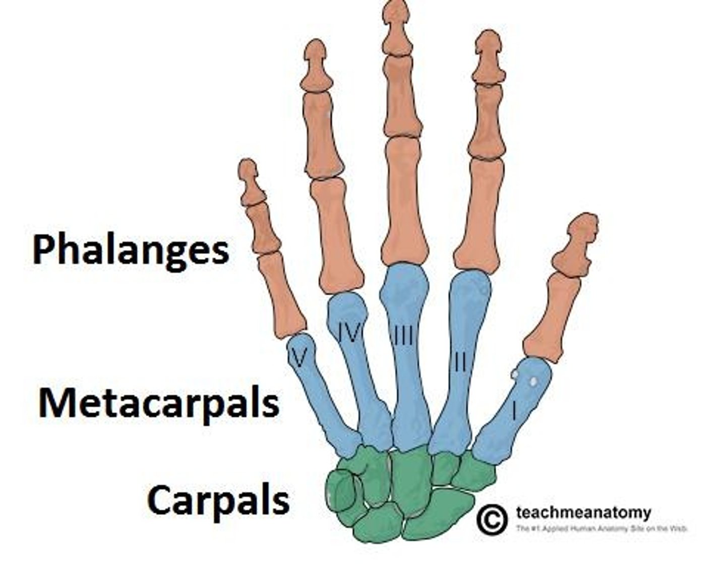
Phalanges
Fourteen bones of the fingers
Cervical vertebrae
Set of seven (C1-C7); form the neck
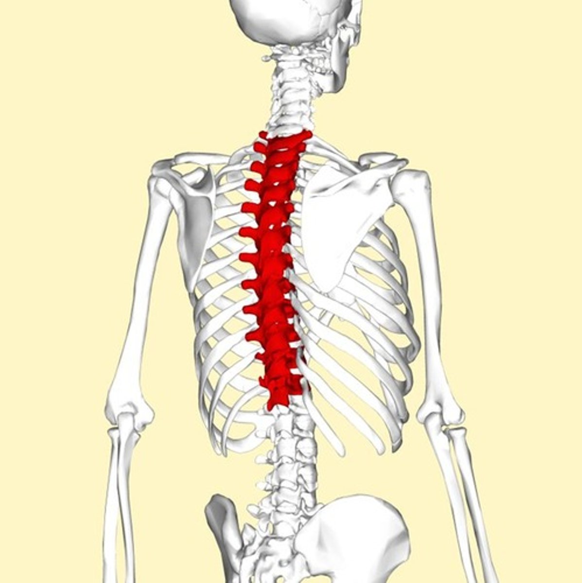
Thoracic vertebrae
Set of 12 (T1-T12); a pair of ribs attached to each
Lumbar vertebrae
Set of five (L1-L5); largest and strongest vertebrae
Sacrum
Forms lower portion of the back; five separate bones at birth that fuse together
Coccyx
Known as tailbone; forms end of the spine
Ilium
Forms back and sides of pubic bone
Ischium
Forms lower posterior portion of pubic bone
Pubis
Forms anterior portion of pubic bone
Pubic symphysis
Cartilaginous joint uniting left and right pubic bones
Acetabulum
Large circular cavity in each side of the pelvis; articulates with head of femur to form hip joint
Femur
Largest bones in the body
Patella
Kneecap
Tibia
Shinbone; larger anterior weight-bearing bone of lower leg
Fibula
Smaller of the two bones of lower leg
Talus
Articulates with tibia and fibula
Calcaneus
Heel bone; largest of tarsal bones
Metatarsals
Five bones that form part of the foot to which toes are attached
Hallux valgus
Abnormal enlargement of joint at the base of the great toe; also known as a bunion
Osteoarthritis
Most commonly associated with aging; characterized by wearing away of articular cartilage within joints
Gout
Type of arthritis characterized by deposits of uric acid crystals in joints, usually beginning with the big toe
Rheumatoid Arthritis
Chronic autoimmune disorder attacking joints and other organs
Herniated Disk
Breaking apart of intervertebral disk resulting in pressure on spinal nerve roots
Osteoporosis
Marked loss of bone density with increase in bone porosity; frequently associated with aging
Multiple Myeloma
Cancer occurring in blood-making cells found in red bone marrow
Osteoporosis-Related Fractures
Fractures associated with weakened bone due to osteoporosis.
Compression fracture
Compression of bone on itself.
Colles' fracture
Fracture at lower end of radius.
Osteoporotic hip fracture
Broken hip due to osteoporosis; may occur spontaneously or as a result of a fall.
Closed fracture
Bone is broken; no open wound in skin.
Open fracture
Bone is broken; open wound in the skin.
Transverse fracture
Occurs straight across the bone.
Oblique fracture
Fracture is at an angle across the bone.
Comminuted fracture
Bone is splintered.
Greenstick fracture
Bone is bent and partially broken; seen frequently in children.
Pathologic fracture
Due to weakened bone; not due to trauma.
Spiral fracture
Due to twisting action.
Stress fracture
Due to overuse.
Fat embolus
May form with fracture of long bone; fat cells from yellow bone marrow are released into the blood.
Crepitation
Grating sound when ends of broken bone move together.
Callus
Bulging deposit around the area of the break; tissue eventually becomes bone.
Bone Density Testing
Screening test for osteoporosis or other conditions related to loss of bone mass.
Orthotic
Mechanical appliance designed to control, correct, or compensate for impaired limb function.
Prosthesis
Substitute for a diseased or missing body part.
Arthrodesis
Surgical fusion of two bones.
Arthroscopic surgery
Minimally invasive procedure for treating interior of joint.
Chondroplasty
Surgical repair of damaged cartilage.
Total knee replacement
All parts of the knee are replaced.
Partial knee replacement
Only part of the knee is replaced.
Total hip replacement
Restores damaged hip to full function.
Hip resurfacing
Placing of metal cap over head of femur to restore function of the hip.