Cardiac Sono: Parasternal Views
 Call Kai
 Learn
Learn Practice Test
 Spaced Repetition
 Match
Match Flashcards
 Knowt Play
1/24

Earn XP
Description and Tags
Test 3 Study Guide
Name | Mastery | Learn | Test | Matching | Spaced | Call with Kai | Chat |
|---|
No analytics yet
Send a link to your students to track their progress
25 Terms
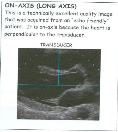
On Axis Images
Produced when the probe is Perpendicular (90) to the structures being imaged.
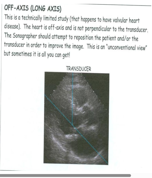
Off Axis Images
The structures (heart) are not perpendicular to the transducer.
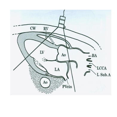
PLAX views
LAX LV
LAX RVIT
LAX RVOT
Place patient in LLD position. Place probe in the Left 3rd or 4th Intercostal space, adjacent to the Sternum.
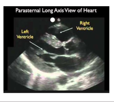
PLAX LV
Chambers visualized: RV, LV, LA
Valves Visualized: AoV (RCC is anterior, NCC is posterior and LCC is
not visualized) , MV
Walls visualized: RVFW , IVS, LVPW
Vessel visualized in the long axis: Aortic Root (AoR) - proximal 3-
4cm of the ascending aorta.
Desc. Ao, coronary sinus (is seen in the atrioventricular groove-
posterior to the MV annulus).
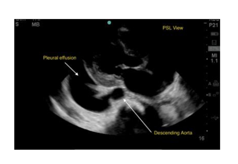
Pleural Effusion
A Pleural Effusion will be seen only Posterior to the Descending Aorta.
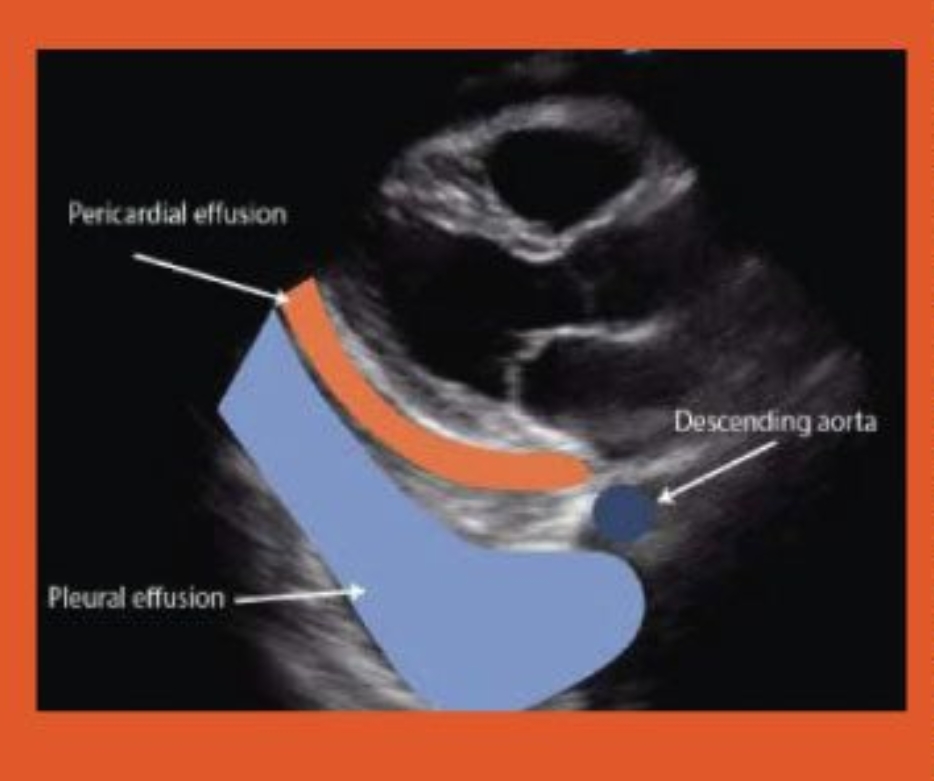
Pericardial Effusion
A Pericardial Effusion can be seen between the LA and the DAO.
Additional structures in PLAX
Descending Aorta is Posterior to the LA in cross section
Descending Thoracic Aorta
Oblique Sinus of the Pericardium lies between the LA the Descending Thoracic Aorta .
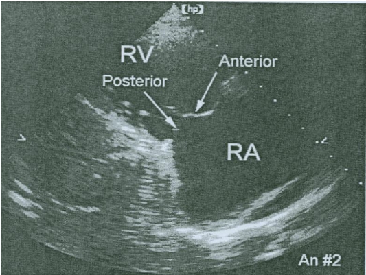
LAX RVIT
Chambers visualized: RV, RA
Valve visualized: TV (Posterior and Anterior Leaflets. Septal and Ant are seen in the PSAX)
The only view that the Posterior Leaflet of the TV is seen.
From the LV PLAX angle the probe toward the patient’s Right Hip.
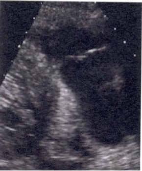
PLAX RVOT
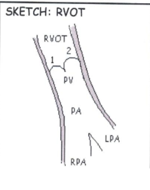
Chamber visualized: RV
Valve visualized: PV
Vessels: PA and bifurcation
Notch pointed at Right Shoulder but Angled towards Left Shoulder. Angle the probe more Superior and Lateral from the PLAX LV.
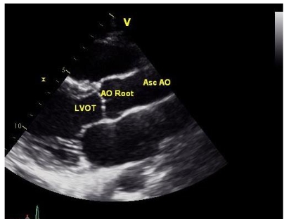
TTE PLAX View
The AOV annulus, Sinuses of Valsalva, Sinotubular Junction, and Proximal Ascending Aorta (AAO) are best visualized from this window.
From the PLAX LV move the probe closer to the Sternum, (usually) up an intercostal space.
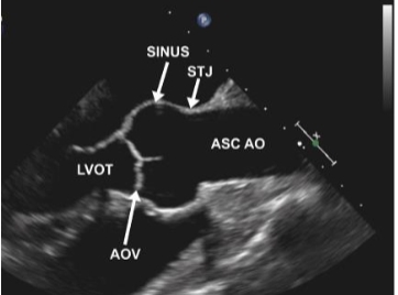
PLAX AO
• AOV Annulus
• Sinus of Valsalva
• STJ – Sinotubular
junction
Normal Tubular Configuration of the Aorta.
Proximal AAO
LAX RVIV/ RVOV
• The IVC is seen entering the RA (inferior to the
coronary sinus.
A prominent Eustachian valve is sometimes seen at
the junction of the IVC and RA
Chiari Network
The Right Atrial Appendage is rarely seen in a TTE
RVIT/RVOT
The IAS (interatrial septum is not well visualized in the RVIT view, but careful angulation between the PLAX of the LV and RVIT allows examination of the IAS (including the fossa ovalis in the central portion and to rule out myxomas/clots).
Moving the transducer toward the base and then angling laterally, a long axis view of the RVOT, pulmonic valve and pulmonary artery can be obtained.
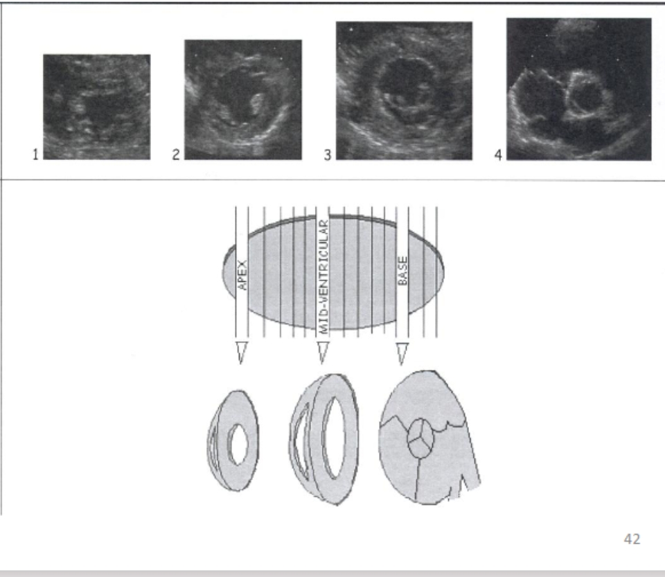
PSAX Views
– Apex (PSAX AP)
– Mid ventricle
• Papillary muscles (PSAX PM)
• Chordae tendineae
– Mitral valve (PSAX MV)
– Base (PSAX base)
• Aortic valve (PSAX AoV)
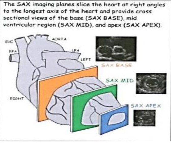
PSAX
Rotate Probe Clockwise 90 degrees from the PLAX and angulate the Probe Superiorly and Inferiorly along the long axis of the heart to obtain imaging planes.
• Chambers visualized: RA, RV, LA, LV
• Valves visualized: MV, TV, AoV, PV
• Walls visualized: septal wall, anteroseptal wall,
anterior wall, anterolateral wall, lateral wall,
inferolateral wall, inferior wall and inferoseptal
wall.
– All walls are seen at different levels:
• Base, Mid, and Apex levels.
PSAX Base AOV Level
The LA appendage can be imaged from this view by a slightly Lateral angulation and Superior rotation of the transducer.
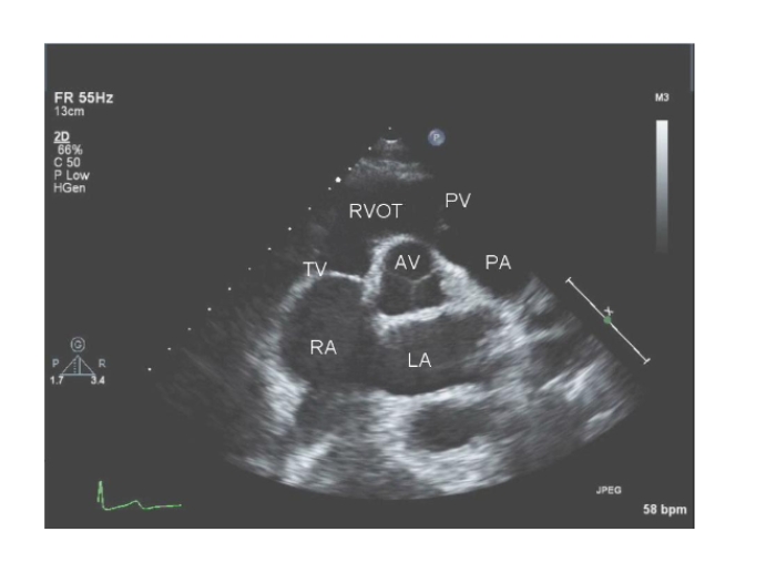
PSAX at MV Level
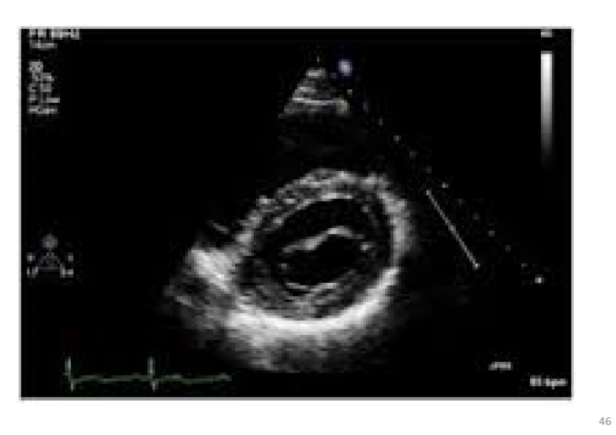
PSAX at PAP Level
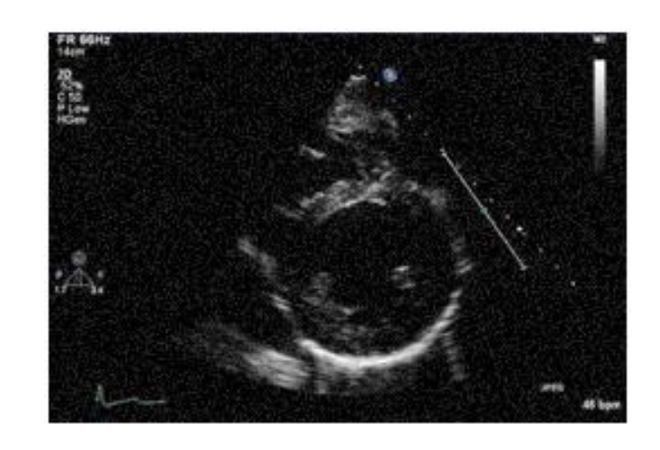
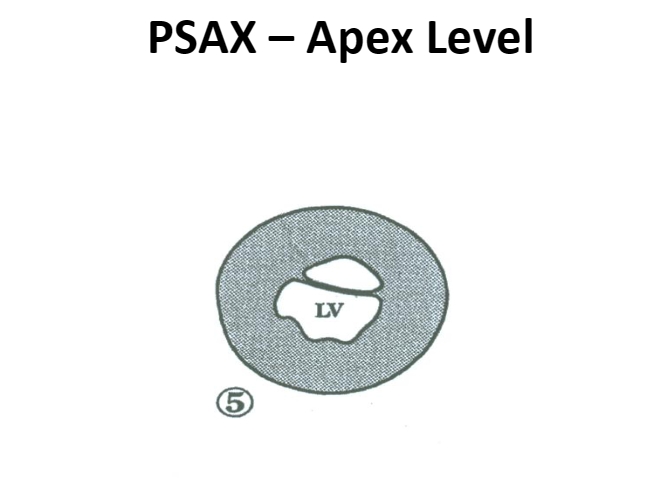
PSAX at Apex Level
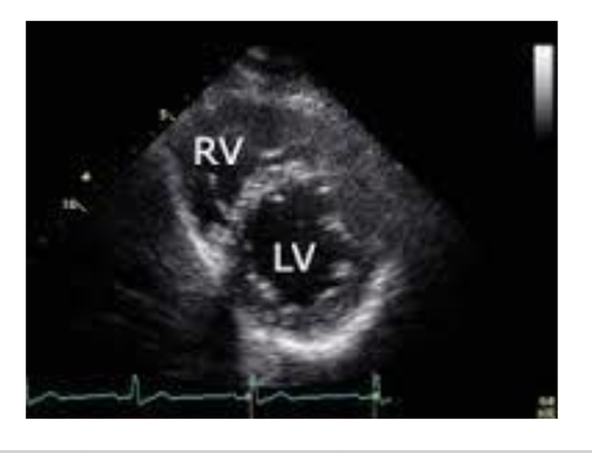
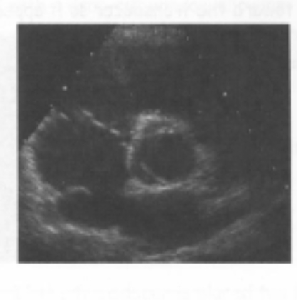
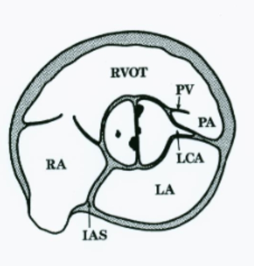
PSAX- AOV Level Structures
AoV – all 3 aortic valve leaflets:
RCC, LCC and NCC
Systole: the aortic leaflets open
to a near circular (triangular)
orifice.
Diastole: the “Y” shaped
coaptation lines of the leaflets is
seen (Mercedes sign).
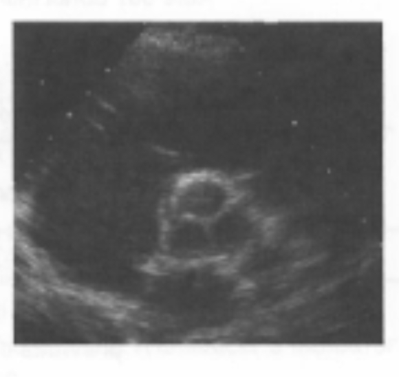
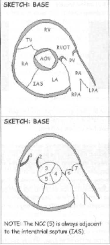
PSAX – AoV level
Chambers and Structures: RA, RV, LA
IAS. LA Appendage.
RV is the most Anterior structure and can be seen wrapping around the Aorta.
PSAX Bicuspid AOV Extra
*Tricuspid vs. bicuspid AoV is most accurately identified in 2D in systole.
– due to a raphe in the position of a normal commissure (the place where two things are joined.) Will have a football shaped opening.
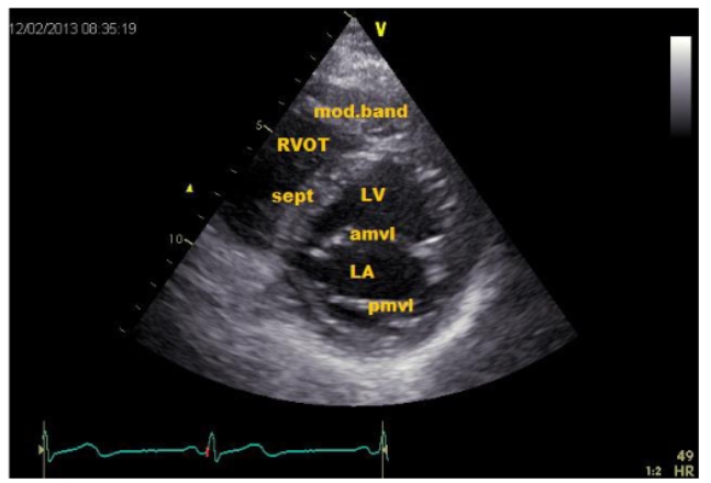
PSAX - MV Level
Structures: The AML and PML (Looks like fish’s mouth in Diastole)
Chordae attaching to the AML, PML and Pap muscle
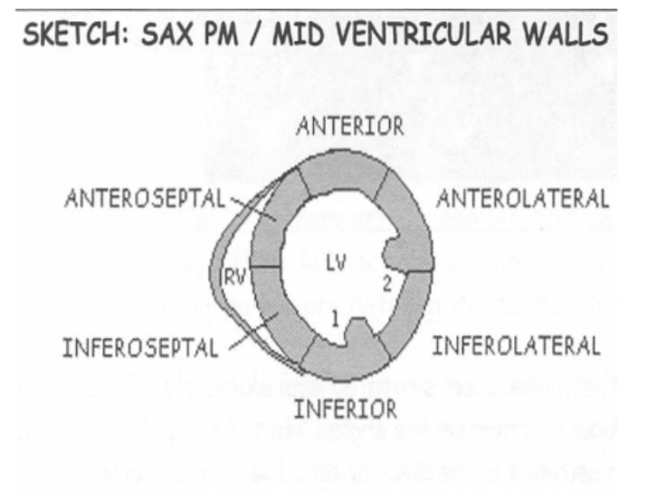
PSAX Mid Level
Structures: RV, LV and Pap Muscles
LV looks like a mushroom
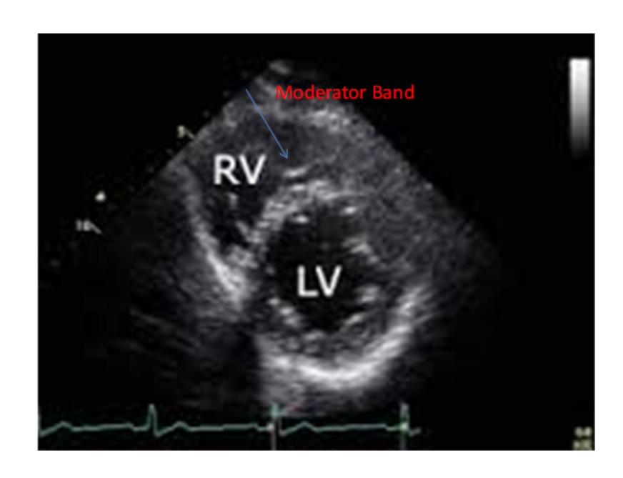
PSAX Apex Level
Structures: RV, Moderator Band and sometimes the LV
The apical segments of the LV myocardium are
NOT seen in standard parasternal views.
However, in some patients, the LV is seen near the Apex by moving the probe Laterally and Angling medially.