C2.2: Neural Signaling | IB Biology HL
1/49
Earn XP
Description and Tags
Name | Mastery | Learn | Test | Matching | Spaced |
|---|
No study sessions yet.
50 Terms
State the function of the following neuron parts: dendrites, axon, cell body, axon terminal.
C2.2.1: Neurons as cells within the nervous system that carry electrical impulses.
Dendrites are multiple shorter fibers that receive signals from other neurons or receptors.
Axons are long single fibers that carries the nerve signal to the axon terminals.
The cell body (soma) is made of the cytoplasm and nucleus and decides if a signal should be sent down the axon.
The axon terminal is where the signal is passed to the next neuron or muscle.
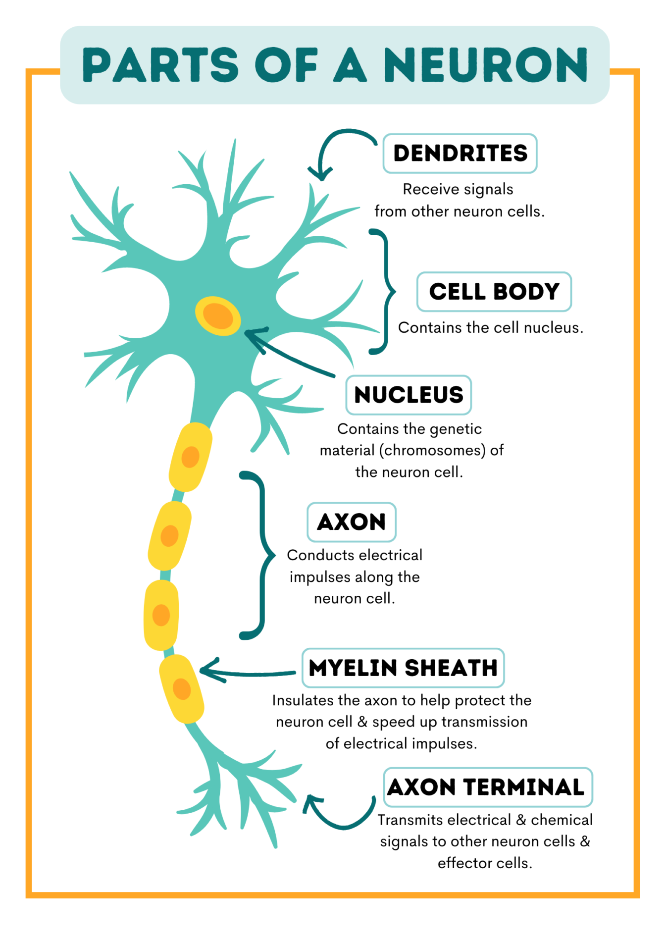
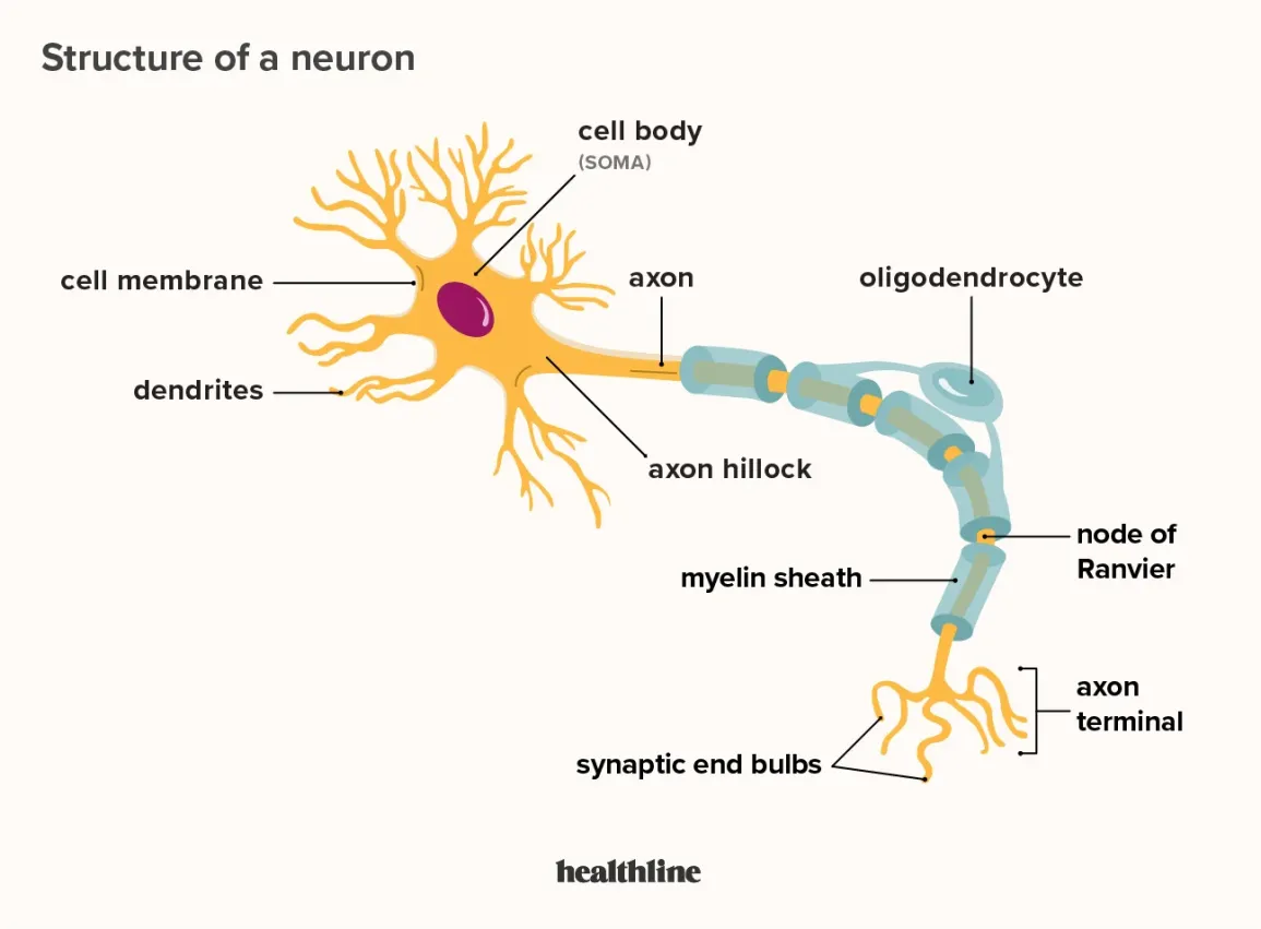
Identify the cell body, axon, and dendrites in diagrams of typical sensory and motor neurons.
C2.2.1: Neurons as cells within the nervous system that carry electrical impulses.
Define membrane potential.
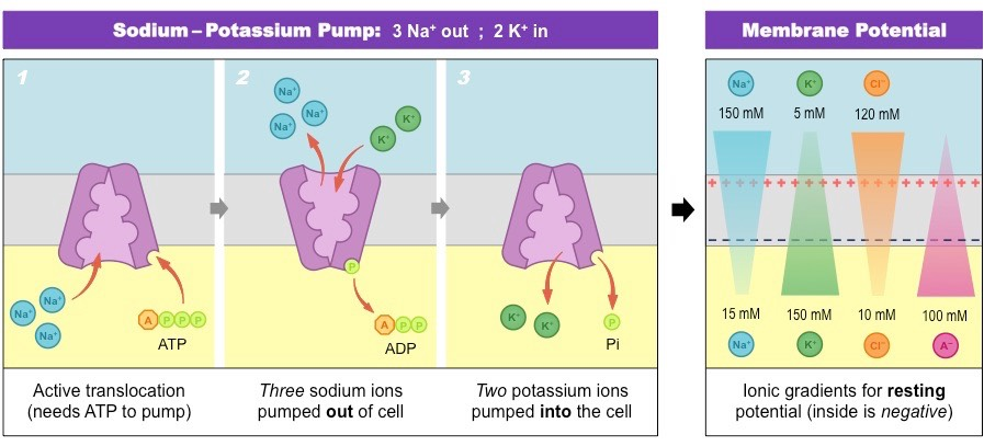
C2.2.2: Generation of the resting potential by pumping to establish and maintain concentration gradients of sodium and potassium ions.
The difference in electric potential between the interior and exterior of a biological cell.
Define resting potential.
C2.2.2: Generation of the resting potential by pumping to establish and maintain concentration gradients of sodium and potassium ions.
The difference in charge between the inside and outside of the cell when the neuron is at rest.
The inside is more negative on the outside than the inside (fewer Na+ outside than K+ inside).
The neuron has a resting potential of about -70 mV.
State the voltage of the resting potential.
C2.2.2: Generation of the resting potential by pumping to establish and maintain concentration gradients of sodium and potassium ions.
The resting potential is -70 mV.
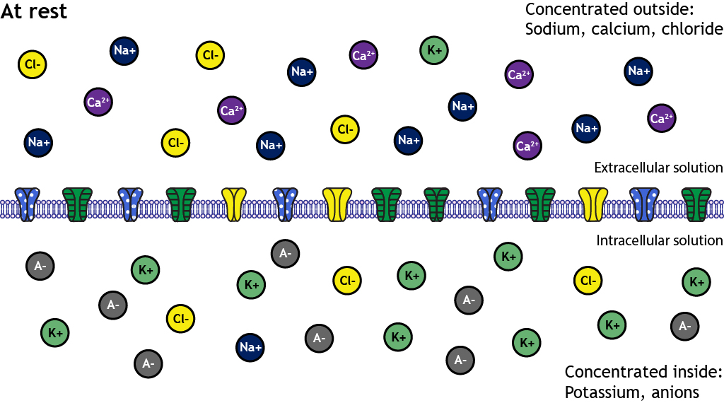
Outline 3 mechanisms that together create the resting potential in a neuron.
C2.2.2: Generation of the resting potential by pumping to establish and maintain concentration gradients of sodium and potassium ions.
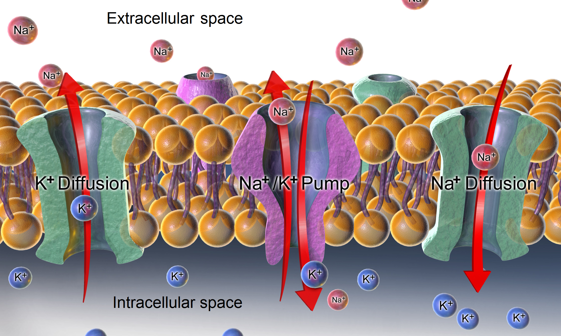
The sodium-potassium pump actively uses energy from ATP to pump 3 Na+ out of the cell for every 2 K+ it brings in. As a result, the inside of the cell is relatively negative compared to the outside.
K+ ions leak out of the cell through the cell membrane.
Negatively charged proteins are within the neuron.
Define nerve impulse.
C2.2.3: Nerve impulses as action potentials that are propagated a long nerve fibers.
An electrical signal that results in a change in electric charge inside the neuron.
Define action potential.
C2.2.3: Nerve impulses as action potentials that are propagated a long nerve fibers.
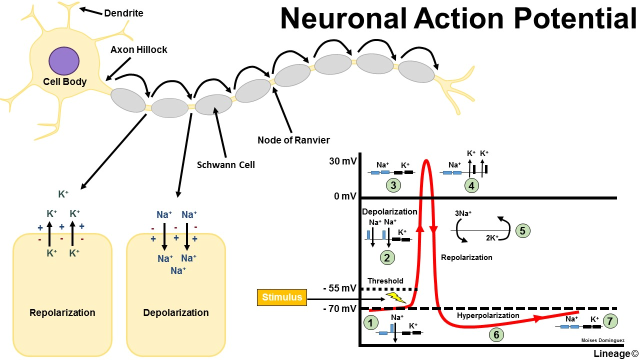
An electrical signal that travels down the neuron & is sent to the next neuron or muscle.
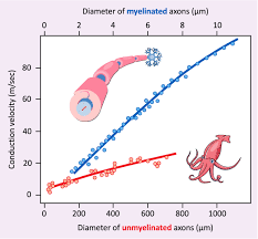
Outline the correlation between conduction speed of nerve impulses & axon diameter.
C2.2.4: Variation in the speed of nerve impulses.
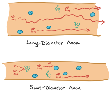
Axons with greater diameter have a faster transmission velocity than those with a smaller diameter (direct relationship).
Larger axon = less resistance to ion flow = faster propagation of action potential
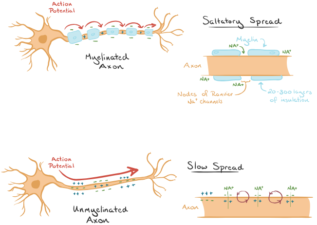
Explain the difference in nerve impulse speed for myelinated & unmyelinated fibers.
C2.2.4: Variation in the speed of nerve impulses.
Nerve impulses travel more quickly in myelinated fibers because the myelin sheaths (made of Schwann cells) insulate the cell, so there is no ion movement in the axon covered by them. The action potentials skip from one node of Ranvier to another.
The in/out movement of ions only occur at nodes, so the impulse travels faster.
State the correlation between conduction speed of nerve impulses and animal size.
C2.2.4: Variation in the speed of nerve impulses.
Body size typically does not significantly affect the speed of its neuron impulses.
However, neurons need to travel further distances due to larger bodies, so larger animals have slower reflexes.
Compare the speed of transmission in giant axons of squid and smaller non-myelinated nerve fibers.
C2.2.4: Variation in the speed of nerve impulses.
The axons in squid are particularly large & unmyelinated. So compared to organisms with smaller non-myelinated nerve fibers (like a human internal organ), the giant squid conducts a nerve impulse much more quickly.
Still, compared to a smaller axon that is myelinated, the myelinated smaller axon is still faster than a giant unmyelinated axon.
Define synapse.
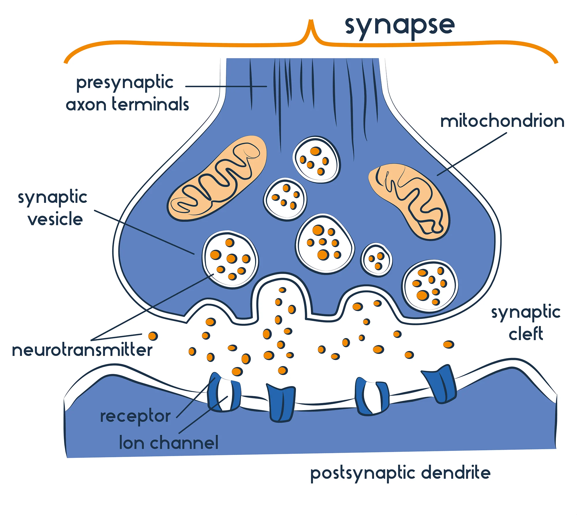
C2.2.5: Synapses as junctions between neurons and between neurons and effector cells.
The junction between 2 neurons or a neuron and a muscle cell.
Define synaptic gap/cleft.
C2.2.5: Synapses as junctions between neurons and between neurons and effector cells.
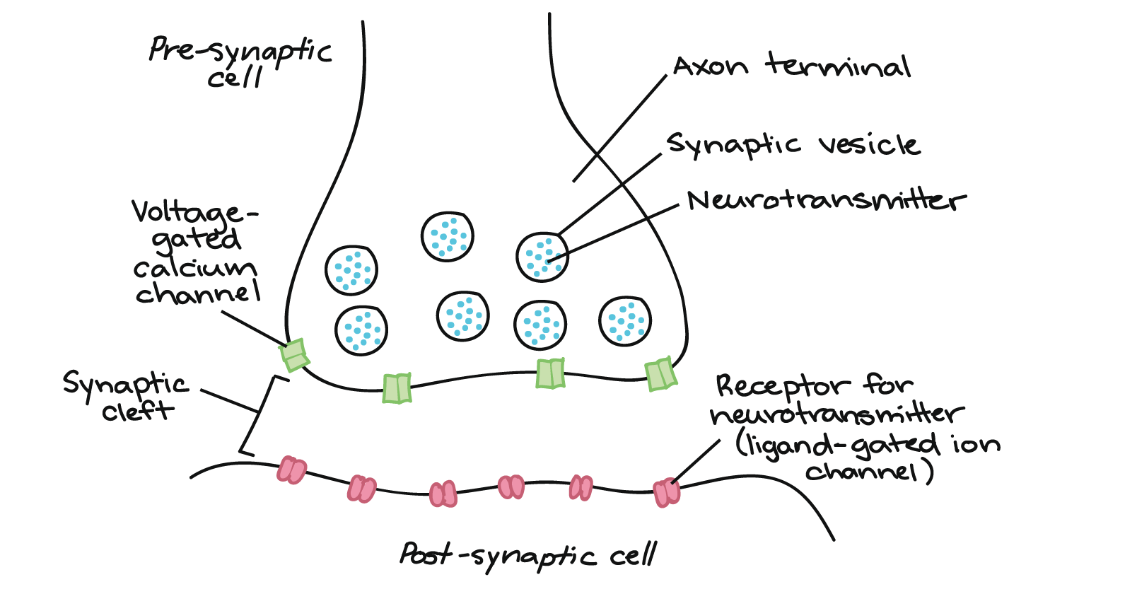
The space between 2 neurons
Define effector.
C2.2.5: Synapses as junctions between neurons and between neurons and effector cells.
A signal that triggers the reaction.
State the direction a signal across a typical synapse.
C2.2.5: Synapses as junctions between neurons and between neurons and effector cells.
A signal can only pass in one direction across a typical synapse.
State the role of neurotransmitters.
C2.2.5: Synapses as junctions between neurons and between neurons and effector cells.
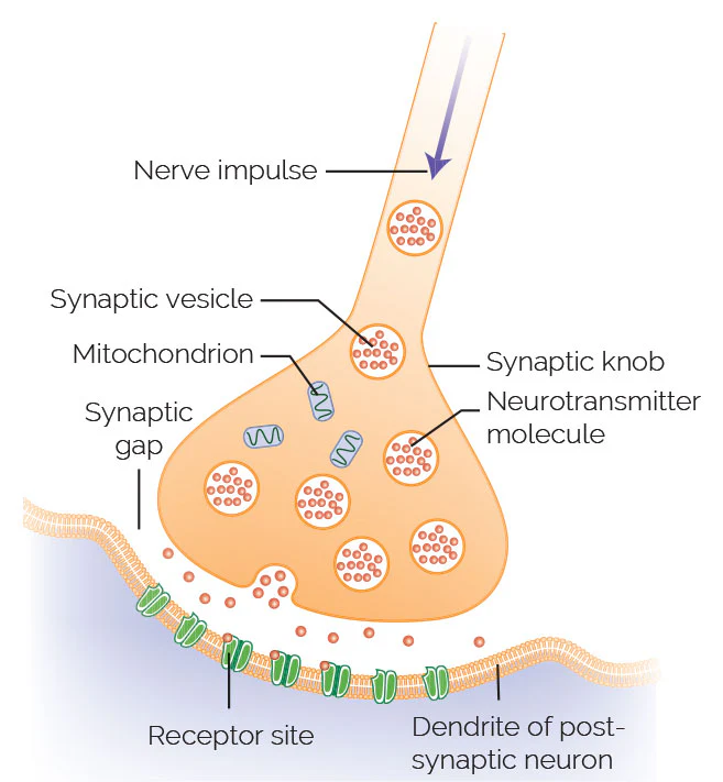
Neurotransmitters are the chemical released from the synaptic terminal buttons from the presynaptic neuron to the postsynaptic neuron.
It carries the signal from one neuron to the other chemically.
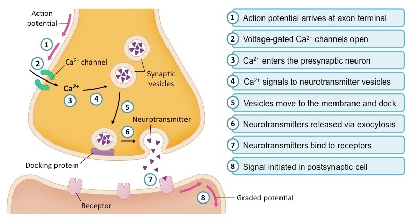
Outline the mechanism of synaptic transmission occurring at a presynaptic cell, including the role of depolarization, calcium ions, exocytosis, and diffusion.
C2.2.6: Release of neurotransmitters from a presynaptic membrane.
When the action potential arrives at the terminal buttons, it is depolarized as Ca2+ ion channels open, and more Ca2+ ions are taken in.
The calcium ions function as a signalling chemical, which activates the pathway that moves vesicles containing the neurotransmitter to fuse with the presynaptic membrane (exocytosis).
The neurotransmitter is released from the fused vesicles & diffuses into the synaptic cleft.
State the role of calcium ions.
C2.2.6: Release of neurotransmitters from a presynaptic membrane.
Calcium ions function as a chemical signal triggering exocytosis of neurotransmitters from the presynaptic cell.
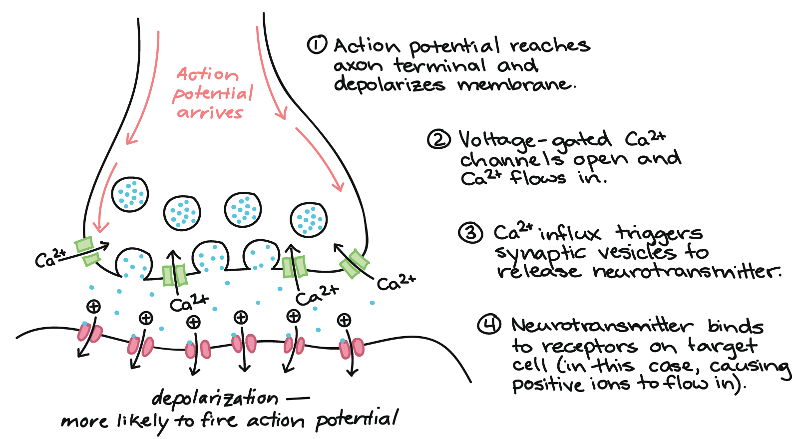
Outline the mechanism of synaptic transmission occurring at a post-synaptic cell, including the role of neurotransmitters, diffusion, receptors, gated ion channels, threshold potential and action potential.
C2.2.7: Generation of an excitatory postsynaptic potential.
After the neurotransmitter is released from fused vesicles into the synaptic cleft, it binds with protein receptors on the postsynaptic neuron membrane.
This binding opens ligand-gated ion channels, and Na+ diffuses into the neuron.
As Na+ diffuses into the neuron, it becomes depolarized until it reaches the threshold potential, and an action potential is initiated to move down the postsynaptic neuron.
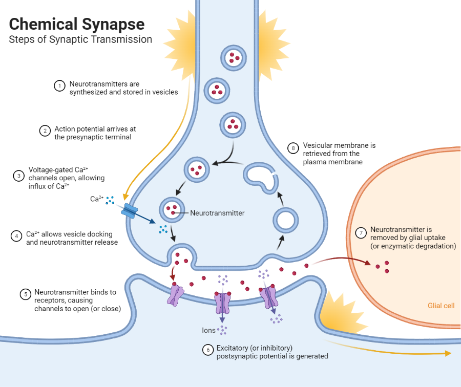
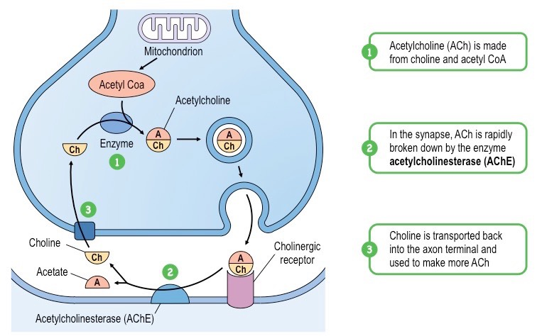
In the synaptic cleft, neurotransmitters are released from the synaptic cleft. Ion channels are closed to Na+ ions, neurotransmitters are degraded by enzymes, and neurotransmitter fragments diffuse back across synaptic cleft to be reassembled in the terminal buttons.
State the role of acetylcholine.
C2.2.7: Generation of an excitatory postsynaptic potential.
Acetylcholine is a very common neurotransmitter in both invertebrates & vertebrates. It is used as the neurotransmitter in many synapses, including between neurons & muscle fibers to stimulate muscle contraction.
Outline the digestion of acetylcholine by acetylcholinesterase.
C2.2.7: Generation of an excitatory postsynaptic potential.
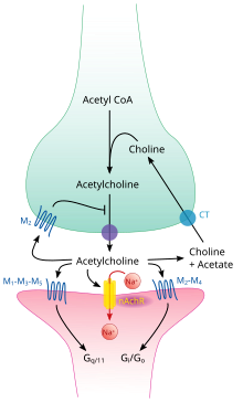
Acetylcholinesterase is an enzyme that breaks down the neurotransmitter acetylcholine.
In many synaptic clefts, they break down acetylcholine into fragments so that the transmission of the action potential occurs only once (and ACh doesn’t continue stimulating contraction).
State when an action potential is initiated.
C2.2.8: Depolarization and repolarization during action potentials.
An action potential is initiated only when the threshold potential (-55 mV) is reached.
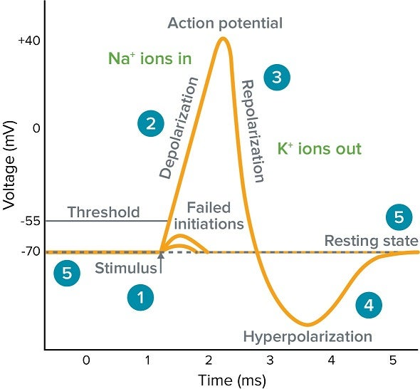
Define depolarization.
C2.2.8: Depolarization and repolarization during action potentials.
The process of the membrane potential becoming more positive (relative to the outside of the cell).
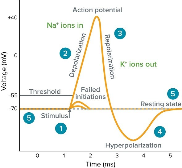
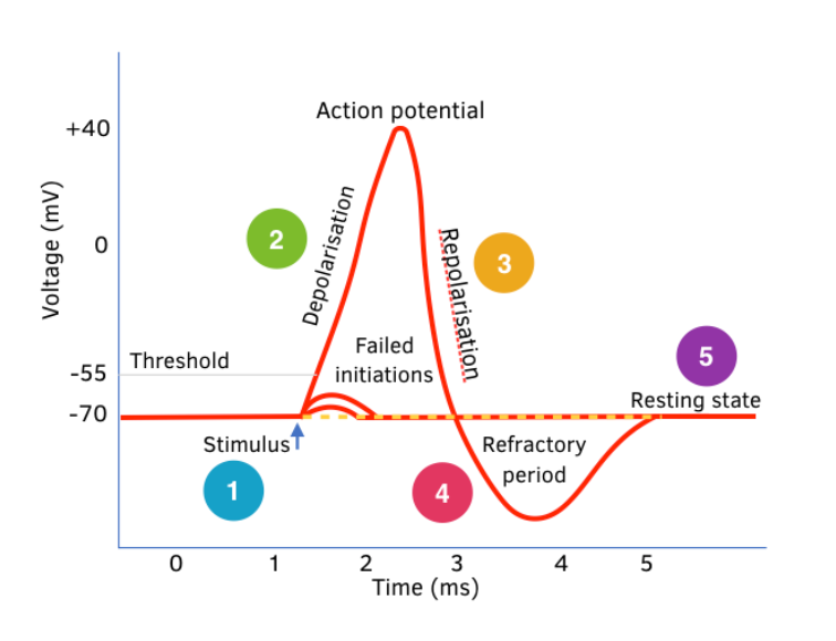
Outline the mechanism of depolarization during an action potential using voltage gated ion channels.
C2.2.8: Depolarization and repolarization during action potentials.
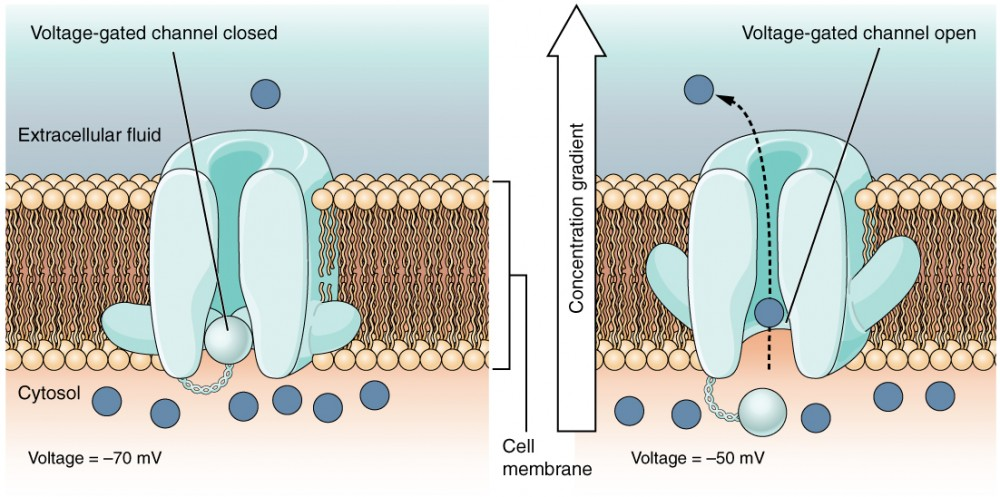
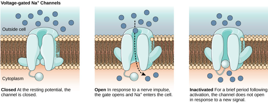
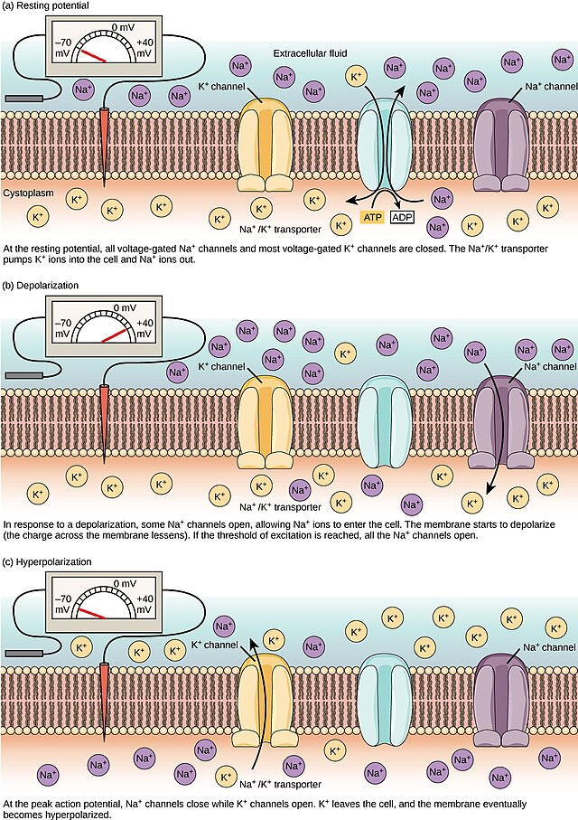
When the threshold potential is reached, voltage-gated sodium channels open. As a result, positive Na+ flow into the cell via facilitated diffusion.
This depolarizes the cell, as the neuron becomes more positively charged (raised up to about +30 mV).
This is “depolarization”, or the “rising phase”.
Define repolarization.
C2.2.8: Depolarization and repolarization during action potentials.
The process of the membrane potential becoming more negative (relative to the outside of the cell wall).
Outline the mechanism of repolarization during an action potential using voltage gated potassium channels.
C2.2.8: Depolarization and repolarization during action potentials.
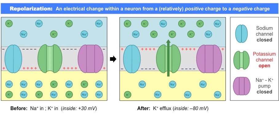
After the peak of about +30 mV, voltage-gated potassium channels open. As a result, K+ ions flow out of the neuron through facilitated diffusion.
This makes the neuron less positive, and the membrane potential becomes more negative. It eventually drops back down to -70 mV.
This is the repolarization, or falling, stage.
Describe the movement of sodium ions in a local current.
C2.2.9: Propagation of an action potential along a nerve fiber/axon as a result of local currents.
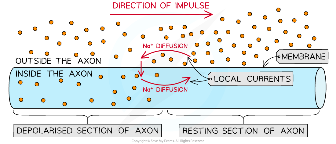
Sodium ions enter the membrane through a channel during the depolarization phase of the action potential.
The sodium ions entering the cell diffuse to neighboring sides of the channel (local current).
The Na+ ions move through the local current to bring their positive charge to the neighboring regions, so the neighboring membrane potential rise from the resting potential of -70 mV (until it rises to above -50 mV). The voltage-gated sodium channels open if it reaches that threshold.
The process is repeated, causing a wave of action potentials along the neuron membrane.
State that local currents cause each successive part of the axon to reach the threshold potential.
C2.2.9: Propagation of an action potential along a nerve fiber/axon as a result of local currents.
Local currents cause each successive part of the axon to reach the threshold potential.
Explain how the movement of sodium ions propagates an action potential along an axon.
C2.2.9: Propagation of an action potential along a nerve fiber/axon as a result of local currents.
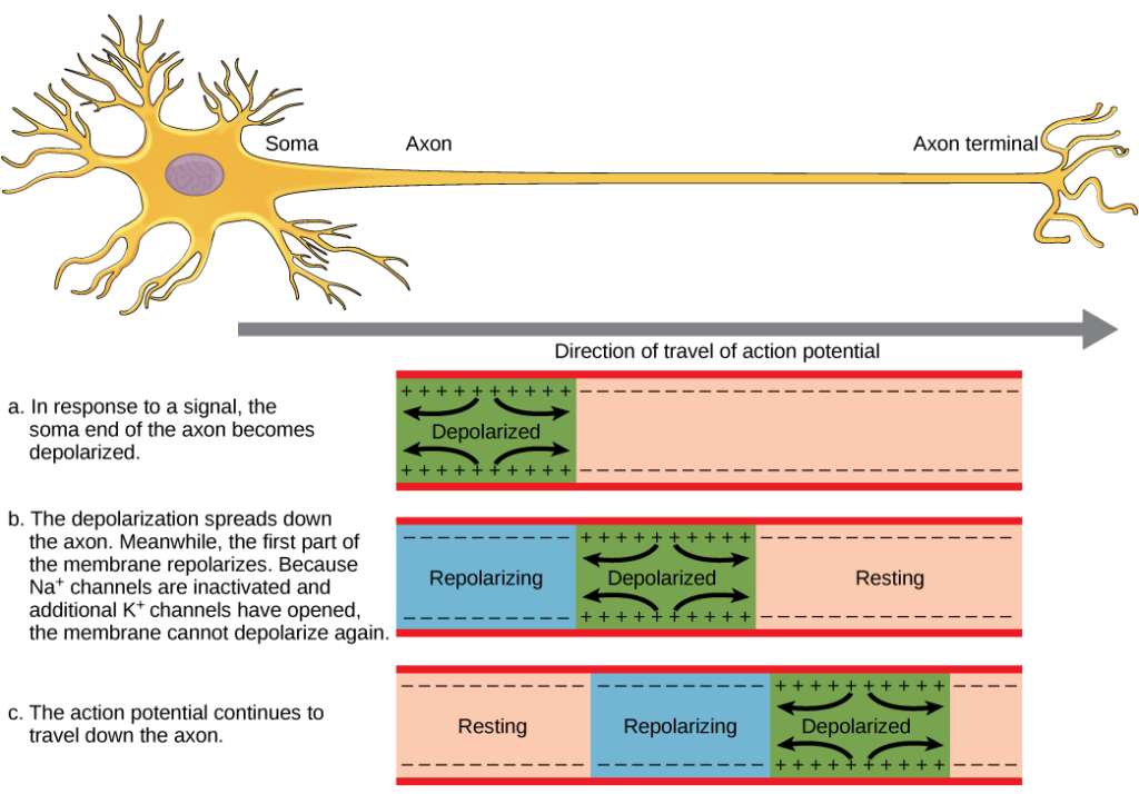
The sodium ions only propagate the local action potential down the axon because the parts behind have already been depolarized and are hyperpolarized in the refractory period (it will not be depolarized again).
Outline the cause and consequence of the refractory period after depolarization.
C2.2.9: Propagation of an action potential along a nerve fiber/axon as a result of local currents.
In hyperpolarization, too many K+ ions leave the cell, so the neuron becomes more negative than usual. As a result, the neuron is less likely to fire a signal for a short time (refractory period).
This allows the action potential to only travel down the axon even though the local current goes both ways, as the part behind is in the refractory period and will not be depolarized.
Outline the use of oscilloscopes in measuring membrane potential.
C2.2.10: Oscilloscope traces showing resting potentials and action potentials.
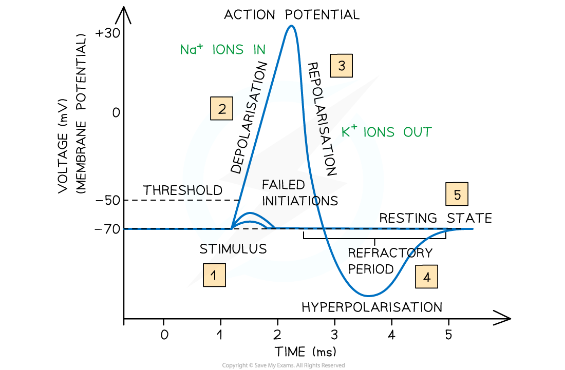
Oscilloscopes are instruments that graphically display electrical signals as waveforms, so they visualize changes in voltage over time.
They can measure the membrane potential in a neuron as the voltage changes over time.
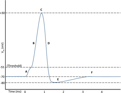
Annotate an oscilloscope trace to show the resting potential, action potential (depolarization and repolarization), threshold potential and refractory period.
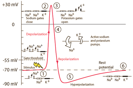
C2.2.10: Oscilloscope traces showing resting potentials and action potentials.
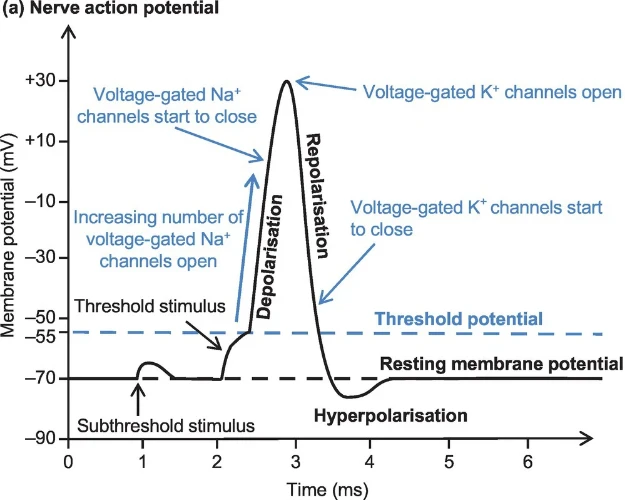
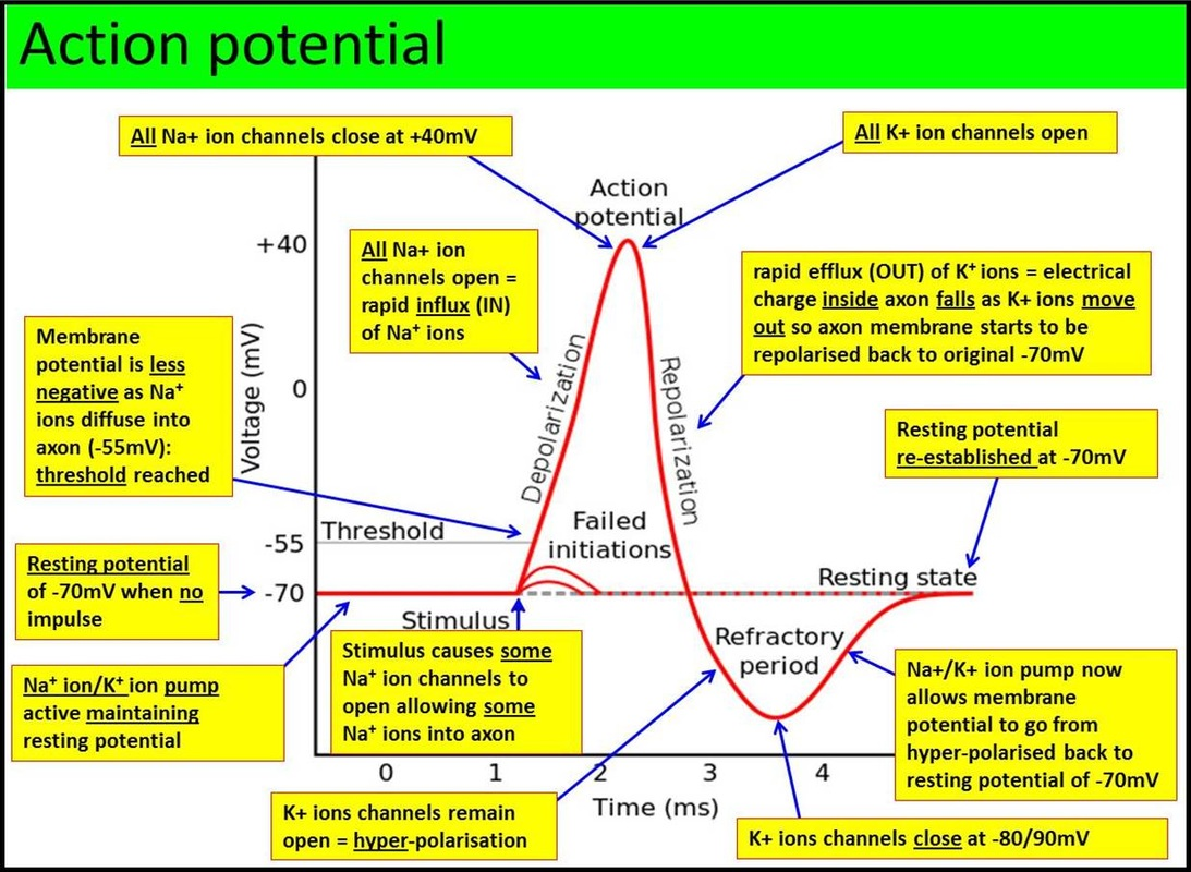
Deduce the number of nerve impulses per second from an oscilloscope trace.
C2.2.10: Oscilloscope traces showing resting potentials and action potentials.
To find the number of nerve impulses per second, find the period of an action potential (from peak to peak). That is the time it takes for one action potential. Then take the reciprocal of that to find the frequency.
Describe the structure of a myelinated nerve fiber.
C2.2.11: Saltatory conduction in myelinated fibers to achieve faster impulses.
Myelinated neurons have layers of myelin around their axons. Myelin sheaths are made of Schwann cells, and they don’t allow for movement of ions.
The gaps between the myelin sheath are known as nodes of Ranvier.
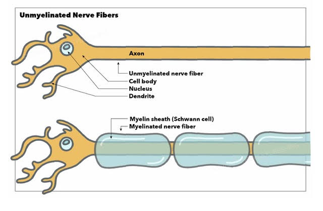
Outline how the myelination of neurons allow for Saltatory conduction, in which they effectively jump from node to Ranvier to node of Ranvier.
C2.2.11: Saltatory conduction in myelinated fibers to achieve faster impulses.
In myelinated neurons, Saltatory conduction occurs, as action potentials jump from node to node down the axon. K+ and Na+ ion channels are clustered in the nodes of Ranvier.
The axon potentials only happen at the nodes because the myelin insulates the rest of the axon.
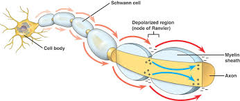
State where Na+ and K+ channels are clustered in myelinated neurons.
C2.2.11: Saltatory conduction in myelinated fibers to achieve faster impulses.
Na+ and K+ ion channels are clustered in the Nodes of Ranvier in myelinated neurons.
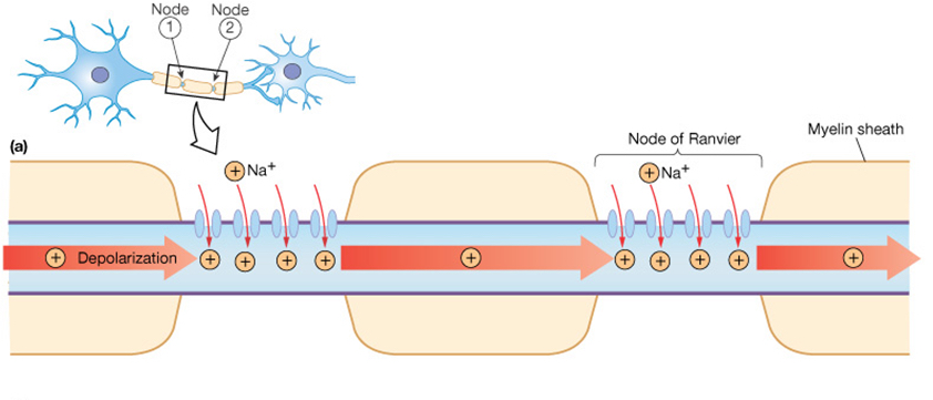
Outline the benefits of Saltatory conduction.
C2.2.11: Saltatory conduction in myelinated fibers to achieve faster impulses.
Saltatory conduction is faster because it doesn’t have to travel as long of a distance.
It also uses less energy compared to regular conduction because only pumps at the nodes are working.
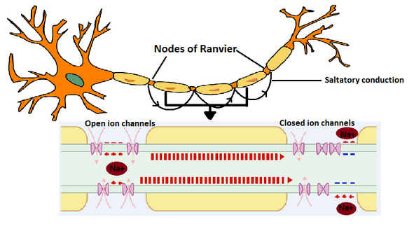
Define exogenous chemicals.
C2.2.12: Effects of exogenous chemicals of synaptic transmission.
Chemicals that come from outside living things.
They can interfere with synaptic transmission.
Outline the effects of neonicotinoids on synaptic transmission.
C2.2.12: Effects of exogenous chemicals of synaptic transmission.
Neonicotinoids are a group of chemicals that are used in pesticides.
They have a similar structure to nicotine and acetylcholine, so they bind permanently to nicotinic cholingeric receptors on the postsynaptic membrane of insects like bees. This blocks synaptic transmission and results in paralysis and death of the insects.
They don’t affect humans as much because ACh receptors in humans have a different structure from those in insects, so they are less toxic to humans.
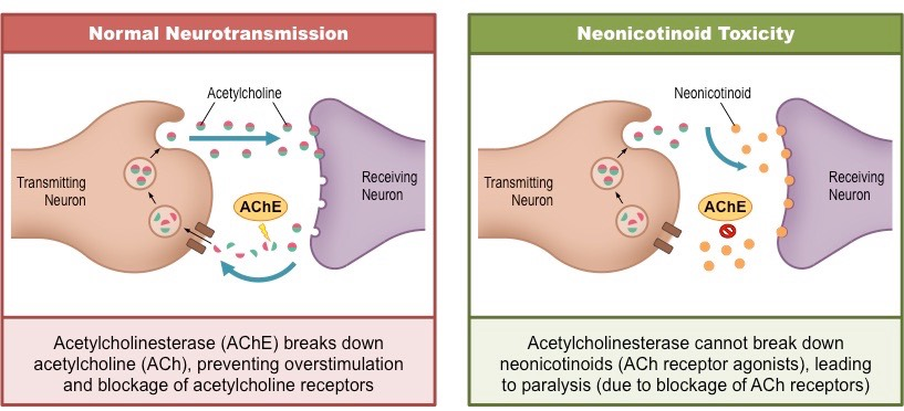
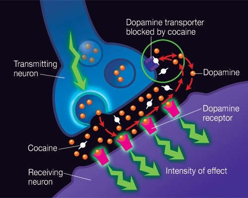
Outline the effects of cocaine on synaptic transmission.
C2.2.12: Effects of exogenous chemicals of synaptic transmission.
Dopamine is a neurotransmitter that controls feelings of pleasure and motivation. Dopamine transporters remove dopamine from the synaptic cleft.
But the drug cocaine blocks these transporters. As a result, dopamine is not removed from the synaptic cleft, and high levels of dopamine remain in the synaptic cleft. Thus, dopamine receptors continually receive signals, and the euphoric feeling is continued.
Outline the consequence of hyperpolarization by inhibitory neurotransmitters.
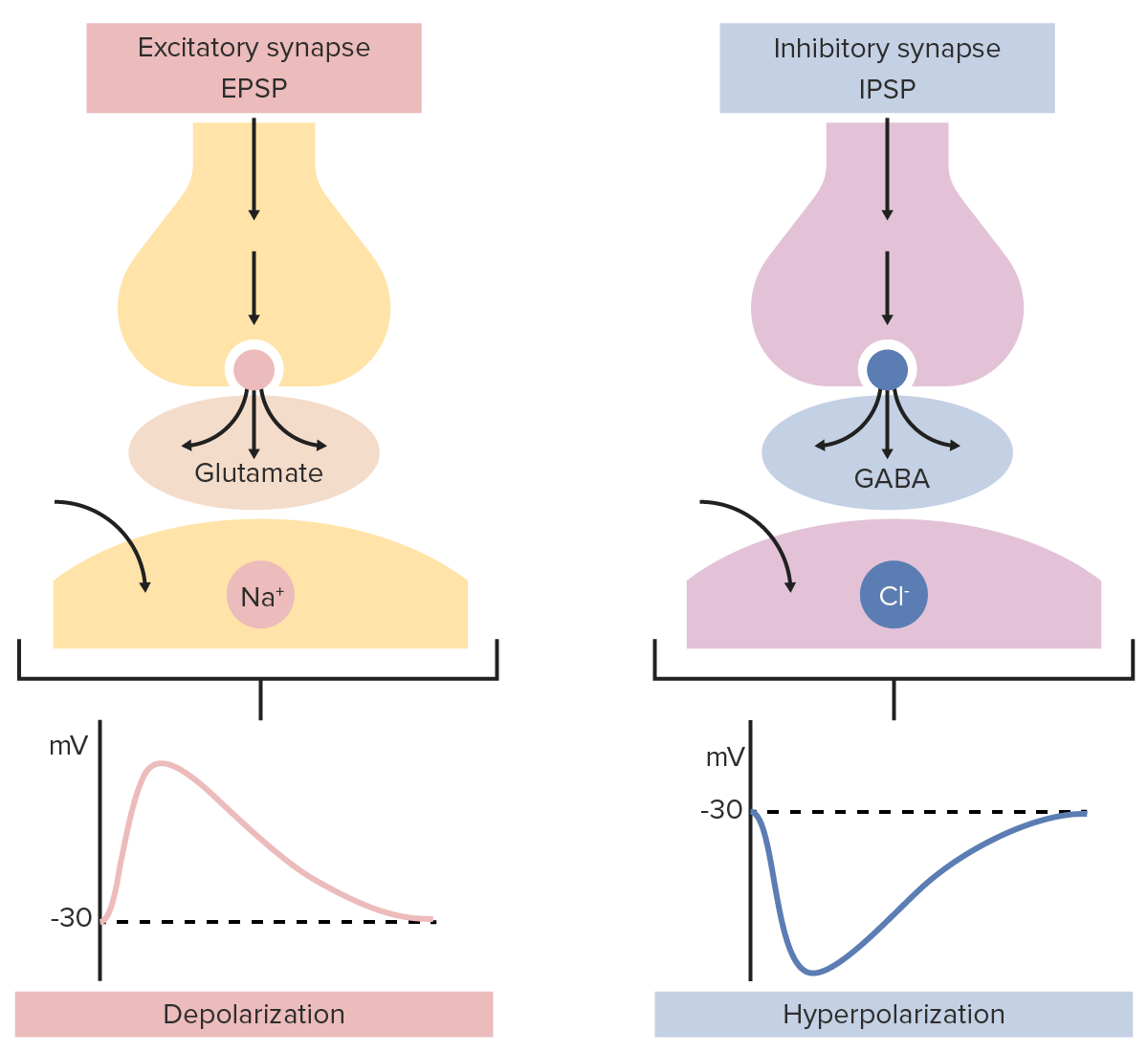
C2.2.13: Inhibitory neurotransmitters and generation of inhibitory postsynaptic potentials.
Inhibitory neurotransmitters bind to receptors on the neuron. This allows negative ions to enter, so the postsynaptic membrane becomes hyperpolarized (more negative).
Inhibitory neurotransmitters decrease the chance of an action potential in the postsynaptic neuron.
Compare excitatory and inhibitory neurotransmitters.
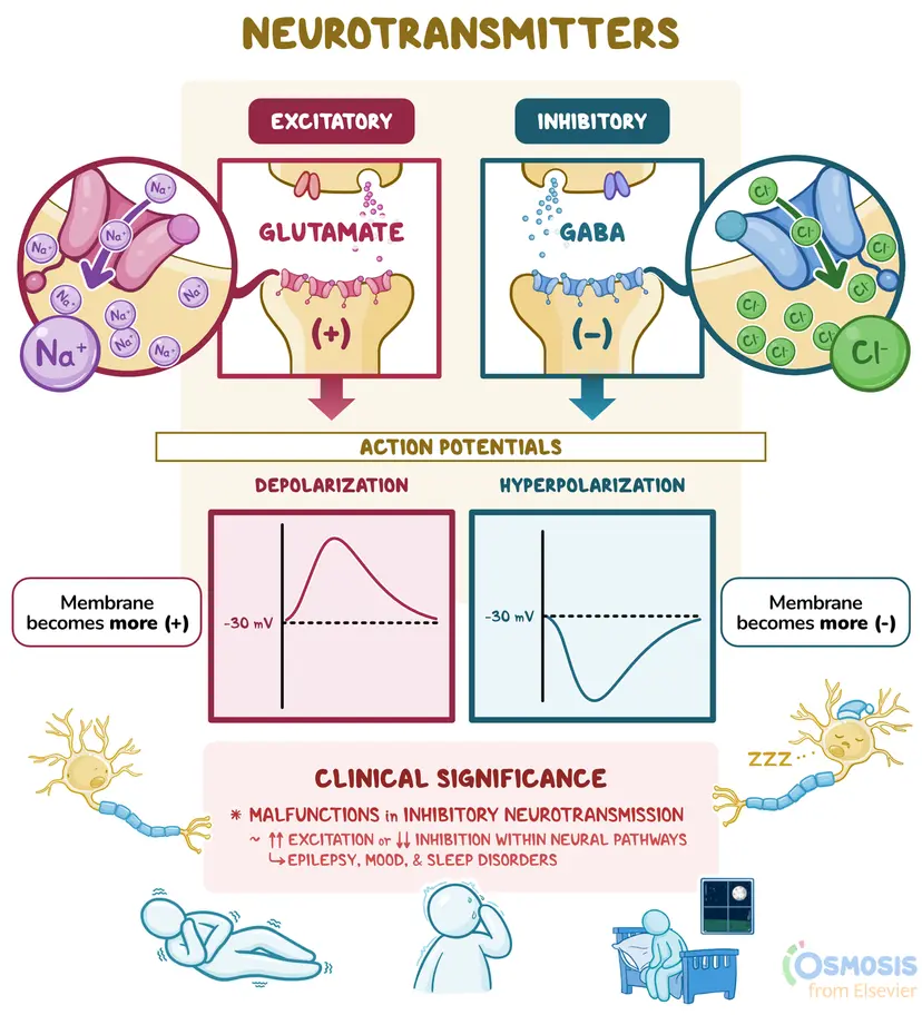
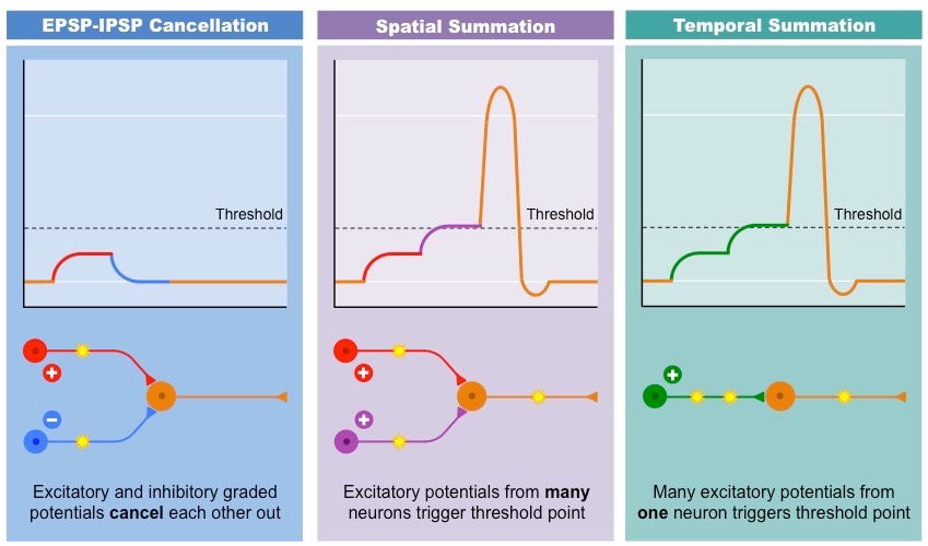
C2.2.14: Summation of the effects of excitatory and inhibitory neurotransmitters in a postsynaptic neuron.
Inhibitory neurotransmitters decrease the chance of an action potential in the postsynaptic neuron, while excitatory neurotransmitters increase the chance of an action potential.
Both neurotransmitters open channels to create their effect. Inhibitory neurotransmitters bind to receptors to allow negative ions to enter, but excitatory neurotransmitters open positive Na+ ion channels. Inhibitory neurotransmitters hyperpolarize the neuron, while excitatory neurotransmitters depolarize the neuron.
Describe the effects of excitatory and inhibitory neurotransmitters on the ability of a postsynaptic cell to reach its threshold potential.
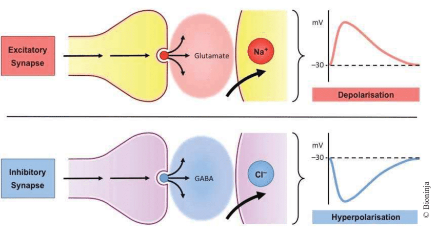
C2.2.14: Summation of the effects of excitatory and inhibitory neurotransmitters in a postsynaptic neuron.
Excitatory neurotransmitters make the postsynaptic cell more positive, so it depolarizes the neuron. This makes it easier for the cell to reach its threshold potential.
Inhibitory neurotransmitters make the postsynaptic cell more negative, so it hyperpolarizes the neuron. This makes it more difficult for the cell to reach its threshold potential.
Define summation.
C2.2.14: Summation of the effects of excitatory and inhibitory neurotransmitters in a postsynaptic neuron.
The total effect of neurotransmitters on a neuron, including excitatory and inhibitory neurotransmitters.
The summation determines whether the neuron will generate an action potential in an all-or-nothing manner
Describe the mechanism by which environmental stimuli are able to activate nerve endings in the skin, including the role of receptor proteins, ions channels, and threshold potential.
C2.2.15: Perception of pain by neurons with free nerve endings in the skin.
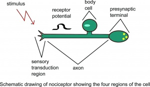
Nociceptors are pain receptors in the skin. They respond to stimuli such as high temperature, acid, and chemicals.
When stimulated, Na+ channels on nociceptors open, so Na+ flow in. If it reaches the threshold potential, an action potential is created and travels to the brain. There, pain is felt.
List stimuli which can trigger a pain response.
C2.2.15: Perception of pain by neurons with free nerve endings in the skin.
High temperature
Acid
Chemicals (such as capsaicin in chili peppers)
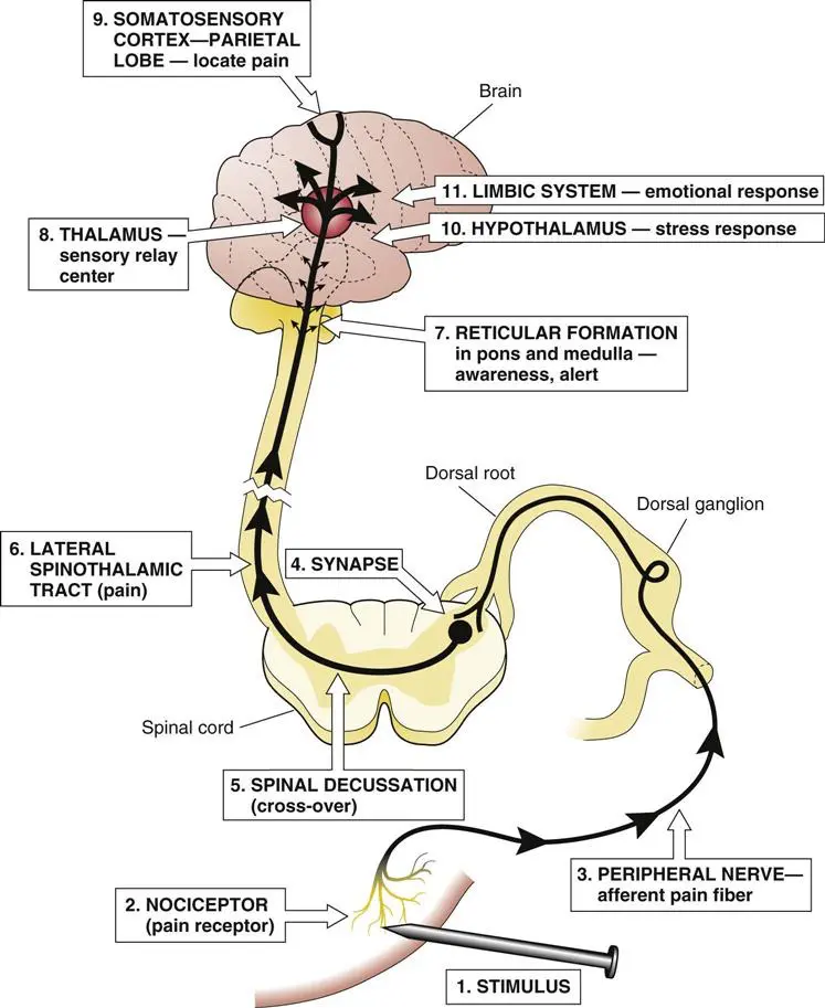
Outline the flow of information during the pain response.
C2.2.15: Perception of pain by neurons with free nerve endings in the skin.
Nociceptors generate an action potential from a stimulus. The nociceptors pass this signal to other neurons until it reaches the brain. There, the brain perceives the signals as pain.
Define emergent property.
C2.2.15: Perception of pain by neurons with free nerve endings in the skin.
An attribute, quality, or characteristic that emerges when parts interact with a larger whole (not made by any individual part).
New properties emerge at each level of biological organization.
Outline consciousness as an emergent property.
C2.2.15: Perception of pain by neurons with free nerve endings in the skin.
Consciousness is the state of being awake and aware.
No single neuron or part allows the brain to be conscious, but it is only possible from collective interactions of neurons in the brain. Consciousness is not a property present in an individual neuron, but only when neurons interact together.