Pathophysiology Flashcards for DIVAS
 Call Kai
 Learn
Learn Practice Test
 Spaced Repetition
 Match
Match Flashcards
 Knowt Play
1/51

Earn XP
Description and Tags
hi sydeny
Name | Mastery | Learn | Test | Matching | Spaced | Call with Kai | Chat |
|---|
No analytics yet
Send a link to your students to track their progress
52 Terms
etiology
the study of disease
pathology v.s. pathophysiology
pathology is study of disease and and pathophysiology is the study of the change in physiology because of disease; study of suffering
what are the 3 common causes of disease?
congenital: get it during development as a baby (fetal alcohol syndrome)
genetics: in your DNA
acquired: get it after birth (literally most everything; tuberculosis, AIDS
sign
something a doctor can observe
elevated temp, fast heartrate, low O2
symptom (acute v.s. chronic)
sensation the patient can feel and describe
headache, pain, throbbing, nausea
acute: develop rapidly and usually only last a little bit
chronic: can happen slowly and stay around for a long time
findings
signs and LAB TESTING
diagnosis
identifying a disease based off of signs and symptoms
determines disease, cause may sometimes be unknown
this leads to potential treatment and/or therapy
pathogenesis
the development of disease
idiopathic
etiology (disease) is unknown
iatrogenic
treatment for something is causing disease or treatment for said disease is making it worse
local disease
in one spot, like a boil on the neck
systemic
all over the body, like toxic shock or full body swelling
focal
disease confined to one area in an organ
diffuse
disease spread throughout the organ
sequelae (plural) sequela (singular)
condition resulting from previous disease that can be temporary or permanent
ex. snakebite results in permanent muscle atrophy
therapy
treatment for improving/removing disease, or just for control
prognosis
a prediction of how a disease and the process of healing will unfold
plasma membrane/cell membrane (how each thing is transported big idea)
selectively permeable (water, gasses, small ions pass freely)
nutrients in, waste out
studded w/ glycoprotein receptors that do immune responses, signaling, and pathogen docking, adhesion, and invasion
some molecules transported w/ the usual stuff
big molecules are moved by:
endocytosis: brings materials into the cell by engulfing them
exocytosis: pushes materials out of the cell by fusing internal vesicles with the membrane
mitochondria
produces ATP via. glycolysis and the krebs cycle (aerobic metabolism) = sugar + water = CO2, water, ATP
endoplasmic reticulum
rough ER = granular, has ribosomes that do protein synthesis
smooth ER = agranular, no ribosomes, contains enzymes for detoxification and construction of lipids (phospholipids, triglycerides, steroid hormones
golgi complex
stores and modifies proteins from the ER and creates vesicles to send them out
lysosome
contains lots of enzymes to break down cell debris and pathogens
binds and fuses with phagosomes to digest debris
nucleus
contains genetic material
contains info for making proteins and regulation of cell activities
cytoskeleton
had actin for ameboid movement (crawling) and has microtubules which form flagella and cilia
neurofibrils only in neurons, they are filaments that provide structure and support
3 causes of cell injury:
trauma - physical injury due to physical agents
temp, radiation, pressure, parasites bacteria and viruses, immune defect
intoxication - the cell has too much of something that impairs function
exogenous - poison from outside like smoke
endogenous - poison generated from the inside, usually from genetic diseases and metabolic malfunctions or from ineffective waste removal
deficiency - when the cell lacks something it needs to function
primary nutritional deficiency: you don’t eat enough of what you need
secondary nutritional deficiency: you do eat enough of what you need but your body can’t use it right
what are the 3 changes in cells due to injury
hydropic change: cells slow down and Na starts to creep in because the cell isn’t working hard enough to keep the pump regulated (1/3 energy goes there). Cl follows and then water follows and the cell swells up
fatty changes: usually seen in liver, which converts glucose to fat. can also be seen in muscle tissue and pancreas. usually a cell ships out its fat. a sick cell can keep making fat but not ship it out to adipose tissue.
residual bodies: trash that wasn’t thrown out. trash can be undigested stuff or waste. sick cells accumulate this because they can’t properly perform exocytosis
hypoxia
cell switches to aerobic respiration because it doesn’t have enough oxygen. they’ll burn fat instead of glucose and may even eat protein and lipids to make energy
hypertrophy
cells get bigger because of stress. happens in the heart if blood flow is being restricted
hyperplasia
cells multiply to meet production requirements
atrophy
cells shrink so they need less nutrients and O2
disuse atrophy: you don’t use it, you lose it
pressure atrophy: pressure is on something long enough to just make it shrink or degrade
metaplasia
a cell type is changed into something else in response to stress to try and be more protective or efficient
in smokers, cell type changes in the lungs to better protect against the irritants
dysplasia
when a cell becomes weird. they differ in size, shape, and organization. can be an indicator of cancer
apoptosis
programmed cell death, genetically regulated
cell will sense damage and will kill itself. ex. sunburn will cause so much cell damage the cell kills itself
organelle changes
a cell can increase in a certain organelle to be more efficient. like muscles might make themselves have more mitochondria so they can have more energy.
3 types of nuclear change
karyolysis; nucleus is gone, basically dissolved
pyknosis: nucleus shrinks
karyorrhexis: nucleus breaks into fragments
what are the 3 outcomes if a cell becomes injured
full recovery
recovers but below normal function
doesn’t recover and undergoes necrosis (cell death)
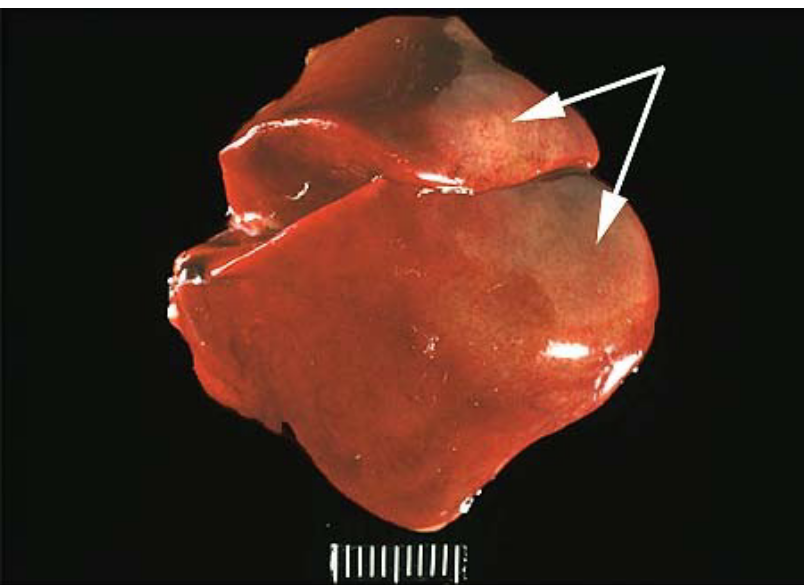
coagulation necrosis
type of cell death where the cell kind of fossilizes. the architecture stays the same but it becomes firm and pale from a lack of nutrients.
influx of calcium and loss of protein, decrease in pH
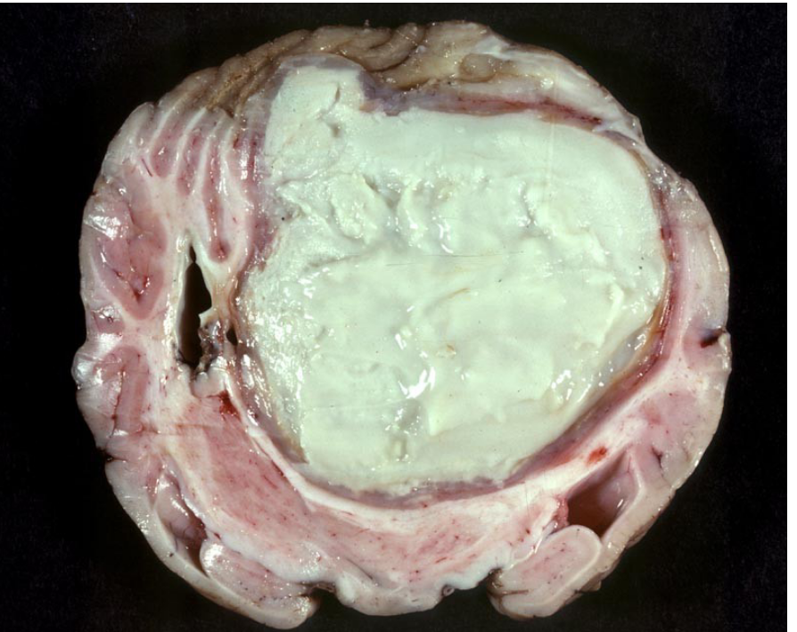
liquefication necrosis
usually occurs in soft tissues with no collagen, like the brain
liquid pools of dead cells

caseous necrosis
looks like cottage cheese
associated with tuberculosis
can be confused with coagulation necrosis but idk why
said to be usually walled off in a granulomatous lesion.
gangrene
death of tissue because of inadequate blood flow (ischemia) or anerobic bacteria (clostridium). there is dry, wet, and gas that leads to putrefaction (the process of decay or rotting in a body or other organic matter) and gas production.
dry: caused by frostbite or ischemia
wet: bacterial infection and swelling
gas: anaerobic bacteria making gas, happens in wat battlefields where this bacteria can infect
calcification
injured areas have a lot of calcium, which shows up in x-rays, called dystrophic necrosis, where calcium crystals form at necrotic spots
ischemia (brain, liver/kidney, heart)
lack of blood flow, therefore not enough oxygen
brain: 3-5 min leads to cell death unless cooled
liver/kidney: hour at body temp, 10 hours on ice
heart: 10 min at body temp, after 20min-1 hour, can’t recover. few hours on ice
fibroblasts: can tolerate anoxia (very low to no O2) very well, helps in tissue repair
intoxication susceptibility in tissues
nephrotoxins: affect kidneys most (fun fact 5 liters of blood go through the kidneys at least 60 times a day, and 20% of blood goes to the kidneys
livertoxins: tetrachloride is broken down making free radicals which damages cells, mitochondria, and molecules
ionizing/nonionizing radiation: makes water molecules with extra electrons, damages DNA, strongly affects rapid dividing cells
viral infections
polio: nervous system
hepatitis: liver
ebola: most organs
papilloma virus: skin, epithelia
how can you determine cell injury?
blood tests and body fluid tests
pH decrease in urine = renal failure
decrease in oxygen = respiratory failure
increase in liver enzymes, decrease in albumin in blood = liver failure
increase in troponins, LDH, CK, potassium in blood: heart failure
electrocardiogram
EKG/ECG determines heart activity and damage
electroencephalogram
brain activity
electromyogram
muscle contraction, conduction at neuromuscular junction
biopsy
take a sample of tissue and examine it for damage or changes
can be done histology wise or gross examination
MRI
magnetic resonance imaging
uses radio waves for soft and hard tissues
CT or CAT
computed tomography, uses x rays, soft and hard tissues
PET
position emission tomography, uses radioactive tracer, can be used with a CT/CAT scan, mostly soft tissue