Renal Infections
 Call Kai
 Learn
Learn Practice Test
 Spaced Repetition
 Match
Match Flashcards
 Knowt Play
1/63

There's no tags or description
Looks like no tags are added yet.
Name | Mastery | Learn | Test | Matching | Spaced | Call with Kai | Chat |
|---|
No analytics yet
Send a link to your students to track their progress
64 Terms
acute pyelonephritis is an
inflammation of the kidney or kidneys
bacteria can spread to the kidney through the
bloodstream or, more commonly, from the lower urinary tract
bacteria spreading through the bloodstream or the lower urinary tract is referred to as an
ascending infection
with an ascending infection, the infection begins in the
bladder and refluxes up through the ureters and into the kidney
acute pyelonephritits is most commonly encountered in —and is treated with antibiotics
women
complications of acute pyelonephritis include the development of
a renal abscess, pyonephrosis, xanthogranulomatous pyelonephritis, emphysematous pyelonephritis, and chronic pyelonephritis
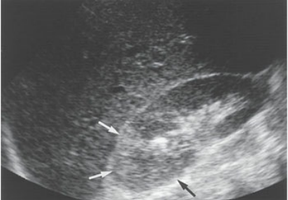
Acute pyelonephritis. Acute renal infection with interstitial hemorrhage produces hyperechoic focal swelling in the upper pole of this kidney (arrows).
clinical findings of Acute pyelonephritis
flank pain
bacteriuria
pyuria
leukocytosis
dysuria
urinary frequency
sonographic findings of Acute pyelonephritis
may appear normal
renal enlargement
focal areas of altered echotexture
compression of the renal sinus
yonephrosis describes the condition of —, also referred to as purulent material, within the collecting system of the kidney
having pus
the accumulation of pus is most likely caused by some —, as seen in many cases of pyelonephritis
obstructive process or infection that leads to urinary stasis
clinical findings of pyonephrosis
pyuria
bacteriuria
fever
flank pain
leukocytosis
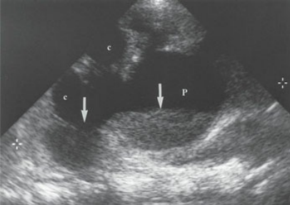
Pyonephrosis. Pus (arrows) is noted within the dilated calices (c) and renal pelvis (P) in this kidney (between calipers).
sonographic findings of pyonephrosis
hydronephrosis
pus and debris appear as internal layering and low-level echoes within teh dilated collecting system
chronic pyelonephritis is the
recurrent kidney infections or chronic obstruction may lead to scarring of the calices and renal pelvis
chronic pyelonephritis can lead to
xanthogranulomatous pyelonephritis and end-stage renal disease
children with a history of — are at increased risk for developing chronic pyelonephritis
vesicoureteral reflux (VUR)
A scar will appear as an
echogenic area that extends from the renal sinus through the renal parenchyma
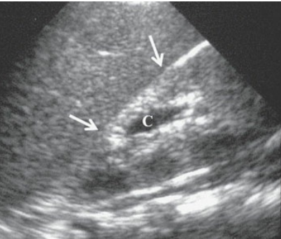
Chronic pyelonephritis. Longitudinal image of a 10-year-old boy showing a small echogenic left kidney with a dilated calyx (C) and overlying parenchymal atrophy (arrows).
clinical findingd of chronic pyelonephritis
flank pain
bacteriuria
pyuria
leukocytosis
dysuria
urinary frequency
history of VUR
sonographic findings of chronic pyelonephritis
small echogenic kidneys that have lobulated borders
renal scar appears as an echogenic area within the kidney that extends from the renal sinus through the renal parenchyma
a renal abscess can occur in regions of the kidney
affected by pyelonephritis or be located adjacent to the kidney
a perinephric abscess is a collection of
purulent material that has leaked through the capsule into the tissue surrounding the kidney
gas development within the abscess produces
dirty shadowing or reverberation artifact
clinical findings of a renal or parinephric abscess
flank pain
high fever
symptoms of pyelonephritis (pyuria, hematuria, and flank pain)
leukocytosis
sonographic findingd of a renal or perinephric abscess
can appear anechoic, hypoechoic, or complex
gas shadows or dirty shadowing may be present within the mass
emphysematous pyelonephritis is a
rare, and yet life-threatening, complication of pyelonephritis
although emphysematous pyelonephritis may be the result of a long-standing urinary tract obstruction, it is found more often in patients who have
diabetes mellitus or who are immunocompromised
the term emphysematous denotes the
formation of air within an organ
with emphysematous pyelonephritis, bacterial formation allows gas to accumulate within the
renal parenchyma
— is the most common culprit of emphysematous pyelonephritis
escherichia coli infection
clinical findingd of emphysematous pyelonephritis
flank pain
diabetes mellitus
immunocompromised patient
fever
leukocytosis
sonographic findings of emphysematous pyelonephritis
gas or air within the renal parenchyma
dirty shadowing or reverberation artifact coming from the renal parenchyma
xanthogranulomatous pyelonephritis is uncommon and is typically caused by a
chronic urinary tract obstruction and subsequent infection
the word part “xantho” means
yellow
with xanthogranulomatous pyelonephritis, — are often present
a staghorn calculus, hydronephrosis, and perinephric fluid
clinical findings of Xanthogranulomatous pyelonephritis
dull or persistent flank pain
pyuria
hematuria
fever
leukocytosis
sonographic findings of Xanthogranulomatous pyelonephritis
hydronephrosis
staghorn calculus
perinepheric fluid collection
the most common cause of fungal urinary tract infections is
Candida albicans
— are at increased risk for developing a fungal infection within their kidney
immunocompromised patients
patients with a history of — are more likely to suffer from renal fungal disease
diabetes mellitus, intravenous drug abuse, and infants who have longstanding, indwelling catheters
when — are noted within the collecting system, the diagnosis of a renal fungal disease can be made
fungal balls
clinical findingd of renal fungal disease
immunocompromised person
diabetes mellitus, IV drug abuse or long-standing indwelling catheter
infant with an indwelling catheter
flank pain
fever
chills
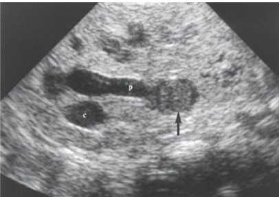
Fungus ball. Transverse image of the kidney in a 2-month-old infant with Candida albicans showing a dilated renal pelvis (p) and calices (c) that contain echogenic urine and a mobile fungal ball (arrow).
sonographic findingd of renal fungal disease
fungal balls appear as hyperechoic, nonshadowing, mobile structures within the renal collecting system
glomerulonephritis can be caused by a
distant infection, such as strep throat, or an autoimmune reaction
some conditions, such as —, have glomerulonephritis as a characteristic feature
lupus
glomerulonephritis can lead to
significant glomerular damage, and the kidneys can slowly shut down secondary to diminished filtration capabilities
glomerulonephritis can be
acute or chronic
in the acute stage of glomerulonephritis, the kidney may appear
normal, enlarged, and have an overall increase in echogenicity
the renal pyramids may appear more prominent with
acute glomerulonephritis
chronic glomerulonephritis, the result of a long-standing infection, can lead to
end-stage renal disease
clinical findings of glomerulonephritis
recent throat infection (acute)
smoky urine
hematuria
proteinuria
fever
hypertension
azotemia
sonographic findings of acute glomerulonephritis
enlarged kidney(s) with increased echogenicity
prominent renal pyramids
sonographic findingd of chronic glomerulonephritis
small, echogenic kidneys
glomerulonephritis can be caused by a
distant throat infection like strep throat
the most common renal parasitic infection is from
schistosomiasis
schistosomiasis
is a worm that enters humans by penetrating the skin
schistosomiasis is most often seen in the
bladder, causing thickening of the bladder wall
the kidney can also succumb to —, with the development of a hydatid cyst
Echinococcus granulosus
Hydatid disease is most often found in
sheep- and cattle-raising countries such as the Middle East, Australia, and the Mediterranean
clinical findingd of parasitic urinary tract infectiion
hematuria
flank pain
pyuria
dysuria
possible recent travel out of the country (hydatid cyst: middle east, austrailia, and mediterranean)
sonographic findings of parasitic urinary tract infection
schistosomiasis: bladder wall thickening
hydatid cyst: depends on the stage of its maturation, as it may appear completely anechoic, contain a daughter cyst with internal debris, or as a complex mass with calcifications