PAS 301: Human Anatomy - Axial Skeleton 1 Flashcards
 Call Kai
 Learn
Learn Practice Test
 Spaced Repetition
 Match
Match Flashcards
 Knowt Play
1/64

Earn XP
Description and Tags
Vocabulary flashcards covering bone shapes, skeletal divisions, cranial landmarks, vertebral components, spinal curvatures, sternum parts, and rib classifications.
Name | Mastery | Learn | Test | Matching | Spaced | Call with Kai | Chat |
|---|
No analytics yet
Send a link to your students to track their progress
65 Terms
Flat Bones
Thin and curved bones, such as those of the brain case and the ribs.
Long Bones
Bones that have a greater length than width, such as the femur and humerus.
Short Bones
Small, cube-shaped bones, such as the carpals in the wrist and tarsals in the foot or ankle.
Irregular Bones
Uniquely shaped bones, such as the vertebrae and bones of the face.
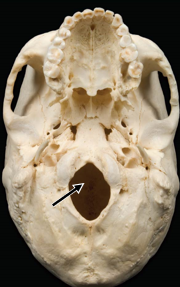
Foramen
A hole through bone that allows for the passage of nerves, arteries, and veins.
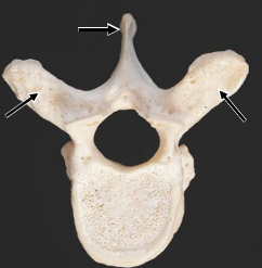
Process
A bony projection that acts as an attachment for muscles, tendons, and ligaments, or forms bumps, holes, grooves, and passages for nerves and vessels.
Axial Skeleton
The division of the skeletal system comprising 80 total bones, which includes the skull, vertebral column, and rib cage.
Appendicular Skeleton
The division of the skeletal system comprising 126 bones.
Cranium
The brain case consisting of 8 bones that contains and protects the brain.
Sutures
Jagged connection points between skull bones that fuse with age as the suture lines vanish.
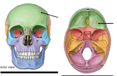
Frontal Bone
Bone of the braincase that protects the brain anteriorly and supports the brain inferiorly from inside the skull.
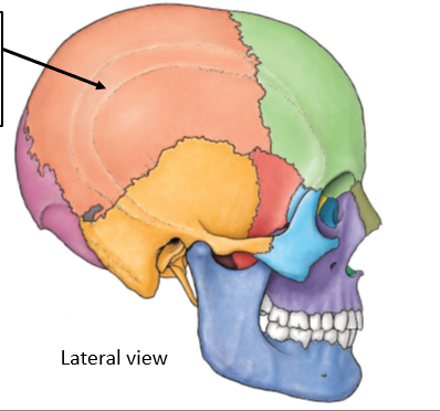
Parietal Bones
Bones that protect the brain laterally, superiorly, and posteriorly.
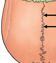
Sagittal Suture
The cranial suture located between the two parietal bones.
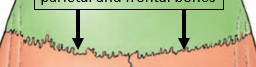
Coronal Suture
The cranial suture located between the parietal and frontal bones.
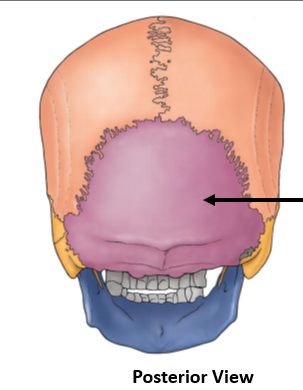
Occipital Bone
Bone that protects the brain posteriorly and inferiorly.
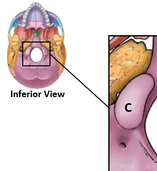
Occipital Condyles
Rounded projections on the occipital bone that articulate with the first vertebra (atlas).
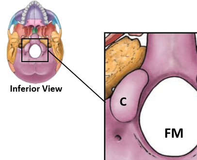
Foramen Magnum
The large opening in the occipital bone that serves as a hole for the spinal cord.
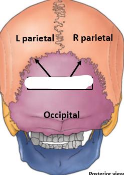
Lambdoidal Suture
The cranial suture located between the occipital bone and the parietal bones.
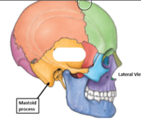
Temporal Bone
Bone that protects the brain laterally and features the mastoid process, mandibular fossa, and external auditory meatus.
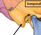
Mastoid Process
A rounded projection on the temporal bone located posterior to the ear.
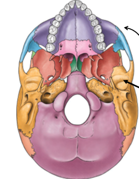
Mandibular Fossa
A landmark on the temporal bone that provides articulation with the mandible.
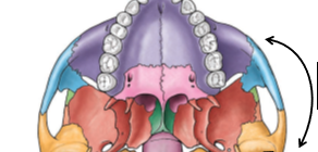
Zygomatic Arch
A bony arch formed by the temporal bone and the zygomatic bone.
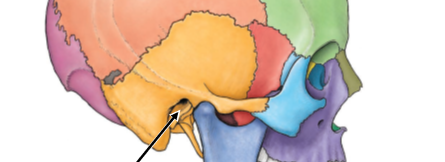
External Auditory Meatus
The outer ear opening located on the temporal bone.

Ethmoid Bone
Bone located at the center of the skull that provides a passage for the first cranial nerve.
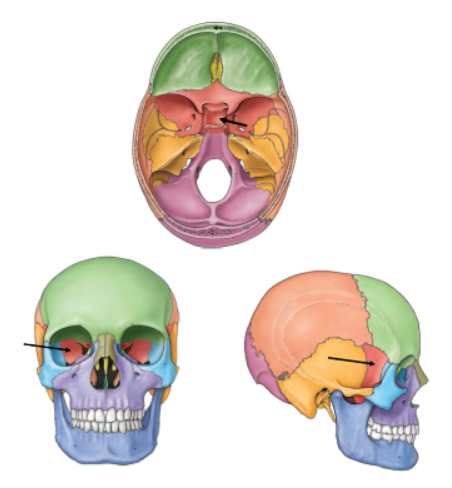
Sphenoid Bone
Bone sitting in the middle of the skull, housing the pituitary gland, and visible in the posterior orbit and lateral aspect of the skull.

Maxilla
Facial bone comprising the upper jaw and upper teeth.
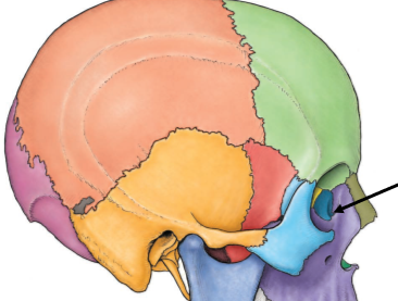
Lacrimal Bone
Facial bone providing a passage for tears into the nasal cavity.
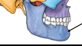
Mandible
The lower jaw bone containing lower teeth that articulates with the temporal bone.

Hyoid Bone
A neck bone in line with C3 that forms a palatable surface landmark, has no articulation with other bones, and is involved in swallowing as well as jaw and tongue movements. Horseshoe shaped.
Lordosis
A normal spinal curvature that curves anteriorly (present in cervical and lumbar regions), or a pathologically extreme curvature. Looks like )
Kyphosis
A normal spinal curvature that curves posteriorly (present in thoracic and sacral regions), or a pathologically extreme curvature. Looks like (
Scoliosis
A clinical malformation of the spine characterized by a side-to-side curvature.

Posterior Arch (Neural Arch)
The major vertebral component that encloses the spinal cord and has narrow processes extending posteriorly and laterally.
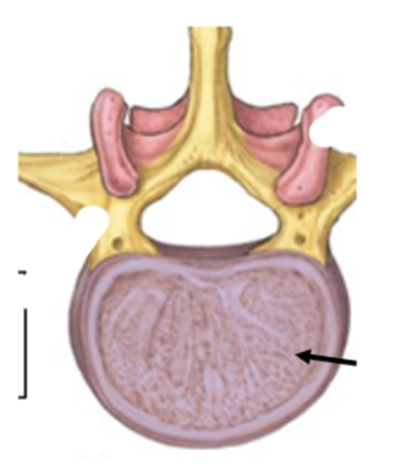
Vertebral Body
The primary support structure of a vertebra, possessing a smooth and curved anterior surface.
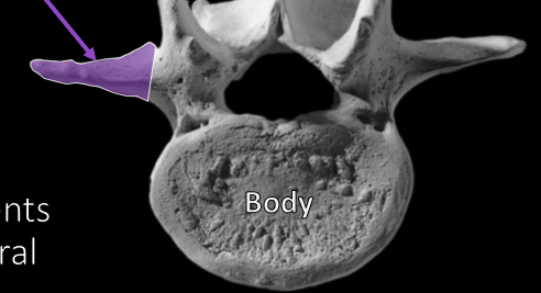
Transverse Process
A minor component of a vertebral element that projects laterally.
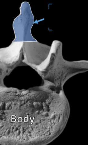
Spinous Process
A minor component of a vertebral element that projects posteriorly.
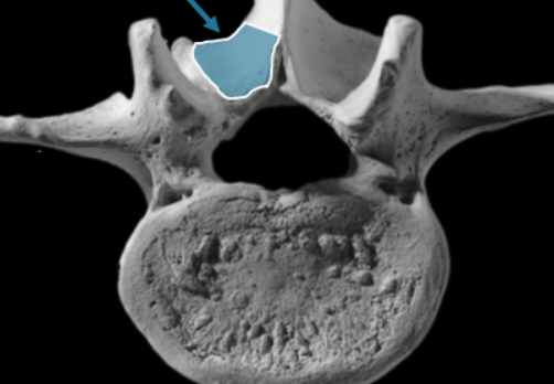
Lamina
The bony structure that connects the spinous process and transverse processes of a vertebra.
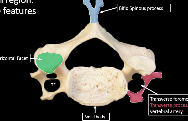
Transverse Foramen and Unique Features in Cerivical Region
An opening through the transverse process of a cervical vertebra through which the vertebral artery passes.
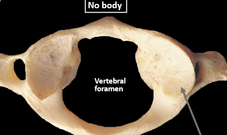
Atlas (C1)
The first cervical vertebra, which lacks a body and spinous process (having a posterior tubercle instead), and features facets that articulate with the occipital condyles of the skull.
Axis (C2)
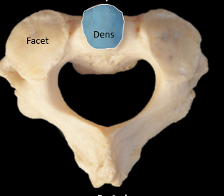
The second cervical vertebra, characterized by a thumb-like projection called the dens that articulates with C1.
Dens
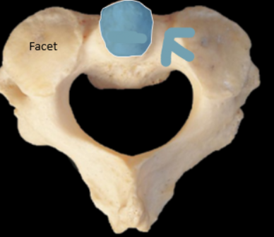
A thumb-like projection on the C2 (axis) vertebra that articulates with C1 (atlas).
Manubrium
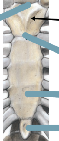
The triangular upper portion of the sternum.
Sternal Angle (of Louis)
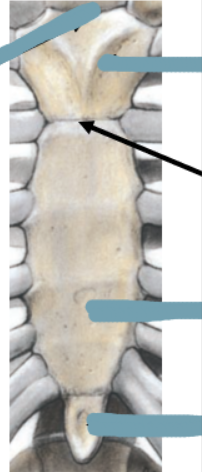
The joint between the manubrium and body of the sternum, marking the level where the trachea bifurcates.
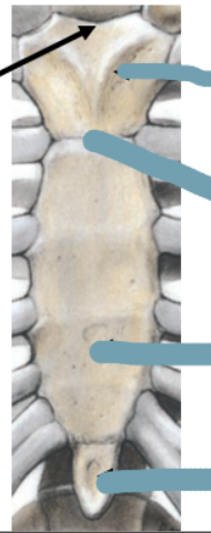
Suprasternal Notch
The superior depression of the sternum, also known as the jugular notch.
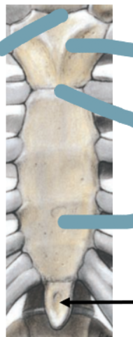
Xiphoid Process
The small inferior projection of the sternum.
True Ribs
Rib pairs 1 through 7, which have a direct connection to the sternum via a single costal cartilage.
False Ribs
Rib pairs 8 through 10, which have an indirect connection to the sternum through the costal cartilage of rib number 7.
Floating Ribs
Rib pairs 11 through 12, which have no attachment or connection to the sternum.
Cranial Bones
Bones that make up the skull, including the frontal, parietal, occipital, temporal, ethmoid, and sphenoid bones.
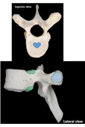
Thoracic Vertebrae
The twelve vertebrae in the mid-back region, labeled T1 to T12, which articulate with the ribs.
Lumbar Vertebrae
The five larger vertebrae in the lower back, labeled L1 to L5, which bear the most weight.
Sacral Vertebrae
Five fused vertebrae in the lower back forming the sacrum, connecting the spine to the pelvis.
Cervical Curve
The anterior curve of the cervical spine, developed as an infant begins to lift its head.
Thoracic Curve
The posterior curve of the thoracic spine, which is present from birth and accommodates the thoracic organs.
Lumbar Curve
The anterior curve of the lumbar spine that develops when a child begins to walk.
Sacral Curve
The posterior curve of the sacral spine, formed by the fusion of the sacral vertebrae.

Nasal Bones
The bone located to where the nose meets the skull, forming the bridge of the nose.
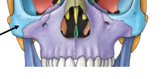
Zygomatic Bones
The pair of bones that form the prominence of the cheeks and the lateral walls of the eye sockets.

Palantine Bones
The bones that form the back part of the hard palate of the mouth and contribute to the floor of the eye orbits.
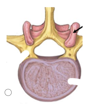
Articular Components
Where the vertebrae articulate inside of the spine
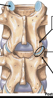
Articular Processes
Each vertebra contains 2 Superior and Inferior Processes and a Facet.
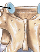
Superior Facet
The superior facet is the smooth, flat articular surface on the superior articular process of a vertebra that forms a joint with the inferior facet of the vertebra above.
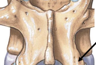
Inferior Facet
Flat surface on the lower part of a vertebra that connects with the vertebra below it.
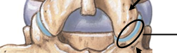
Articulating Facets
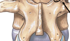
Superior Articular Process