exam 2 anatomy review
 Call Kai
 Learn
Learn Practice Test
 Spaced Repetition
 Match
Match Flashcards
 Knowt Play
1/133

Earn XP
Description and Tags
diseases that have not been taught yet - rickets and osteomalacia, osteopoorsis, myasthenia gravis
Name | Mastery | Learn | Test | Matching | Spaced | Call with Kai | Chat |
|---|
No analytics yet
Send a link to your students to track their progress
134 Terms
Shingles
VZV that causes chicken pox lays dormant in sensory neurons
Kyphosis
Hump condition rounded upper back
Lordosis
Exaggerated lumbar curve
Scoliosis
Lateral side ways curve
Osteoporosis
Degenerative disease of isolated joints of the articular cartilage cause of excessive wear
Inflammation secondary cartilage erosion follows
Rheumatoid arthritis
Systemic chronic inflammation disorder on multiple joints ankylosis can occur destroying articular cartilage and adjacent bone.
Gout
build up of urate crystals in joints that causes acute inflammatory reaction
Unknown cause but could be linked to chronic renal disease (kidney cannot excrete urate crystals
Bursitis
inflammation of the bursae a synovial lined sac
Carpal Tunnel Syndrome
Entrapment of the median nerve as it passes through the connective tissue sheath causing numbness, pain and tingling
Muscular Dystrophy
group of muscle disorders characterized by progressive muscle weakness and degradation
Duchenne Muscular Dystrophy
genetic defect in Dystrophin (ac cessory protein that stablizes the thin filaments)
Albinism
Defects in melanocytes differentiation (the are not produced) or defects in melanin production - there are different forms of this with different defects
Vitiligo
autoimmune mediated destruction of melanocytes
Malignant Melanomia
neoplasia of melanocytes. can spread to the dermis, lymph nodes and blood
Acanthosis
Abnormal epidermal thickening, usually stratum spinosum (level of epidermis)
rete pegs: epidermal extentions into papillary are abnormally long
Associated with chronic inflammatory condition, insulin resistance, obesity
Hyperkeratosis
thickening of the corneum and stratum granulosum
Parakeratosis
cells of corneum retain nuclei, normally occurs in oral cavity, may occur with some skin conditions
Psoriasis
Chronic Inflammatory Conditions
Excessive proliferation of the keratinocytes and accelerated upward cellular migration
Cell reaches surface layers before the are filled with keratin
Scaly white patchs and red inflamed areas
Acne
Chronic disease involving hair follicle blockage and inflammation
Breakdown of sebaceous lipids into fatty acids irritates skin resulting in HYPERPROLIFERATION of epidermal cells
excess epidermal cells block the hair follicle causing of excess sebum and inflammation
genetic, excess androgens, bacteria contribute the the development of acne
Intervertebral Foramen
opening wear spinal nerve exits through the vertebral column, formed by the articulation of adjacent vertebrae.
Where and why are spinal tap given there
Spinal taps are given at L3 because the cauda equina the tail of dorsal and ventral nerve roots extend here to reach the lower exrematies . It will avoid hitting the spinal cord here
cauda equina
the of the spinal cord that floats and reaches the lower extrematies
What does the Spinal Cord (SC) made up of?
Inner gray matter where the neurons are located
Outer white matter ( axons running up and down SC)
motor neurons are located in the Grey matter Ventral horns and the lateral horns
No sensory neurons in here
Dorsal Root and Cranial nerve sensory ganglia
where the pseudounipolar ( only found int PNS Sensory Ganglia) neurons due to them being GENERAL sensory neurons

Ventral Root
Carries motor neurons
this and dorsal root make up the spinal root 31 pairs that carry axons to the tissue of the outer body)
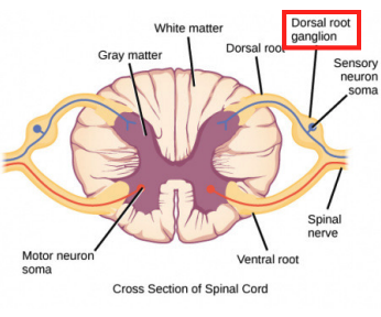
Dorsal Root
Carries central process (axon) of pseudounipolar neurons( which extend through the spinal nerve to innervate the skin and skeletal muscle)
this and the ventral root make up the spinal nerve ( 31 pairs that carry axons to the tissue of the outer body)
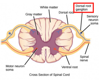
Dorsal Ramus
gives rise to the nerves that innervate the back proper
Ventral Ramus
gives rise to nerves that innervate the anterior body and limbs
gives rise to nerve roots that branch and join forming collection of nerves or a nerve plexus - branches of this innervate (sends axons to) limbs and body wall
Cervical Plexus
C1 - C5
Innervate the neck
Brachial Plexus
C5 -T1
Innervate the upper limbs
Thoracic Spinal Nerve
T2 -T11
does not form a plexus as it runs along the ribs
Lumbar Plexus
T12 - L4
innervates the lower limbs
Sacral Plexus
L4 - S4
Innervate the pelvi and gluteal region
Dorsal Root Ganglia
Pseudounipolar general sensory neurons are located in this ganglia _______
Sympathetic Chain Ganglia and Prevertebral (preartotic) ganglia
Sympathetic Motor neurons are located in this ganglia _______
Cranial Nerve Motor Ganglia and Intramural Ganglia
Parasympathetic Motor neurons are located in these ganglia _____
Somatic Tissues are made of/ found in
Skeletal muscle and skin which are found in the outer body
Visceral Tissues are made of/found
Found in the outer and inner body and include
Gland: found in the skin (outer body)
Smooth Muscle: found in the blood vessals and arrector pili (raise skin hair)
Cardiac Muscle (inner body)
What nerves Innervate the outer body wall?
spinal nerves are made of motor and sensory axons
Skin needs sensory innervations
Skeletal muscle needs motor innervation and sensory innervation
Visceral Tissue needs sympathetic innervations
What tissue in the outer body need innervations?
The 3 components (axons) of a spinal nerve
These are the 3 components of what?
Motor axons to skeletal muscle
Sensory Axons (peripheral processes of pseudounipolar neurons) to skin and skeletal muscle
Sympathetic motor post ganglionic axons to viscera of the outer body
Spinal Cord Ventral Horns
Where the NCB of a spinal nerve motor axon to skeletal muscle located?

Dorsal Root Ganglia
Where the NCB of a spinal nerve sensory axon located?

Autonomic nervous system (sympathetic and parasympathetic)
Visceral tissue recieve MOTOR innervation from this?
Parasympathetic system
control rest and digest and sexual arousal - controlled by the ANS
Sympathetic System Pathway
controls the bodies fight or flight response - is controled by the ANS
innervates ALL VISCERAL TISSUE
sympathetic preganglionic neurons ALWAYS located on the lateral horn in the T-L levels
axon travels through ventral horn and root → then to short nerve called the SYMPATHETIC CHAIN GANGLIA
post-ganglionic axon goes through a spinal nerve to get to the outer body → goes through the ramus communicans to the join spinal nerve and travels through branches of the SN to innervate the outerbody viscera

Autonomic Nervous System
its pre-ganglionic neuron is located in the CNS and its axon synapsis on the ganglionic neuron (post-ganglionic) in the motor ganglion in the PNS
Post-ganglionic neuron synapsis on glands, smooth muscle or cradiac muscle (visveral tissue)

Spinal Nerve Components
1. Motor to skeletal muscle
2. Sensory to skin and muscle
3. Sympathetic motor to blood vessels, sweat glands, arrector pili

Sympathetic Pathways
For internal organs, ____________ preganglionic axons will synapse in either the sympathetic chain or the preaortic (prevertebral) ganglia. The postganglionic axons don’t use spinal nerves to get to the organs but go through special nerves to get to the organs

Parasympathetic Pathway
cranial nerves and pelvic nerves provide _______ innervation to internal organs
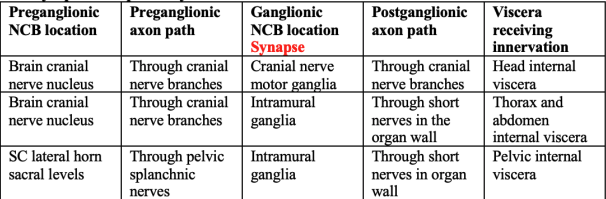
Organs from HEAD to LOWER ABDOMINAL - preganglionic neuron is in the BRAIN and synapse on either the CRANIAL NERVE MOTOR GANGLIA or INTRAMURAL GANGLIA IN THE ORGAN WALLS
Internal organs in the PELVIS preganglionic neuron is located on LATERAL HORN of the SC at Sacral Levels
preganglionic axon travels through PELVIC SPLANCHNIC NERVES to reach the intramural ganglia in organ walls where it synapses on the ganglionic neurons
Spinal Cord lateral horn thoracic/lumbar levels
Where are the NCBs of all sympathetic pregangliionic axons located?
Sympathetic Chain Ganglia or preaortic ganglia
Where are the NCBs of sympathetic postganglionic axons located?
Sympathetic chain ganglia (1)
Where is the NCBs of a sympathetic postganglionic axon that innervates body wall viscera located? (1)
Sympathetic Chain Ganglia (2)
Where is the NCB of a sympathetic postganglionic axon that innervates head and thoracic visceral organ located (2)
Preaortic Ganglia
Where is the NCB of a sympathetic postganglionic axon that innervates the abdominal & pelvic viscera located?
In cranial nerve nuclei in the brain
Where are the NCBs of parasympathetic preganglionic axons that innervate viscera in the head on down to the lower abdomen located?
In the cranial nerve motor ganglia located in the head or in intramural ganglia in the walls of organs
Where are the NCBs of parasympathetic postganglionic axons that innervate viscera from the head on down to the lower abdomen located?
The NCB of preganglionic neurons are located in the spinal cord lateral horn sacrel levels
The NCBs of ganglionic neurons are located in the intramural ganglia
For parasympathetic innervation of the pelvic viscera, where are the preganglionic and ganglionic neurons located?
Pelvic splanchnic Nerve
What nerve carry parasympathetic preganglionic axons to pelvic viscera?
Fontanelles
this separates the skull bones at birth
it is a dense CT
at birth the skull bones start as “islands” that fuse together as the baby grows - intramembranous bone formation
Spine weight and size
from top (superior) to bottom (inferior) SC decreases in diameter and the verterbral canal decreases in diameter to match with it
the weight loa increases as you go from superior to inferior therefore the vertebral centra increases in size with the sacrum fuse for greatest stability

Atlas and Axis Articulation
are special vertebrae
Atlas (sit directly under skull) has no spinous processes which allows us to move our head around
Axis has a large projection called a dens that rotates INSIDE the vertebral canal of the atlas. dens his held together a special ligament
this is why it is important to support a babies head so we do not damage this because it can be critical

Sacrum, inferior ilium and superior pubic bone and symphysis
What are the borders of the pelvic inlet?

Sacrum, coccyx, ischium and inferior pubic bones and symphysis
What are the borders of the pelvic outlet?
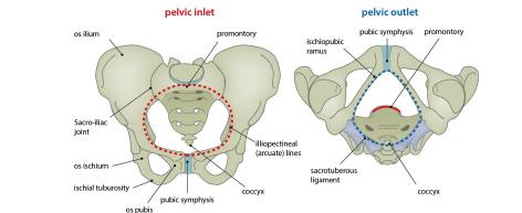
Female Adaptations for Childbearing
A wider more circular pelvic inlet
Sacrum is shorter and wider with flater curvature
Coccyx tilts posteriorly
Lager pubic bone
The inferior angle between the opposite pubic bones is farther apart
the illium projects farther laterally
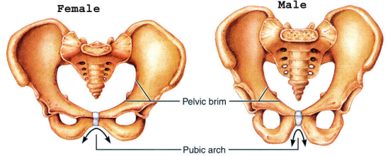
Synarthroses Joint
Immovable (sutures of skull)
Amphiarthroses Joint
Slightly moveable (pubic symhysis, vertebral)
Diarthroses Joint
Freely moveable (synovial joints - articular cartilage on the ends of the bones: hyaline cartilage)
Joint Capsule: outer dense CT, inner synovial membranesurrounding a joint and enclosing the synovial cavity, providing stability and secreting synovial fluid.
Synovial membrane: inner surface consists of synovial cells that secrete a viscous fluid.
Synovial fluid: combination of synovial cell secretion, capillary ultrafiltrate and WBCs
In some joints, the synovial lined sac is called a bursa. Inflammation of the bursa: bursitis
Synarthrosis: No movement
What type of joint are the skull sutures?
Pubic symphysis, intervertebral joints
Give an example of an amphiarthrosis Joint
The synovial membrane consists of cells that secrete synovial fluid that lubricate the joint
In a diarthrosis joint or synovial joint, what lines the joint capsule and what is its functions?
Skeletal Muscle Characteristics
Cell Features: Long cells, multinucleated
Locations: Outer Body muscles
Myosin, actin, Tropomyosin: Yes has it
Troponin: Has it
Special Features:
Red Fibers: lots of myosin, slow sustained cintractions
White Fibers: less myosin, rapid contraction, easily fatigue
Mitotic Capacity: adult cells cannot divide
Hypertrophy when stressed: Yes
Striated: Yes
Cardiac Muscle
Cell Features: Short branching cells, central nucleus
Locations: Heart
Myosin, actin, Tropomyosin: has it
Troponin: Has it
Special Features:
Intercalated discs which are intracellular attachments thatfacilite rapid synchronized contraction
Mitotic Capacity: adult cells cannot divide
Hypertrophy when stressed: Yes
Striated: Yes
Smooth Muscle
Cell Features: Wispy Cells with long nuclei
Locations: Walls of blood vessels, hollow organs, arrector pili
Myosin, actin, Tropomyosin: Yes has it
Troponin: DOES NOT HAVE IT SO THE MECHANISM OF CONTRACTION IS DIFFERENT FROM SKELETAL AND CARDIAC MUSCLE
Special Features:
Dense Bodies: Cytoskeletal proteins that attach to the thick and thin filaments and the cell membranes
Mitotic Capacity: HIgh Regenerative Capacity
Hypertrophy when stressed: Yes
Striated: No
Flex elbow
Location of Muscle: Anterior Arm
Origin: Humerus
Insertion: ulna, radius
Its action is?
Extend Wrist
Location of Muscle: posterior forearm
Origin humerus
Insertion: metacarpals
Its action is?
Adduct Shoulder
Location of Muscle: Back
Origin vertebrae
Insertion: scapula
Its action is?
Flex Vertebral Column
Location of Muscle: Anterior Vertebral Column
Origin Anterior Surface of Vertebrae
Insertion: Vertebrae and the base of skull
Its action is?
Adduct Hip
Location of Muscle: Medial Thigh
Origin pubis
Insertion: femur
Its action is?
Plantarflex Ankle
Location of Muscle: Posterior leg
Origin Femur, tibia, fibula
Insertion: Tarsals, metatarsals
Its action is?
Myofilaments
Thick and thin filaments
Skeletal and cardiac muscle cells are filled with many overlapping _____________
Smooth Muscle: ___________ criss-cross rather than overlap

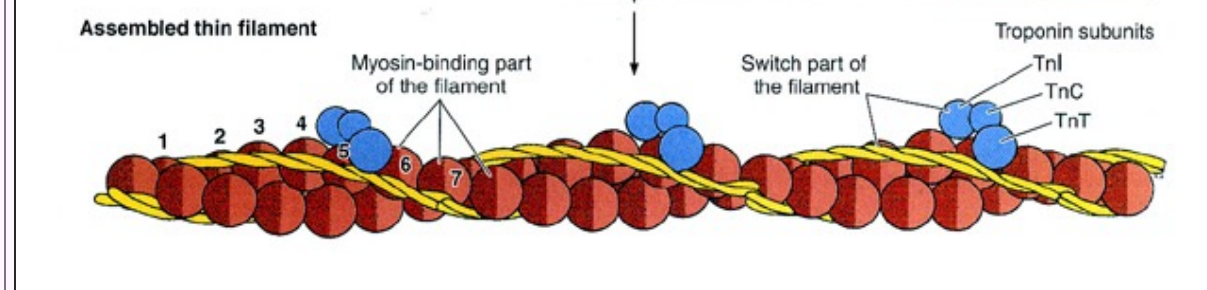
thick filaments and the 2 binding sites
————- are composed of myosin molecules that have globular heads which bind for ATP and a binding site for Actin of the thin filament. The 2 sites can’t be occupied by actin and ATP at the same time

What are the 3 components of thin filaments?
F- actin
Tropomyosin
Troponin: 3 subunits → TnC binds to calcium, TnT bind to tropomyosin, TnL inhibits actin-myosin interaction
What is a sarcrimere?
The smallest unit of muscle contraction: the distance between the two Z lines
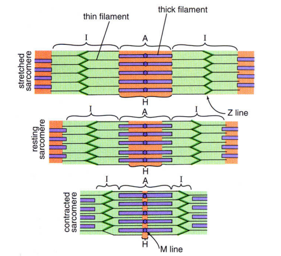
A band
dark band, zone occupied by the thick filament and overlapping part of thin filament
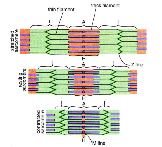
I Band
·light band, zone occupied by only thin filaments
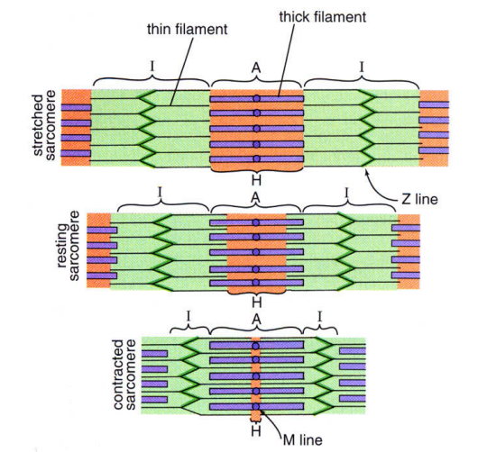
Z line
In the center of the I band, point of attachment between adjacent thin filaments

At rest/contract what do the thick and thin filament do?
They partially overlap. When a muscle contracts, the thin filament slides across the thick filament towards the middle of the thick filament

sarcoplasmic reticulum
It stores and releases Calcium

T-Tubule
It is an extension of the muscle cell plasma membrane (sarcolemma) that dips down into the muscle cell in between the sarcoplasmic reticulum. A triad is 1 T-tubule and adjacent sarcoplasmic reticulum
Accessory Linker Proteins
Stabilize and maintain alignment of actin and myosin and link myofibrils to each other and the external lamina
If there is a defect in accessory linker proteins, the thick and thin filaments are unstable and the muscle fiber can tear when contracted (cause of the Muscular dystrophies)
Neuromuscular Junction
1 nerve fiber (axon) may innervate 1 muscle fiber or branch to many muscle fibers
• 1 nerve fiber and all muscle fibers it innervates is called a motor unit.
Motor neuron axons terminate as motor endplates and the plasma membrane is the presynaptic membrane

Muscle Contraction
• Relaxed muscle: myosin - actin interaction is blocked.
• Neural stimulation causes depolarization of the muscle plasma membrane.
• The depolarization spreads to the T-tubules and the adjacent sarcoplasmic reticulum.
• Stored calcium is released from the sarcoplasmic reticulum
Motor neurons release the neurotransmitter, acetylcholine into the synaptic gap/cleft.
• The muscle fiber plasma membrane (postsynaptic membrane) is thrown into junctional folds and has acetylcholine receptors.
Calcium floods around the myofibrils and binds to TnC of troponin.
• Troponin shifts, tropomyosin shifts and this exposes binding site on actin.
• ATP bound to the myosin head hydrolyzes into ADP + phosphate
• Myosin binds to actin forming a “crossbridge”.
• Myosin pulls actin towards the center of the thick filament

Muscle Relaxation
New ATP binds to myosin causing release of myosin from actin and the cross-bridge is broken.
• Rigor mortis (stiff death) sustained contraction after death due to lack of ATP. Without new ATP, the myosin head remains linked to actin.
Proprioception
It is our skeletal muscle position sense. It is a general sensory function so muscles must have special sensory receptors.
What receptors convey proprioception in skeletal muscle?
Special stretch receptors: the muscle spindle in muscle and the Golgi tendon organ in the tendon attached to the muscle.
Stratum Basale
In space 1
highest proliferation of the epithelial cells
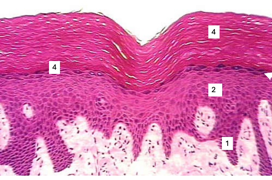
Stratum spinosum
in space 2
keratin production
the desmosome attachments between cells gives them a spiny appearance

Stratum Granulosum
in space 3
cells contain keratohyalin granuales and keratinosomes
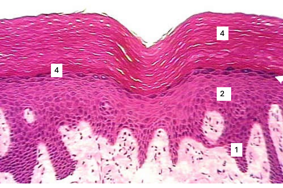
Stratum Lucidum
bottom 4
cells are dead
have lost there nucleus and organelles
