Lecture 9: The Appendicular Skeleton
1/43
Earn XP
Description and Tags
Vocabulary flashcards covering key bones, joints, and developmental concepts of the appendicular skeleton as presented in Lecture 9.
Name | Mastery | Learn | Test | Matching | Spaced | Call with Kai |
|---|
No analytics yet
Send a link to your students to track their progress
44 Terms
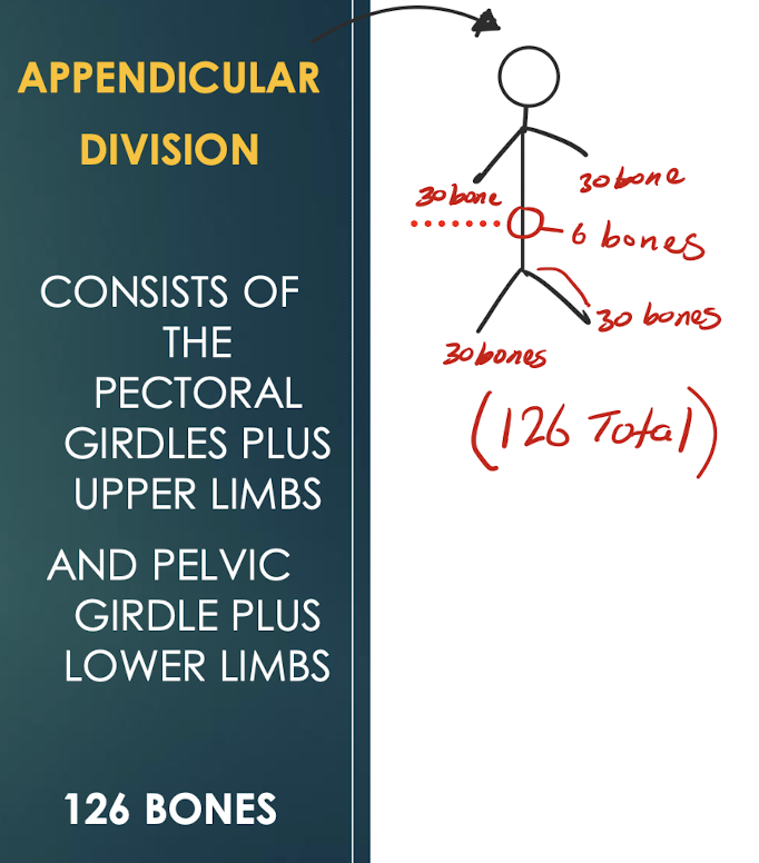
Appendicular Skeleton
Portion of the skeleton consisting of 126 bones of the limbs and their supporting girdles that connect to the trunk.
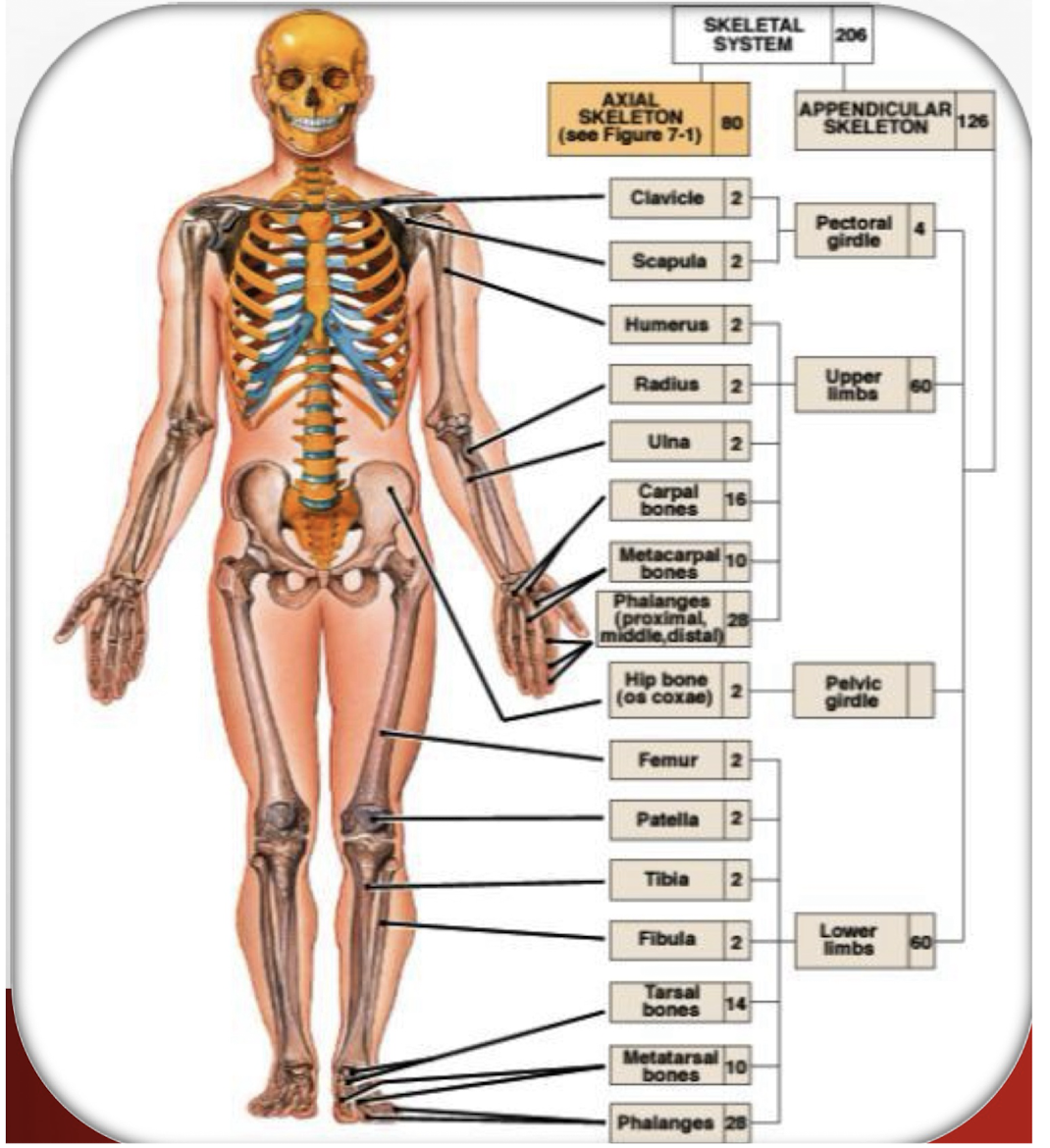

Pectoral Girdle
Set of bones (clavicle and scapula) that attach the upper limb to the axial skeleton; provides a flexible shoulder joint.
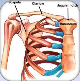
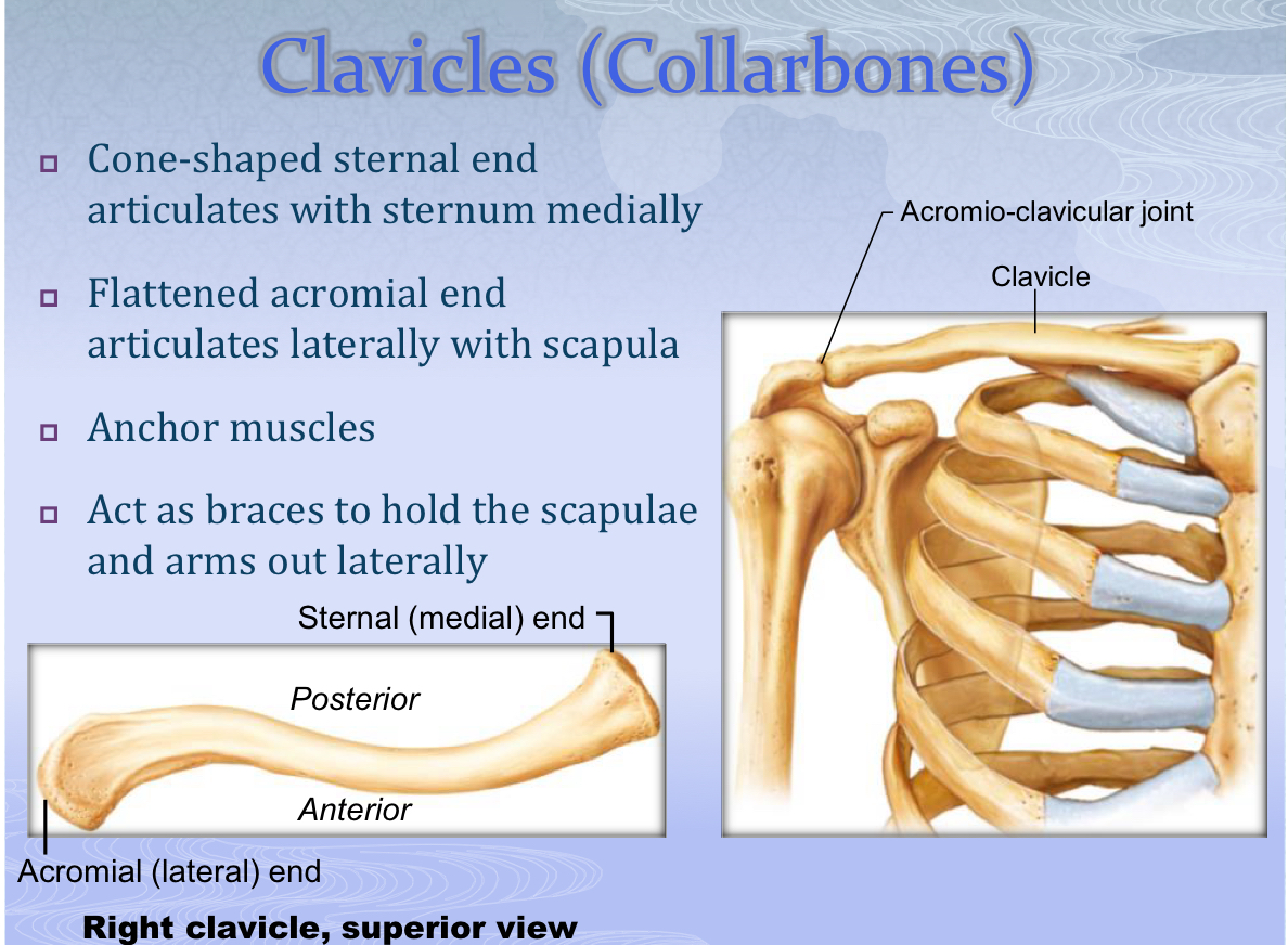
Clavicle (Collarbone)
S-shaped bone whose sternal end articulates with the manubrium and whose acromial end articulates with the scapula; braces the shoulder.
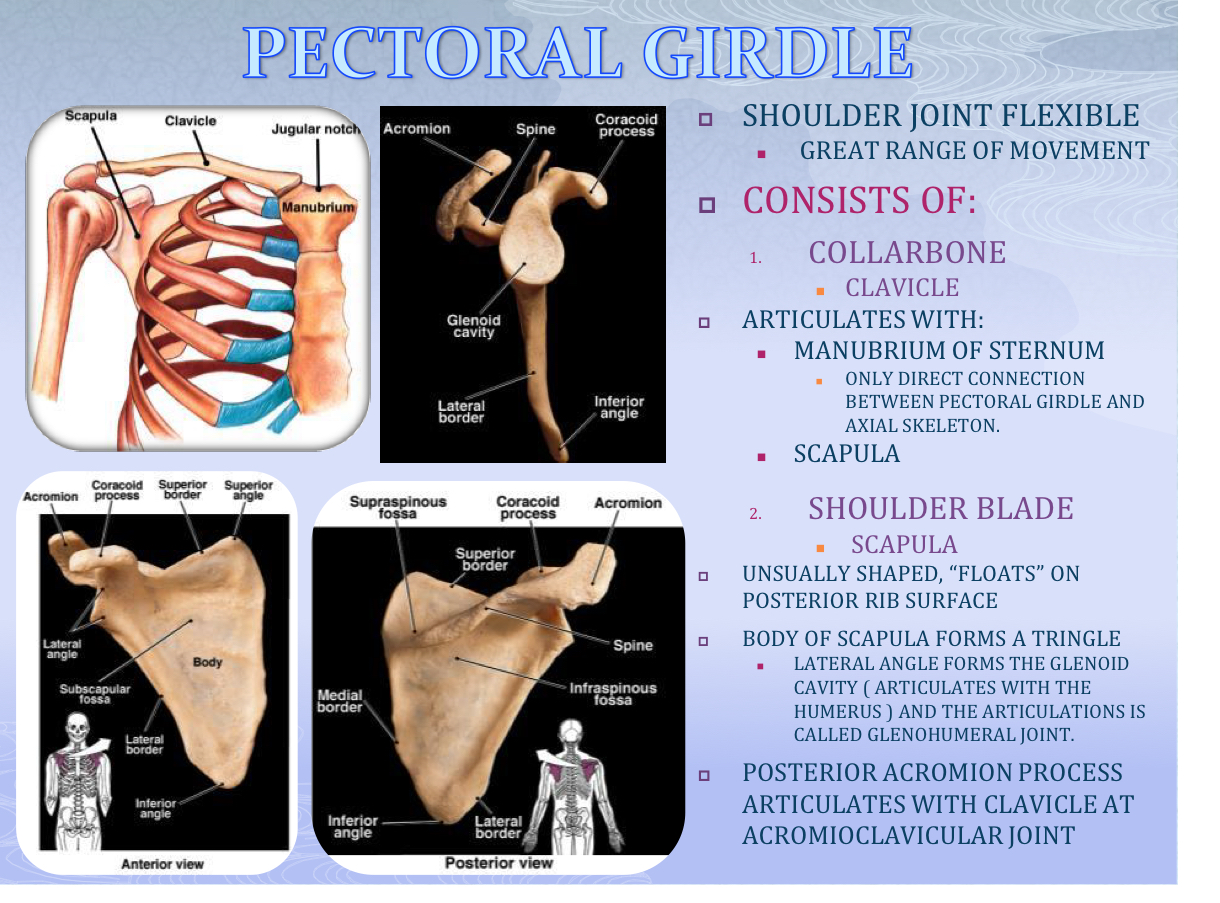
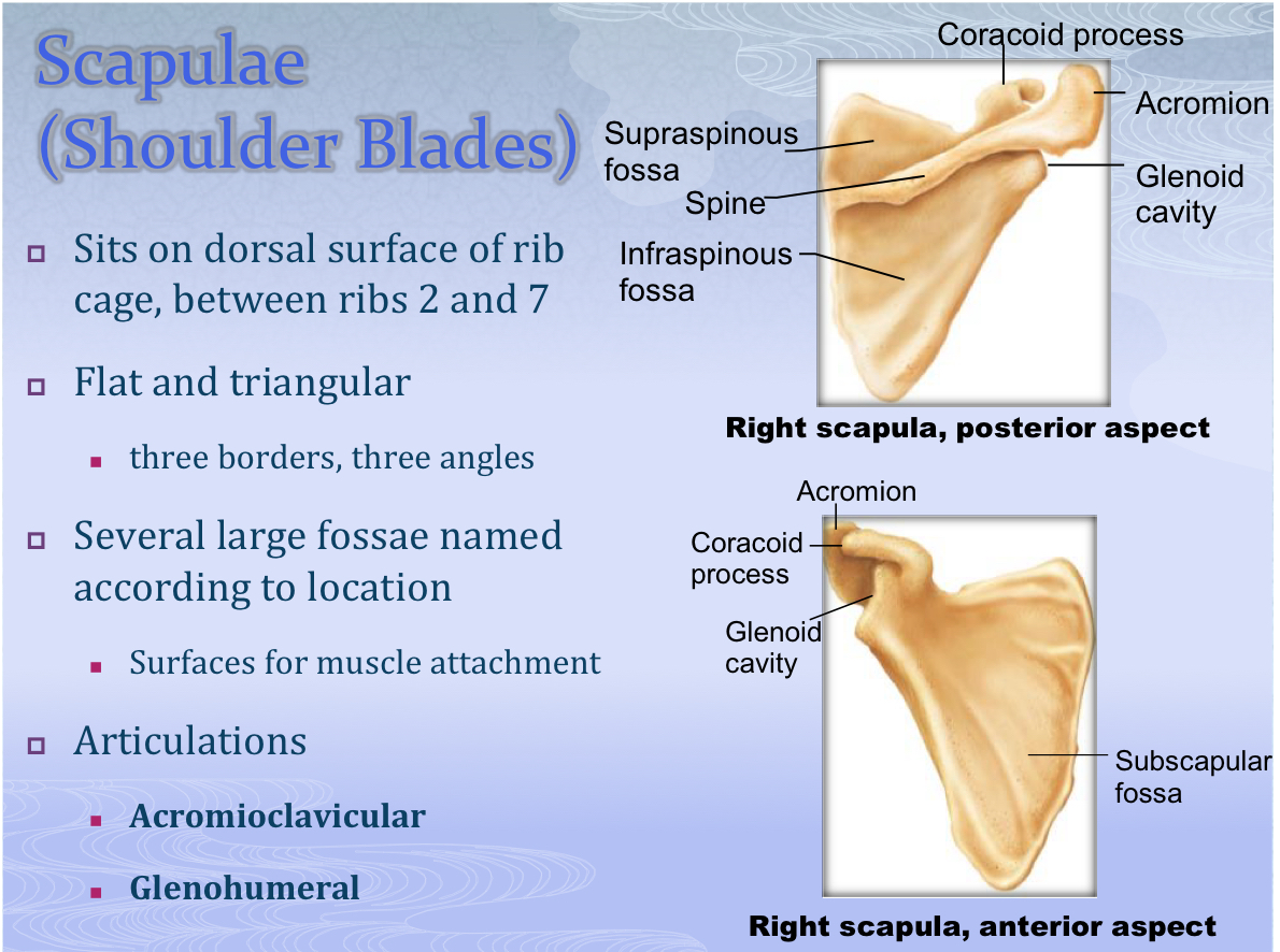
Scapula (Shoulder Blade)
Flat triangular bone that ‘floats’ on the posterior rib surface; features the acromion and glenoid cavity.

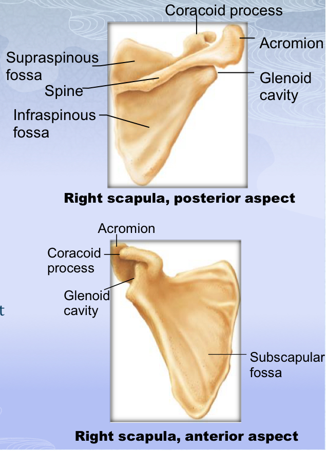
Glenoid Cavity
Lateral scapular depression that receives the head of the humerus to form the glenohumeral joint.

Glenohumeral Joint
Shoulder articulation between the glenoid cavity of the scapula and the head of the humerus.
Acromioclavicular Joint
Articulation between the acromion of the scapula and the acromial end of the clavicle.

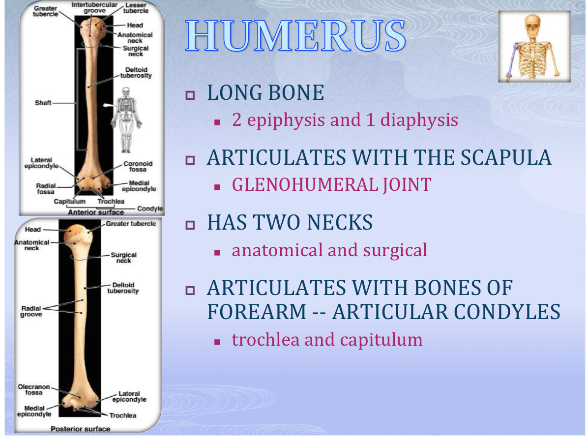
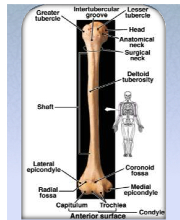
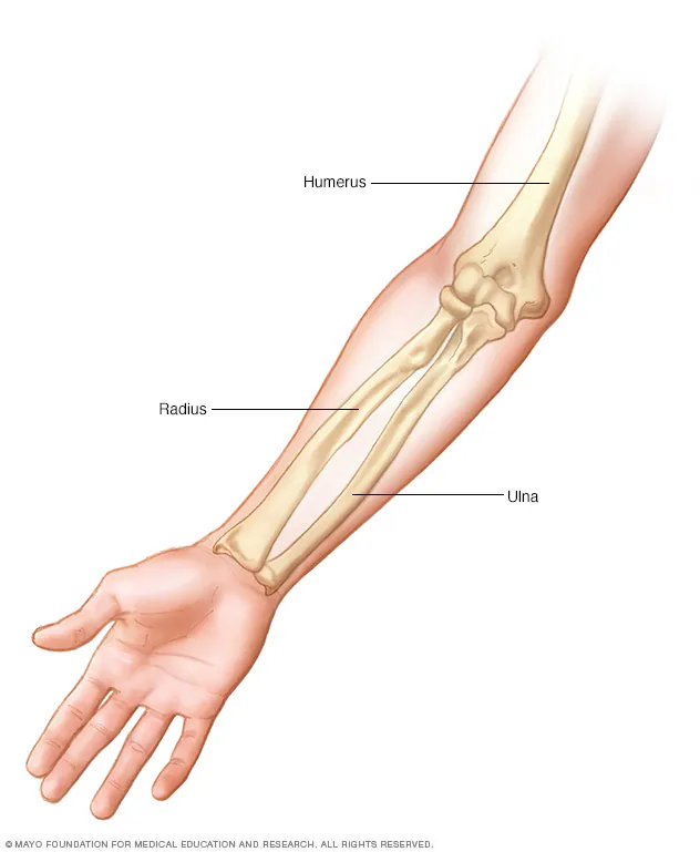
Humerus
Single bone of the brachium; long bone with anatomical and surgical necks; distally presents trochlea and capitulum.


Trochlea
Medial condyle of the distal humerus that articulates with the ulna.

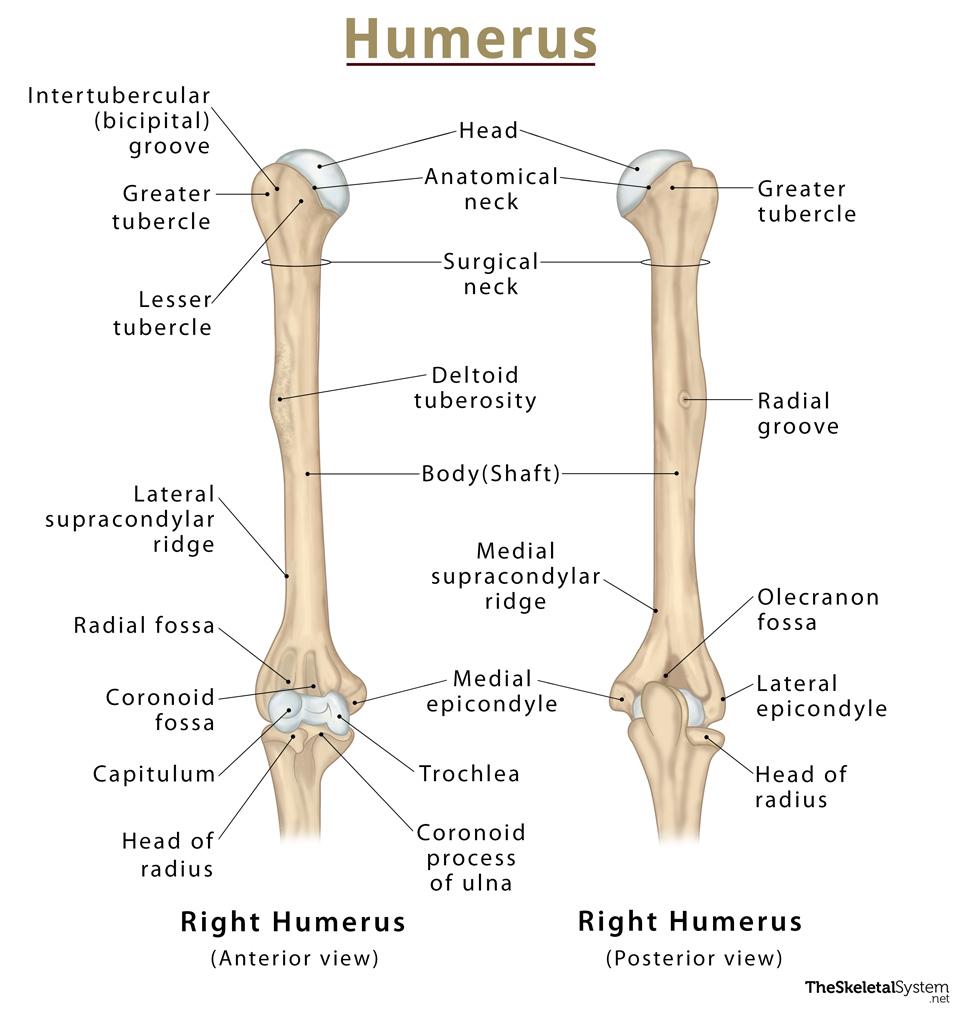
Capitulum
Lateral condyle of the distal humerus that articulates with the head of the radius.

Ulna
Medial bone of the forearm featuring the olecranon and coronoid processes; forms distal radioulnar joint with the radius.

Olecranon Process
Proximal projection of the ulna that forms the elbow’s point and articulates with the humerus.

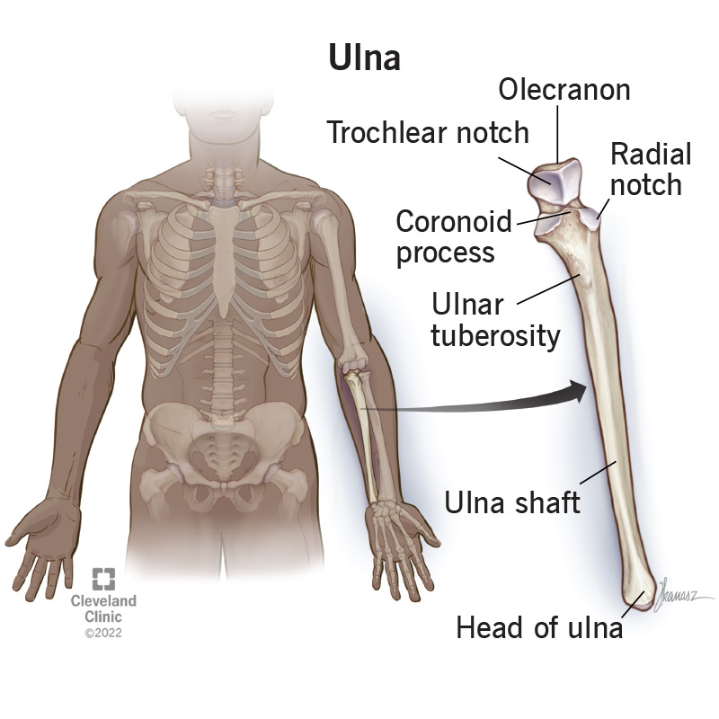
Coronoid Process
Anterior projection of the ulna just below the olecranon; locks into the trochlea during flexion.
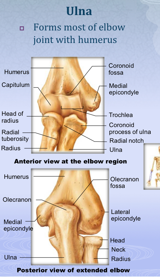
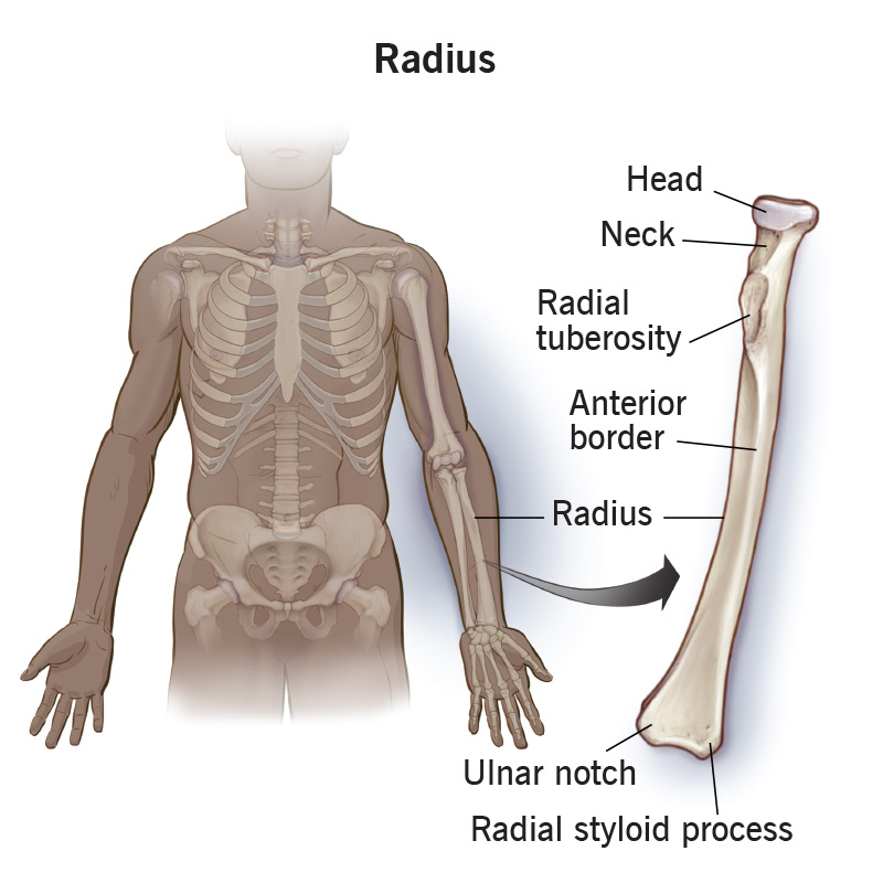
Radius
Lateral forearm bone; its head articulates with the capitulum; widened distally to form most of the wrist joint.

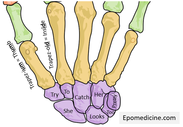
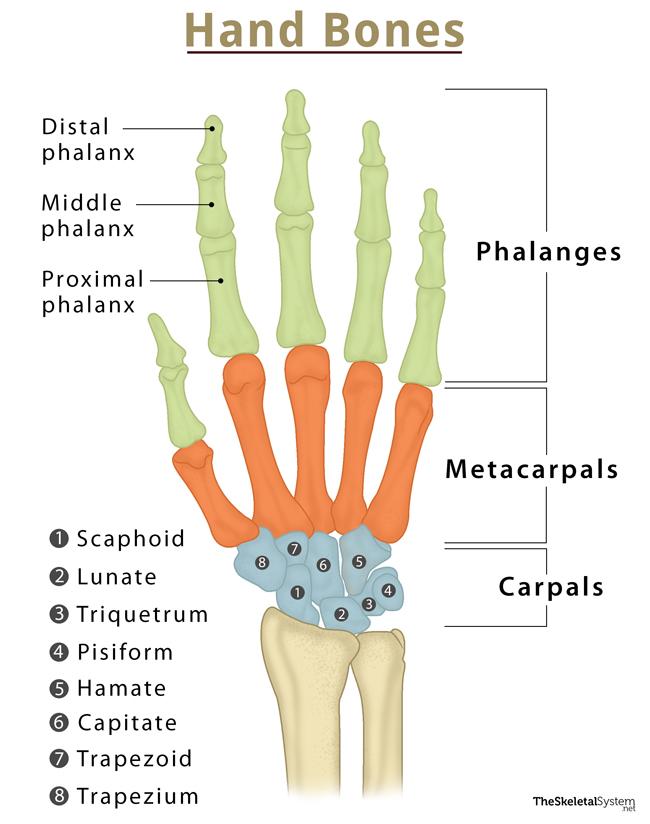
Carpal Bones
Eight wrist bones arranged in two rows: scaphoid, lunate, triquetrum, pisiform (proximal) and trapezium, trapezoid, capitate, hamate (distal).

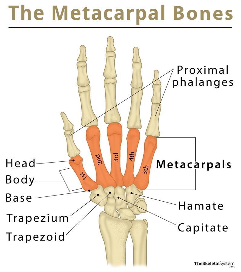
Metacarpals
Five long bones of the palm that articulate proximally with carpals and distally with proximal phalanges.

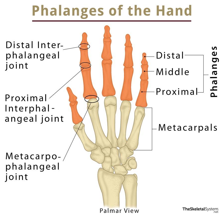
Phalanges (Hand)
Four fingers contain three phalanges each (proximal, middle, distal); the pollex has two (proximal, distal).
Pelvic (Hip) Girdle
Paired coxal bones that attach lower limbs, transmit body weight, and protect pelvic organs; less mobile but more stable than pectoral girdle.
Coxal (Hip) Bone
Bone formed by fusion of ilium, ischium, and pubis.
Ilium
Superior region of the coxal bone; auricular surface articulates with the sacrum at the sacroiliac joint.
Ischium
Posteroinferior part of the hip bone.
Pubis
Anterior part of the hip bone; two pubic bones meet at the pubic symphysis.
Acetabulum
Cup-like lateral socket of the hip bone that articulates with the femoral head forming the coxal joint.
Sacroiliac Joint
Articulation between the auricular surface of the ilium and the sacrum.
Pubic Symphysis
Fibrocartilaginous joint uniting the two pubic bones anteriorly.
Obturator Foramen
Large opening between ischium and pubis for passage of blood vessels and nerves.
Femur
Largest, strongest bone forming the thigh; articulates proximally with acetabulum and distally with tibia and patella.
Patella
Largest sesamoid bone embedded in the quadriceps tendon; posterior surface articulates with femoral condyles.
Tibia
Large medial, weight-bearing bone of the lower leg; has two condyles that meet the femur and inferior surface that forms part of the ankle.
Fibula
Thin, non-weight-bearing lateral bone of the leg; important for muscle attachment; articulates with tibia but not femur.
Interosseous Membrane
Fibrous sheet that connects the shafts of tibia and fibula (and radius and ulna).
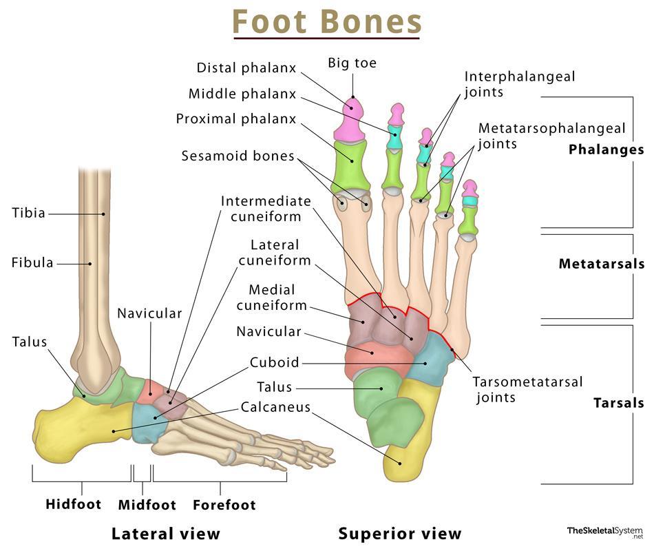
Tarsal Bones
Seven ankle bones: talus, calcaneus, navicular, cuboid, and three cuneiforms (medial, intermediate, lateral).
Talus
Superior tarsal bone that receives body weight and articulates with the tibia.
Calcaneus
Heel bone; largest tarsal bearing body weight with the talus.
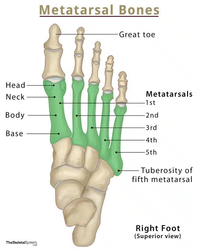
Metatarsals
Five long bones (I–V) forming the distal foot; head of metatarsal I creates the ball of the foot.
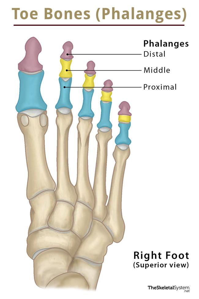
Phalanges (Foot)
Toes contain 14 bones; hallux has two (proximal, distal), other toes have three (proximal, middle, distal).

Hallux
Great toe; contains only proximal and distal phalanges.
Arches of the Foot
Lateral longitudinal, medial longitudinal, and transverse arches that distribute body weight and provide springiness.
Fontanelles
Unossified fibrous membranes between fetal skull bones (anterior, posterior, mastoid, sphenoidal) allowing birth and brain growth.
Cleft Palate
Congenital absence of medial fusion of the right and left halves of the palate.
Primary Curvatures
Thoracic and sacral spinal curves present at birth, giving the infant spine a posteriorly convex C-shape.
Secondary Curvatures
Cervical and lumbar spinal curves that develop after birth (as child lifts head and walks), convex anteriorly.
Sesamoid Bone
Bone formed within a tendon; the patella is the largest example.
Intervertebral Disc Degeneration
Age-related thinning and dehydration of discs, increasing risk of herniation and height loss.