AP Psych Terms I Don't Know (Units 1-3)
1/139
There's no tags or description
Looks like no tags are added yet.
Name | Mastery | Learn | Test | Matching | Spaced | Call with Kai |
|---|
No analytics yet
Send a link to your students to track their progress
140 Terms
evolutionary psychology
(1.1.1) the study of the evolution of behavior and the mind, using principles of natural selection
behavior genetics
(1.1.1) the study of the relative power and limits of genetic and environmental influences on behavior
genome
(1.1.2) the complete instructions for making an organism
central nervous system (CNS)
(1.2.1) the brain and spinal cord
peripheral nervous system (PNS)
(1.2.1) the sensory and motor neurons that connect the central nervous system (CNS) to the rest of the body
sensory neurons
(1.2.1) neurons that carry incoming information from the body’s tissues and sensory receptors to the brain and spinal cord (afferent → carry inward)
motor neurons
(1.2.1) neurons that carry outgoing information from the brain and spinal cord to the muscles and glands (efferent → carry outward)
interneurons
(1.2.1) neurons within the brain and spinal cord; they communicate internally and process information between the sensory inputs and motor outputs
somatic nervous system
(1.2.1) the division of the peripheral nervous system that controls the body’s skeletal muscles. Also called the skeletal nervous system (voluntary movements)
autonomic nervous system (ANS)
(1.2.1) the part of the peripheral nervous system that controls the glands and the muscles of the internal organs (such as the heart). Its sympathetic division arouses; its parasympathetic division calms (autonomic → self-regulating)
spinal cord
(1.2.1) two-way information highway connecting the peripheral nervous system and the brain
dendrite
(1.3.1) a neuron’s often bushy, branching extensions that receive and integrate messages, conducting impulses toward the cell body
axon
(1.3.1) the segmented neuron extension that passes messages through its branches to other neurons or to muscles or glands.
neuron diagram (picture)
(1.3.1)
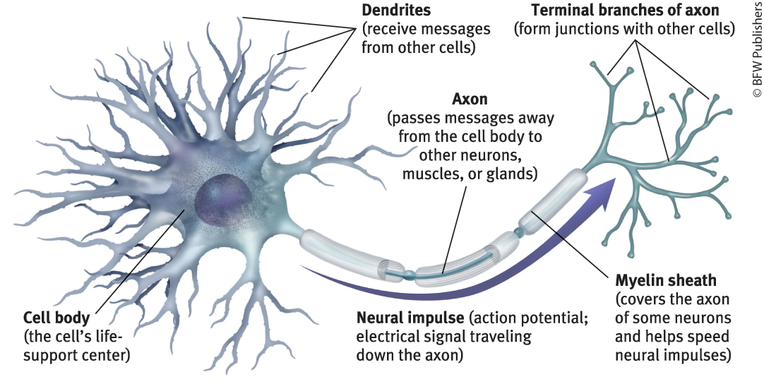
myelin sheath
(1.3.1) a fatty tissue layer segmentally encasing the axons of some neurons; it enables vastly greater transmission speed as neural impulses hop from one node to the next
glial cells
(1.3.1) cells in the nervous system that support, nourish, and protect neurons; they may also play a role in learning, thinking, and memory
acetylcholine (ACh)
(1.3.3) enables muscle action, learning, and memory
dopamine
(1.3.3) influences movement, learning, attention, and emotion
serotonin
(1.3.3) affects mood, hunger, sleep, and arousal
norepinephrine (noradrenaline)
(1.3.3) helps control alertness and arousal
GABA (gamma-aminobutyric acid)
(1.3.3) major inhibitory neurotransmitter
glutamate
(1.3.3) major excitatory neurotransmitter; involved in memory
endorphins
(1.3.3) neurotransmitters that influence the perception of pain or pleasure
substance P
(1.3.3) involved in pain perception and immune response
agonist
(1.3.3) a molecule that increases a neurotransmitter’s action
antagonist
(1.3.3) a molecule that inhibits or blocks a neurotransmitter’s action
endocrine system
(1.3.4) the body’s “slow” chemical communication system; a set of glands and fat tissue that secrete hormones into the bloodstream
epinephrine (adrenaline)
(1.3.4) works with norepinephrine to increase heart rate, blood pressure, and blood sugar, providing a surge of energy to power our fight-or-flight response
pituitary gland
(1.3.4) pea-sized body attached to the base of the brain, the pituitary is important in controlling growth and development and the functioning of the other endocrine glands
oxytocin
(1.3.4) enables orgasm and, in women, labor contractions and milk flow while nursing
feedback system of nervous and endocrine system
brain → pituitary → other glands → hormones → body and brain
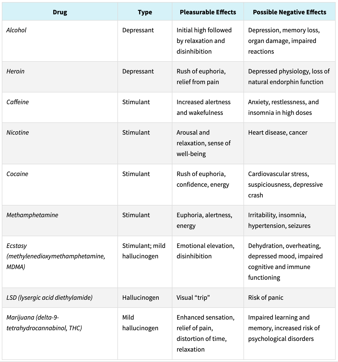
psychoactive drugs
(1.3.5) a chemical substance that alters the brain, causing changes in perceptions and moods
depressants
(1.3.6) drugs that reduce neural activity and slow body functions
barbiturate
(1.3.6) drugs that depress central nervous system activity, reducing anxiety but impairing memory and judgment
opioids
(1.3.6) opium and its derivatives; they depress neural activity, temporarily lessening pain and anxiety
stimulants
(1.3.7) drugs that excite neural activity and speed up body functions
hallucinogens
(1.3.8) psychedelic (“mind-manifesting”) drugs that distort perceptions and evoke sensory images in the absence of sensory input
chart of psychoactive drugs (picture)
(1.3.8)
phrenology
(1.4.1) studying bumps on the skull, believed to reveal a person’s mental abilities and character traits
localization of function
(1.4.1) the idea that various brain regions have particular functions
neuroplacisitiy
(1.4.2) the brain’s ability to change, especially during childhood, by reorganizing after damage or by building new pathways based on experience
lesion
(1.4.3) [LEE-zhuhn] tissue destruction. Brain lesions may occur naturally (from disease or trauma), during surgery, or experimentally (using electrodes to destroy brain cells)
EEG (electroencephalogram)
(1.4.3) an amplified recording of the waves of electrical activity sweeping across the brain’s surface. These waves are measured by electrodes placed on the scalp
MEG (magnetoencephalography)
(1.4.3) a brain-imaging technique that measures magnetic fields from the brain’s natural electrical activity.
CT (computed tomography) scan
(1.4.3) a series of X-ray photographs taken from different angles and combined by computer into a composite representation of a slice of the brain’s structure
PET (positron emission tomography)
(1.4.3) technique for detecting brain activity that displays where a radioactive form of glucose goes while the brain performs a given task
MRI (magnetic resonance imaging)
(1.4.3) a technique that uses magnetic fields and radio waves to produce computer-generated images of soft tissue. MRI scans show brain anatomy
ventricles
(1.4.3) fluid-filled brain areas in some people with schizophrenia
fMRI (functional MRI)
(1.4.3) a technique for revealing blood flow and, therefore, brain activity by comparing successive MRI scans. fMRI scans show brain function as well as structure
hindbrain
(1.4.4) consists of the medulla, pons, and cerebellum; directs essential survival functions, such as breathing, sleeping, and wakefulness, as well as coordination and balance
midbrain
(1.4.4) found atop the brainstem; connects the hindbrain with the forebrain, controls some motor movement, and transmits auditory and visual information
forebrain
(1.4.4) consists of the cerebral cortex, thalamus, and hypothalamus; manages complex cognitive activities, sensory and associative functions, and voluntary motor activities
brainstem
(1.4.5) the central core of the brain, beginning where the spinal cord swells as it enters the skull; the brainstem is responsible for automatic survival functions
medulla
(1.4.5) the hindbrain structure that is the brainstem’s base; controls heartbeat and breathing
thalamus
(1.4.5) [THAL-uh-muss] the forebrain’s sensory control center, located on top of the brainstem; it directs messages to the sensory receiving areas in the cortex and transmits replies to the cerebellum and medulla
reticular formation
(1.4.5) a nerve network that travels through the brainstem into the thalamus; it filters information and plays an important role in controlling arousal
cerebellum
(1.4.5) [sehr-uh-BELL-um] the hindbrain’s “little brain” at the rear of the brainstem; its functions include processing sensory input, coordinating movement output and balance, and enabling nonverbal learning and memory
limbic system
(1.4.6) neural system located mostly in the forebrain — below the cerebral hemispheres — that includes the amygdala, hypothalamus, hippocampus, thalamus, and pituitary gland; associated with emotions and drives
amygdala
(1.4.6) [uh-MIG-duh-la] two lima-bean-sized neural clusters in the limbic system; linked to emotions such as anger and fear
hypothalamus
(1.4.6) [hi-po-THAL-uh-muss] a limbic system neural structure lying below (hypo) the thalamus; it directs several maintenance activities (eating, drinking, body temperature), helps govern the endocrine system, and is linked to emotion and reward
cerebral cortex
(1.4.7) the intricate fabric of interconnected neural cells covering the forebrain’s cerebral hemispheres; the body’s ultimate control and information-processing center
frontal lobes
(1.4.7) the portion of the cerebral cortex lying just behind the forehead. They enable linguistic processing, muscle movements, higher-order thinking, and executive functioning (such as making plans and judgments), also regulates emotions, personality, and behavior
pariental lobes
(1.4.7) the portion of the cerebral cortex lying at the top of the head and toward the rear; it receives sensory input for touch and body position, plays a role in attention, memory, and language comprehension
occipital lobes
(1.4.7) the portion of the cerebral cortex lying at the back of the head; it includes areas that receive information from the visual fields
temporal lobes
(1.4.7) the portion of the cerebral cortex lying roughly above the ears; it includes the auditory areas, each of which receives information primarily from the opposite ear. They also enable language processing
motor cortex
(1.4.7) a cerebral cortex area at the rear of the frontal lobes that controls voluntary movements
somatosensory cortex
(1.4.7) a cerebral cortex area at the front of the parietal lobes that registers and processes body touch and movement sensations
association areas
(1.4.7) areas of the cerebral cortex that are not involved in primary motor or sensory functions; rather, they are involved in higher mental functions such as learning, remembering, thinking, and speaking
corpus callosum
(1.4.9) [KOR-pus kah-LOW-sum] the large band of neural fibers connecting the two brain hemispheres and carrying messages between them
cognitive neuroscience
(1.5.1) the interdisciplinary study of the brain activity linked with cognition
dual processing
(1.5.2) the principle that information is often simultaneously processed on separate conscious and unconscious tracks
blindsight
(1.5.2) a condition in which a person can respond to a visual stimulus without consciously experiencing it
parallel processing
(1.5.2) processing multiple aspects of a stimulus or problem simultaneously
REM sleep
(1.5.5) rapid eye movement sleep; a recurring sleep stage during which vivid dreams commonly occur. Also known as paradoxical sleep, because the muscles are relaxed (except for minor twitches) but other body systems are active. (Sometimes called R sleep)
alpha waves
(1.5.5) the relatively slow brain waves of a relaxed, awake state
stage 1 sleep
(1.5.5) may experience hallucinations, falling, floating, etc.
hypnagogic sensations
(1.5.5) bizarre experiences, such as jerking or a feeling of falling or floating weightlessly, while transitioning to sleep. (Also called hypnic sensations)
delta waves
(1.5.5) the large, slow brain waves associated with deep sleep (stage 3)
sleep spindles
(1.5.5) bursts of rapid, rhythmic brain-wave activity that aid memory processing, occurs during stage 2 sleep
suprachiasmatic nucleus (SCN)
(1.5.6) a pair of cell clusters in the hypothalamus that controls circadian rhythm. In response to light, the SCN adjusts melatonin production, thus modifying our feelings of sleepiness
melatonin
(1.5.6) sleep-inducing hormone found in the hypothalamus
ghrelin
(1.5.8) hunger-arousing hormone, decreases its hunger suppressing partner leptin
cortisol
(1.5.8) stress hormone that stimulates the body to make fat and decrease metabolic rates
REM sleep behavior disorder
(1.5.9) a sleep disorder in which normal REM paralysis does not occur; instead, twitching, talking, or even kicking or punching may occur, often acting out one’s dream
narcolepsy
(1.5.9) a sleep disorder characterized by uncontrollable sleep attacks. The affected person may lapse directly into REM sleep, often at inopportune times
sleep apnea
(1.5.9) a sleep disorder characterized by temporary cessations of breathing during sleep and repeated momentary awakenings
physiological function dream theory
(1.5.9) regular brain stimulation from REM sleep may help develop and preserve neural pathways
activation synthesis dream theory
(1.5.9) REM sleep triggers neural activity that evokes random visual memories, which our sleeping brain weaves into stories
cognitive development dream theory
(1.5.9) dream content reflects dreamers’ level of cognitive development — their knowledge and understanding. Dreams simulate our lives, including worst-case scenarios
prosopagnosia
(1.6.0) face blindness
bottom-up processing
(1.6.0) information processing that begins with the sensory receptors and works up to the brain’s integration of sensory information
top-down processing
(1.6.0) information processing guided by higher-level mental processes, as when we construct perceptions drawing on our experience and expectations
psychophysics
(1.6.1) the study of relationships between the physical characteristics of stimuli, such as their intensity, and our psychological experience of them
absolute threshold
(1.6.2) the minimum stimulus energy needed to detect a particular stimulus 50 percent of the time
signal detection theory
(1.6.2) a theory predicting how and when we detect the presence of a faint stimulus (signal) amid background stimulation (noise). Assumes there is no single absolute threshold and that detection depends partly on a person’s experience, expectations, motivation, and alertness
subliminal
(1.6.2) below one’s absolute threshold for conscious awareness
difference threshold
(1.6.2) the minimum difference between two stimuli required for detection 50 percent of the time. We experience the difference threshold as a just noticeable difference (or jnd)
Weber’s law
(1.6.2) the principle that, to be perceived as different, two stimuli must differ by a constant minimum percentage (rather than a constant amount)
sensory adaptation
(1.6.3) diminished sensitivity as a consequence of constant stimulation
cornea
(1.6.4) the eye’s clear, protective outer layer, covering the pupil and iris