Week 6 lab
 Call Kai
 Learn
Learn Practice Test
 Spaced Repetition
 Match
Match Flashcards
 Knowt Play
1/29

There's no tags or description
Looks like no tags are added yet.
Name | Mastery | Learn | Test | Matching | Spaced | Call with Kai | Chat |
|---|
No analytics yet
Send a link to your students to track their progress
30 Terms
What is the primary function of the four heart valves?
To ensure blood flows in only one direction.
Heart valves prevent what?
Backflow of blood.
What are the valves between the atria and ventricles called?
Atrioventricular, or AV, valves.
What are the valves between the ventricles and great arteries called?
Semilunar valves.
When does the first heart sound, S1, occur?
When the AV valves close.
When does the second heart sound, S2, occur?
When the semilunar valves close.
Which heart sound is produced by ventricular contraction?
The first heart sound, S1.
Which heart sound is produced by ventricular relaxation?
The second heart sound, S2.
How is the second heart sound typically described compared with the first?
It is shorter and more crisp.
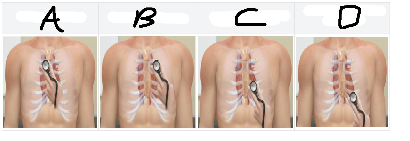
According to the diagram, what label marks the best location to hear the right AV, or tricuspid, valve?
C.
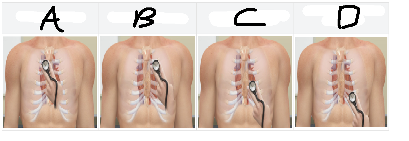
According to the diagram, what label marks the best location to hear the pulmonary valve?
B.
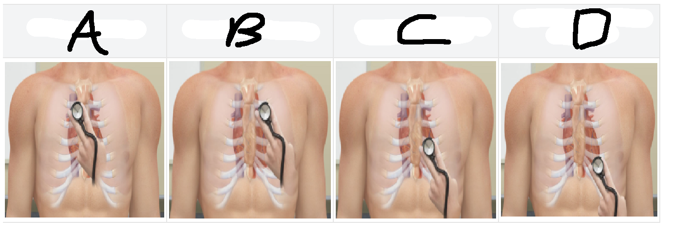
According to the diagram, what label marks the best location to hear the aortic valve?
A.
What does an ECG measure?
The electrical activity of the heart.
What does the P wave represent on an ECG?
Atrial depolarization.
What does the QRS complex represent on an ECG?
Ventricular depolarization.
What does the T wave represent on an ECG?
Ventricular repolarization.
What does the PR, or PQ, interval represent?
The time it takes for the impulse to travel from the atria to the AV node.
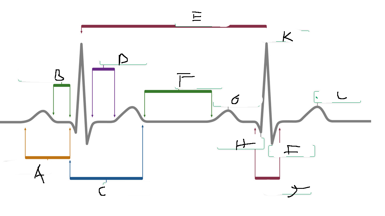
According to the ECG diagram, what label marks the PR interval?
A.
What is the time between two consecutive R waves called?
The R-R interval.
How can heart rate be calculated using the R-R interval?
Divide 60 seconds by the R-R interval.
Depolarization always occurs before what?
Repolarization.
What does an action potential consist of?
Depolarization followed by repolarization.
After repolarization, there may be a brief period of what?
Hyperpolarization.
The cardiac cycle includes both electrical and what events?
Mechanical events.
Where are action potentials initiated?
At the axon hillock.
What type of channels does the axon hillock contain?
Voltage-gated channels.
When do voltage-gated channels open?
When the membrane reaches threshold.
Proper heart function depends on coordinated electrical activity and what other type of activity?
Mechanical activity.
ECG waves correspond to changes in what?
Electrical activity of the heart.
What causes heart sounds?
The closing of valves.