Cranium
1/114
There's no tags or description
Looks like no tags are added yet.
Name | Mastery | Learn | Test | Matching | Spaced |
|---|
No study sessions yet.
115 Terms
a
Which of the following bones is part of the floor of the cranium?
a. Temporal
b. Occipital
c. Frontal
d. Parietal
a
The widest portion of the cranium is found at the level of the:
a. parietal tubercles.
b. right and left pterion.
c. squamous portion of the temporal bone.
d. external acoustic meatus (EAM).
c
What is the name of the joint found between the lateral condylar processes of the skull and the superior articular process of C1?
a. Zygapophyseal joint
b. Intervertebral joint
c. Atlanto- occipital joint
d. Cervico-occipital joint
c
Which cranial bone articulates with all the other cranial bones?
a. Parietal
b. Ethmoid
c. Sphenoid
d. None of the above
c
The slight depression above each eyebrow is termed the:
a. glabella.
b. supraorbital foramina.
c. supraorbital groove.
d. frontal tuberosity.
d
Which of the following cranial bones does not articulate with the parietal bone?
a. Frontal
b. Sphenoid
c. Occipital
d. All of the above articulate with the parietal bone
a
The left mastoid fontanel becomes the _____ in an adult.
a. left asterion
b. left pterion
c. left bregma
d. squamosal suture
c
There are a total of _____ fontanels in an infant.
a. four
b. two
c. six
d. eight
a
The frontal bone articulates with _____ cranial bones.
a. four
b. six
c. two
d. five
b
Which of the following landmarks corresponds with the level of the petrous ridge?
a. External acoustic meatus (EAM)
b. Top of ear attachment (TEA)
c. Squamosal suture
d. Inion
d
The pituitary gland (hypophysis cerebri) is associated with and protected by the _____ bone.
a. temporal
b. ethmoid
c. palatine
d. sphenoid
a
Which cranial bone contains the foramen ovale?
a. Sphenoid
b. Occipital
c. Ethmoid
d. Temporal
d
Which cranial bone contains the cribriform plate?
a. Sphenoid
b. Occipital
c. Temporal
d. Ethmoid
d
Which of the following sutures separates the parietal from the occipital bone?
a. Squamosal
b. Sagittal
c. Coronal
d. Lambdoidal
a
Which of the following terms describes the anterior fontanel found in the adult skull?
a. Bregma
b. Pterion
c. Asterion
d. Lambda
b
Which of the following terms describes the small irregular bones occasionally found in the sutures?
a. Asterion
b. Wormian
c. Sesamoid
d. Squamosal
d
The ethmoid notch is part of which cranial bone?
a. Temporal
b. Ethmoid
c. Sphenoid
d. Frontal
c
Which cranial bone is labeled vi inthis Figure?
(Anatomic structures are labeled i through vi and sutures a through h.)
a. Temporal
b. Occipital
c. Greater wing of Sphenoid
d. Parietal

b
Which bony structure is labeled v?
(Anatomic structures are labeled i through vi and sutures a through h.)
a. EAM
b. Mastoid process
c. Styloid process
d. Zygomatic process
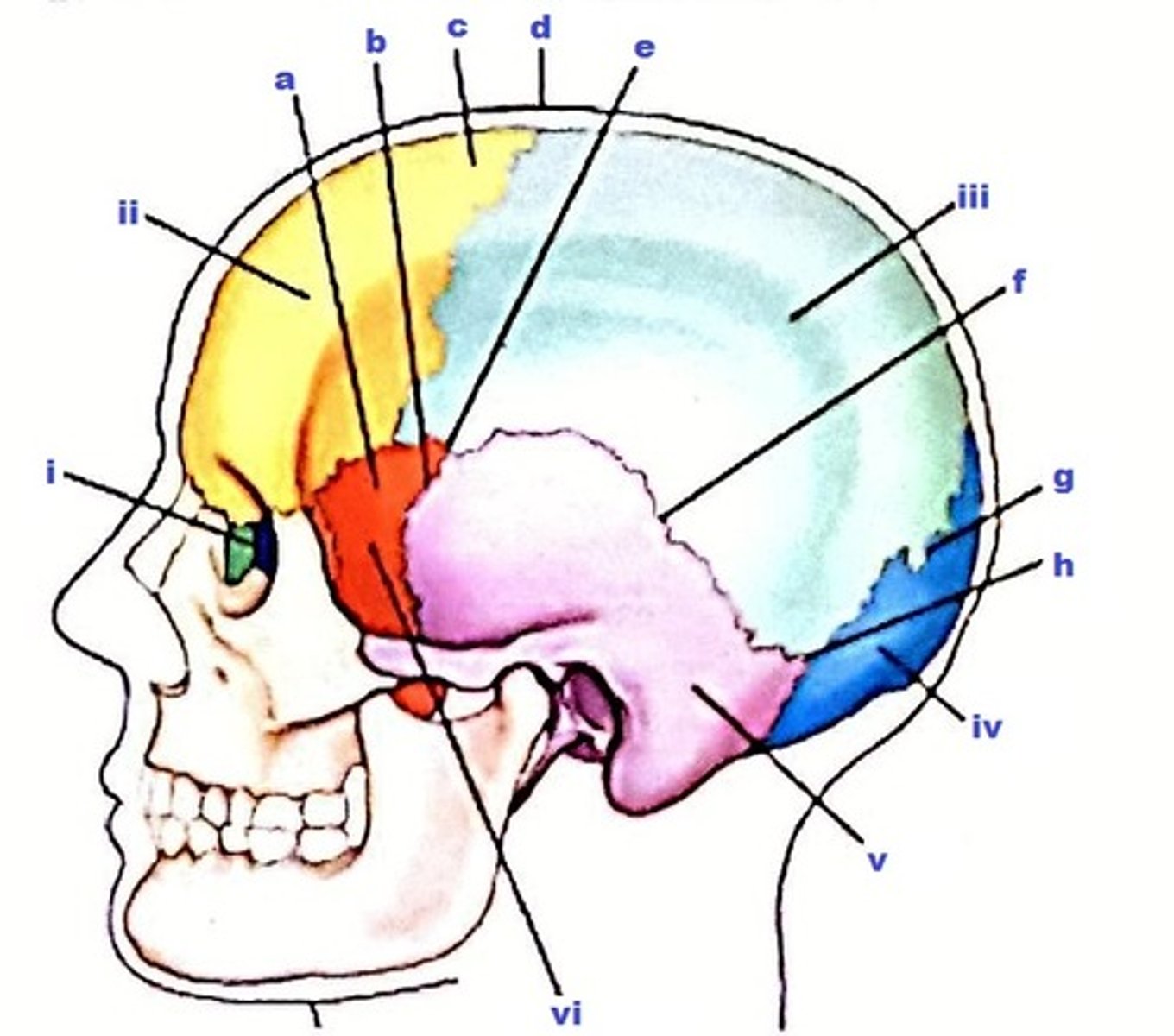
c
The cranial bone labeled i is the:
(Anatomic structures are labeled i through vi and sutures a through h.)
a. sphenoid.
b. lacrimal.
c. ethmoid.
d. palatine.

a
Which cranial suture is labeled c?
(Anatomic structures are labeled i through vi and sutures a through h.)
a. Coronal
b. Squamosal
c. Lambdoidal
d. Sagittal

d
The sutural point or area labeled e is the:
(Anatomic structures are labeled i through vi and sutures a through h.)
a. lambda.
b. asterion.
c. bregma.
d. pterion.

a
The sutural point or area labeled d is termed:
(Anatomic structures are labeled i through vi and sutures a through h.)
a. bregma.
b. lambda.
c. asterion.
d. pterion.

d
The suture labeled f is termed the:
(Anatomic structures are labeled i through vi and sutures a through h.)
a. asterion.
b. lambdoidal.
c. coronal.
d. squamosal.

c
Which of the fontanels is the last to close are about 18 months of age?
a. Sphenoid
b. Mastoid
c. Anterior
d. Posterior
a
Which term describes the small flap of cartilage covering the opening to the ear?
a. Tragus
b. Pinna
c. Acanthion
d. EAM
d
Which one of the following technical considerations is most critical for demonstrating air and/or fluid levels within the cranium?
a. Medium kV
b. Detail image receptor (IR)
c. Short exposure time
d. Horizontal x-ray beam
a
Which division of the temporal bone contains the organs of hearing and equilibrium?
a. Petrous
b. Mastoid
c. Squamous
d. Antrum
c
Which one of the following structures is part of the middle ear?
a. Tragus
b. Cochlea
c. Tympanic cavity
d. Eustachian tube
b
To which aspect of the ear does the eustachian tube attach?
a. External ear
b. Middle ear
c. Inner ear
d. Cochlea
d
The aditus is defined as:
a. a large chamber containing the mastoid air cells.
b. a thin plate of bone separating the mastoid air cells from the brain.
c. a passageway for the auditory nerve.
d. an opening between the equitymapanic recess and the mastoid air cells.
a
The incus auditory ossicle is labeled as:
a. iii
b. CHOICE BLANK
c. iv.
d. ii.
e. vii.
f. CHOICE BLANK

a
The oval window is labeled as:
a. vi.
b. ix.
c. vii.
d. CHOICE BLANK
e. v.
f. CHOICE BLANK

b
The internal acoustic meatus is labeled as:
a. vi.
b. vii.
c. x.
d. ix.

c
The cochlea is labeled as:
a. v.
b. vii.
c. viii.
d. ix.
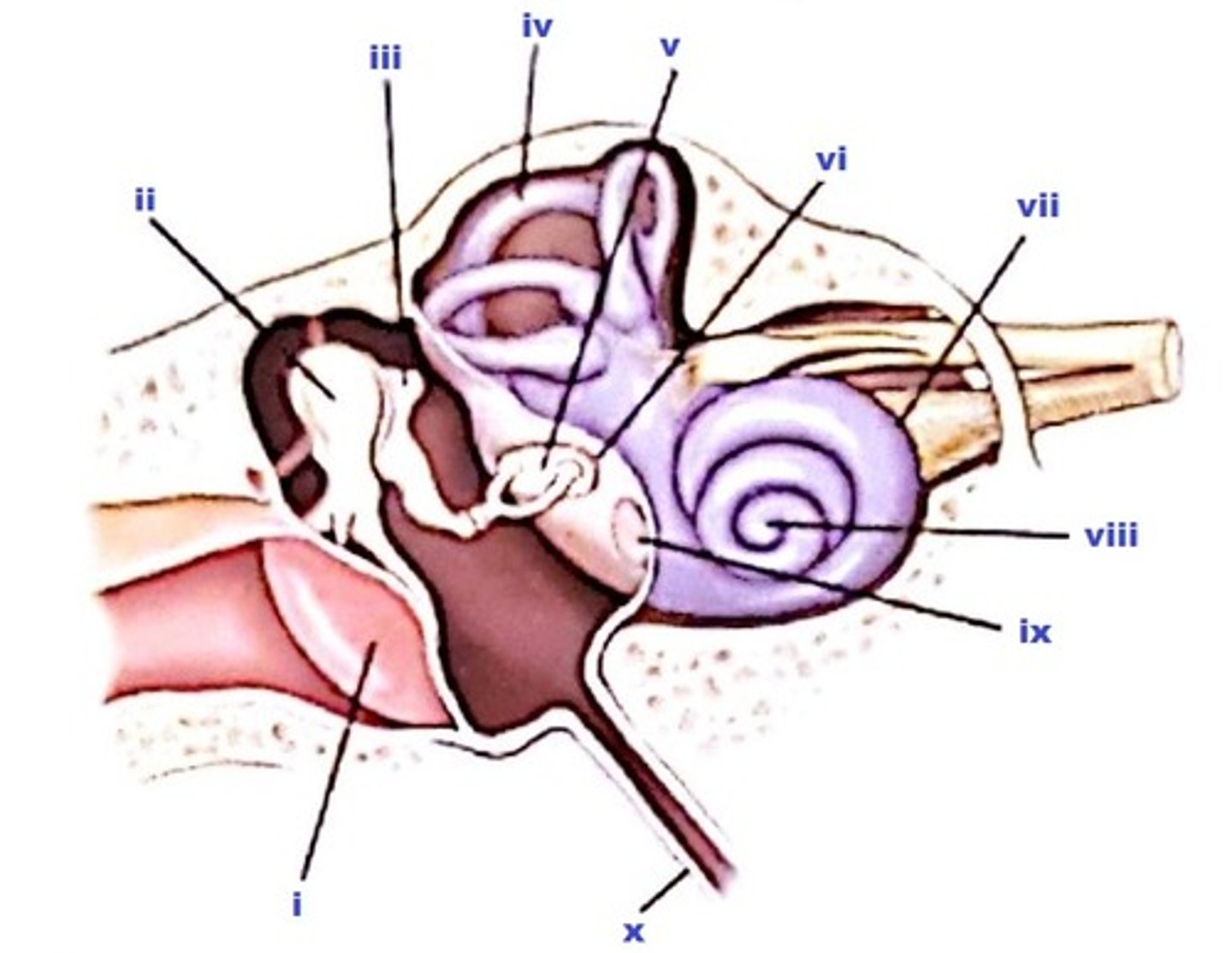
c
The part labeled ii is the:
a. stapes.
b. incus.
c. malleus.
d. cochlea.

d
The part labeled i is the:
a. external acoustic meatus.
b. tegmen tympani.
c. auditory tube.
d. tympanic membrane.

a
The part labeled x is the:
a. eustachian tube.
b. internal auditory canal.
c. tympanic tube.
d. internal auditory meatus.

b
The mastoid air cells communicate with the:
a. inner ear.
b. middle ear.
c. external ear.
d. base of the brain.
c
Which of the following structures of the inner ear is responsible for hearing?
a. Vestibule
b. Semicircular canals
c. Cochlea
d. Round window
a
The osseous labyrinth includes the cochlea, the vestibule, and the semicircular canals.
a. True
b. False
a
The sensory apparatus of both equilibrium and hearing are contained in the internal ear.
a. True
b. False
b
The vestibule is located in the middle ear.
a. True
b. False
a
Which of the following cranial bones make up the majority of the calvarium or skull cap?
a. Parietal
b. Occipital
c. Frontal
d. Sphenoid
e
Select the imaging modality that would best demonstrate the following pathologies.
Paget's disease
a. Nuclear medicine
b. MRI
c. CT
d. Ultrasound
e. Conventional radiography
d
Select the imaging modality that would best demonstrate the following pathologies.
Craniosynostosis (premature suture closing in neonate skull)
a. Nuclear medicine
b. MRI
c. CT
d. Ultrasound
e. Conventional radiography
d
Select the imaging modality that would best demonstrate the following pathologies.
Hydrocephalus (in neonate)
a. Nuclear medicine
b. MRI
c. CT
d. Ultrasound
e. Conventional radiography
a
Select the imaging modality that would best demonstrate the following pathologies.
Early detection of metastases
a. Nuclear medicine
b. MRI
c. CT
d. Ultrasound
e. Conventional radiography
c
Select the imaging modality that would best demonstrate the following pathologies.
Recent bleeding within the brain
a. Nuclear medicine
b. MRI
c. CT
d. Ultrasound
e. Conventional radiography
d
Select the imaging modality that would best demonstrate the following pathologies.
Intracranial hemorrhage in premature infants
a. Nuclear medicine
b. MRI
c. CT
d. Ultrasound
e. Conventional radiography
b
Select the imaging modality that would best demonstrate the following pathologies.
Most sensitive modality for detecting differences between normal and abnormal brain tissues
a. Nuclear medicine
b. MRI
c. CT
d. Ultrasound
e. Conventional radiography
c
Match each of the following radiographic appearance with the corresponding pathologic indication.
Distinctive lesions with moth-eaten appearance, a combination of increased density and destructive irregular border lesions
a. Multiple myeloma
b. Paget's disease
c. Metastases
d. Pituitary adenoma
d
Match each of the following radiographic appearance with the corresponding pathologic indication.
Enlarged sella turcica
a. Multiple myeloma
b. Paget's disease
c. Metastases
d. Pituitary adenoma
a
Match each of the following radiographic appearance with the corresponding pathologic indication.
Radiolucent areas within the bony cranium
a. Multiple myeloma
b. Paget's disease
c. Metastases
d. Pituitary adenoma
b
Match each of the following radiographic appearance with the corresponding pathologic indication.
Destructive stage of bony areas demonstrated by areas of radiolucency followed by reparative stages of sclerotic (radiodense) regions resulting in a "cotton-wool" appearance
a. Multiple myeloma
b. Paget's disease
c. Metastases
d. Pituitary adenoma
c
Match each radiographic appearance with the corresponding pathologic indication.
Bone destruction most commonly involving middle ear
a. Otosclerosis
b. Acoustic neuroma
c. Cholesteatoma
d. Mastoiditis
d
Match each radiographic appearance with the corresponding pathologic indication.
Increased densities replace mastoid air cells
a. Otosclerosis
b. Acoustic neuroma
c. Cholesteatoma
d. Mastoiditis
b
Match each radiographic appearance with the corresponding pathologic indication.
Bone destruction with widened internal auditory canal
a. Otosclerosis
b. Acoustic neuroma
c. Cholesteatoma
d. Mastoiditis
a
Match each radiographic appearance with the corresponding pathologic indication.
Excessive bone formation generally involving both the middle and inner ear
a. Otosclerosis
b. Acoustic neuroma
c. Cholesteatoma
d. Mastoiditis
a
An average-shaped skull with a 47° angle between the petrous pyramids and the midsagittal plane is classified as:
a. mesocephalic.
b. brachycephalic.
c. dolichacephalic.
d. morphocephalic.
c
What is the difference, in degrees, between the infraorbitomeatal and orbitomeatal lines?
a. 10°
b. 15° to 2°
c. 7° to 8°
d. 20° to 25°
b
Lesions of decreased density are termed osteoblastic lesions.
a. True
b. False
a
Which cranial bone possesses the superior nasal conchae?
a. Ethmoid
b. Sphenoid
c. Frontal
d. Temporal
c
Which cranial bone possesses the zygomatic process?
a. Frontal
b. Sphenoid
c. Temporal
d. Ethmoid
b
Which of the following modalities best demonstrates early signs of Paget's disease of the skull?
a. CT
b. Nuclear medicine
c. MRI
d. Sonography
b
An axiolateral oblique projection (Law method) for the temporomandibular joints on a brachycephalic type of skull would require _____ rotation as compared with an average-shaped skull.
a. more
b. less
c. the same
d. Rotation depends on the patient's age.
a
Which one of the following positioning errors most often results in a repeat exposure of a cranial position?
a. Rotation
b. Incorrect central ray placement
c. Slight flexion
d. Slight extension
a
Both CT (computed tomography) and MRI (magnetic resonance imaging) can provide reconstructed images in three planes: axial, sagittal, and coronal.
a. True
b. False
b
The PA axial projection (Haas method) for the cranium requires a CR angle of 25° caudad.
a. True
b. False
a
The submentovertex projection requires that the inferior OML (IOML) is placed parallel to the image receptor.
a. True
b. False
c
Which one of the following cranial projections will best demonstrate a possible basilar fracture?
a. PA axial-Caldwell method
b. Lateral recumbent
c. Horizontal beam lateral
d. AP axial (Townes method)
b
Which positioning line should be perpendicular to the plane of the IR for the AP axial (Towne) projection with a 37° caudad CR angle?
a. OML
b. IOML
c. AML
d. LML
c
Where is the CR centered for a lateral projection of the cranium?
a. EAM
b. Three-fourths inch (2 cm) anterior and 3/4 inch (2 cm) superior
c. Two inches (5 cm) superior to EAM
d. Midway between EAM and nasion
a
Which of the following terms describes the junction of the two nasal bones?
a. Nasion
b. Acanthion
c. Glabella
d. Supraorbital groove
d
The upper and lower teeth are embedded in the :
a. symphysis menti.
b. condyloid processes.
c. palatine processes.
d. alveolar processes.
c
The point of union between both halves of the mandible is termed:
a. gonion.
b. ramus.
c. symphysis menti.
d. mental foramina.
b
Infections involving the upper teeth may involve the frontal sinuses.
d
Which bone is involved with a tripod fracture?
a. Maxilla
b. Ethmoid
c. Temporal
d. Zygomatic
b
Which of the following imaging modalities should NOT be used to rule out a possible metal foreign body in the eye?
a. CT
b. MRI
c. Nuclear medicine
d. Radiography
b
What is the angle between the OML and the plane of the IR for the parietoacanthial (Waters) projection?
a. 40°
b. 37°
c. 42°
d. 15° to 20°
b
The modified parietoacanthial (modified Waters) projection requires more extension of the head and neck as compared with the parietoacanthial (Waters) projection.
a. True
b. False
b
The 15° PA axial (Caldwell) projection produces an unobstructed view of the maxilla.
a. True
b. False
c
Where does the CR exit for a modified parietoacanthial (modified Waters) projection of the facial bones?
a. Nasion
b. Glabella
c. Acanthion
d. Midorbits
d
Which of the following technical factors do NOT apply to lateral nasal bone projections?
a. The technologist should not use automatic exposure control (AEC).
b. The technologist should use a small focal spot.
c. The technologist should use low to medium kV.
d. All of the above apply.
c
Which positioning line is placed perpendicular to the plane of the IR with the true lateral nasal bone projection?
a. OML
b. AML
c. Interpupillary
d. Midsagittal
a
Which of the following projections will best demonstrate the bony nasal septum?
a. Parietoacanthial
b. Lateral nasal bone
c. AP axial projection
d. Lateral facial bone
b
The lateral projection for the nasal bones is generally a unilateral projection. (Both right and left lateral projections are usually not required.)
a. True
b. False
d
The CR must be placed parallel to the _____ positioning line for the superoinferior (tangential) projection of nasal bones.
a. glabellomeatal
b. mentomeatal
c. lips-meatal
d. glabelloalveolar
b
How much skill tilt and rotation are required for the oblique inferosuperior (tangential) projection for zygomatic arches?
a. 10°
b. 15°
c. 25°
d. None
c
What can the technologist do if the patient cannot extend the head and neck adequately for the routine submentovertex projection of the zygomatic arches?
a. Perform the Haas method.
b. Use a short SID to magnify the arches.
c. Angle the CR to place it perpendicular to the IOML.
d. Rotate the skull 15° away from the affected side.
b
Which positioning line is parallel to the IR for the oblique inferosuperior (tangential) projection of the zygomatic arches?
a. Midsagittal plane
b. Infraorbitomeatal
c. Orbitomeatal
d. Glabelloalveolar
d
Which of the following points is NOT true about the oblique inferosuperior (tangential) projection of the zygomatic arches?
a. It requires both rotation and tilt of the skull.
b. A small focal spot should be used.
c. The AEC should not be used.
d. A grid must be used.
a
How much difference is there between the OML and IOML positioning lines?
a. 7° to 8°
b. 8° to 9°
c. 10° to 11°
d. 5° to 6°
b
Which projection best demonstrates the floor of the orbits?
a. Parietoacanthial (Waters)
b. Modified parietoacanthial (modified Waters)
c. PA axial (Caldwell)
d. Lateral facial bones
c
Which positioning line must be used with a 30° caudad angle for an AP axial projection of zygomatic arches?
a. IOML
b. AML
c. OML
d. GAL
d
What is the angle between the midsagittal plane and the IR for a parieto-orbital oblique projection of the optic foramen?
a. 45°
b. 12°
c. 30°
d. 53°
b
Which positioning line is placed perpendicular to the IR for the parieto-orbital oblique projection of the optic foramina?
a. OML
b. AML
c. MML
d. IOML
b
The proper name for the parieto-orbital oblique projection is the Schuller method.
a. True
b. False
a
Optic foramen studies are routinely taken as bilateral projections.
a. True
b. False
b
What is the maximum CR angulation used for the axiolateral oblique projection of the mandible?
a. 10° to 15°
b. 25°
c. 30°
d. 45°