Avian Influenza
1/31
There's no tags or description
Looks like no tags are added yet.
Name | Mastery | Learn | Test | Matching | Spaced | Call with Kai |
|---|
No analytics yet
Send a link to your students to track their progress
32 Terms
Definition
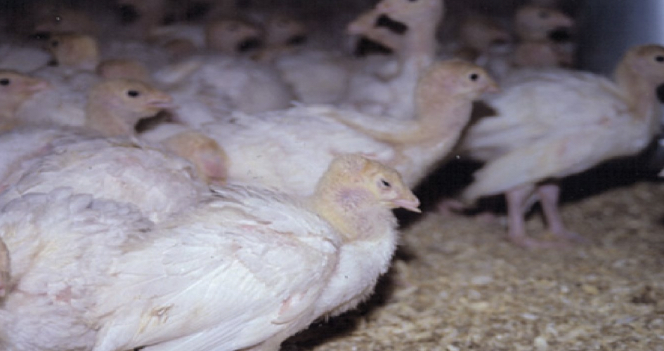
acute highly contagious and infectious viral disease of all species of birds in general, and poultry in particular. AI can occur in all domestic and captive species of birds, affecting the respiratory, digestive and /or nervous system of the birds.
Causative agent
Orthomyxoviridae, genus Influenza virus type A.
Classified into subtypes on the basis of
the antigenic relatedness of the surface glycoproteins haemagglutinin (HA) and neuraminidase (NA). ■ At present, 16 HA subtypes (H1-H16) and 9 NA subtypes (N1-N9) are recognised.
Transmission by
Secretions from infected birds,
wild birds .
contaminated feed, equipment and people.
Sea birds and migratory waterfowls the main reservoir for avian influenza virus.
Natural reservoir
Migratory ducks
Influenza A virus pathotypes
Highly pathogenic avian influenza (HPAI) Low pathogenic avian influenza (LPAI)
Low pathogenic avian influenza (LPAI)
cause a mild, primarily respiratory disease in poultry, unless there is exacerbation by other infections or factors
Highly pathogenic avian influenza (HPAI)
cause an extremely serious disease characterised by generalised infection of the infected poultry, with very high flock mortality (up to 100%)
Low pathogenicity avian influenza is caused by
viruses of all H subtypes (H1-H16)
Higly pathogenic avian influenza is caused by
some viruses of the H5 and H7 subtypes Once introduced into poultry, LPAI virus strains of H5 and H7 subtypes may mutate into HPAI strains. To date only viruses of the H5 and H7 subtypes have been shown to cause HPAI.
The severity of the disease produced by LPAI viruses is greatly influenced by:
the strain of virus, the species and age of host; the immune status of the host against the virus and particularly the presence of other infectious agents such as: Pasteurella spp, Newcastle disease viruses (including vaccine strains), PMV-2, PMV-3, avian pneumovirus, infectious bronchitis virus, E. coli and Mycoplasma spp, immunodeficiency conditions and environmental factors (such as excess ammonia, dust, hot or cold temperatures)
Incubation period
Variable - few hours to days Depends on virulence of the virus and the route of exposure
Mortality & morbidity
LPAI viruses, high morbidity and low mortality rates are typical. Mortality rates are usually less than 5% unless accompanied by secondary pathogens or if the disease is in young birds. With the HPAI viruses, morbidity and mortality rates are very high (50%- 90%) and can reach 100% in some flocks.
Clinical signs
Sudden death.
Marked loss of appetite, reduced feed consumption.
Cessation of normal flock vocalization.
Drops in egg production.
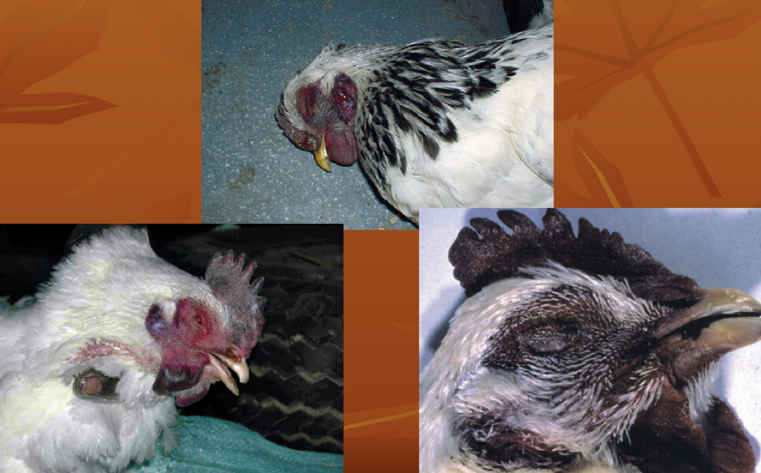
Cyanosis of comb &wattles.
Depression.
Diarrhea (often green).
Dyspnea .
Coughing , sneezing, rales, excessive lacrimation .
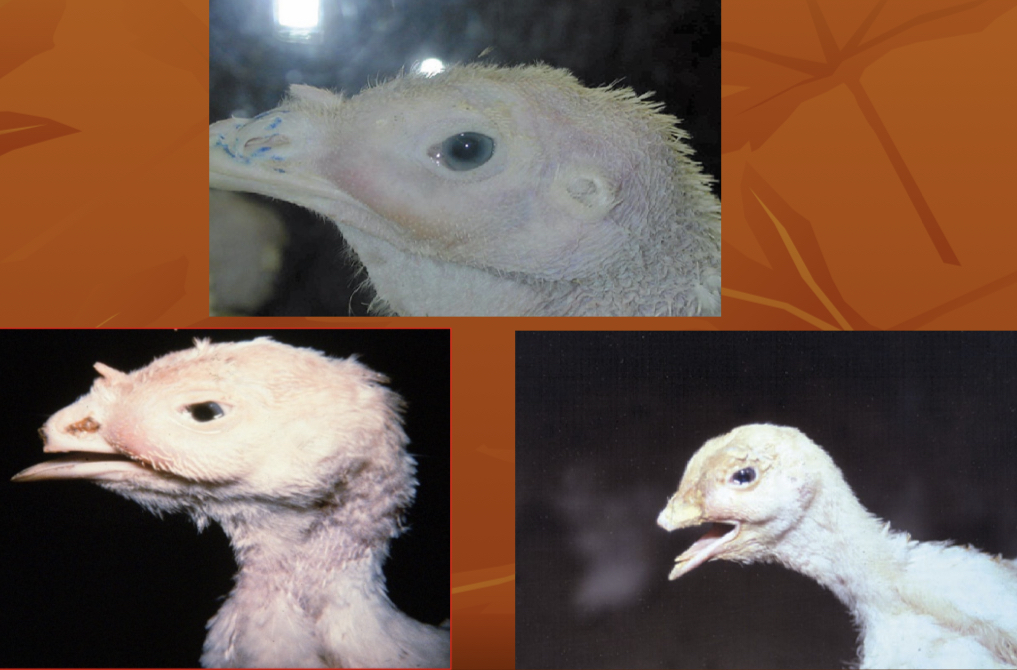
Nasal and ocular discharge.
Swollen face and Sinusitis.
Nervous signs such as paralysis.
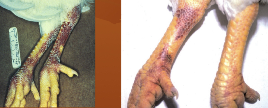
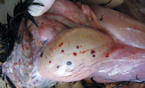
Hemorrhages on the legs.
.
Severe depression
.
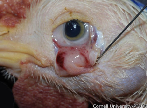
severe conjunctivitis and swelling of the infraorbital sinuses (Sinusitis)
.
congestion and cyanosis of the comb and wattles (unfeathered skin of the head)
.
haemorrhages on the shank
Gross lesions
• Lesions may be absent in cases of sudden death • Severe congestion of the musculature and Dehydration • Subcutaneous oedema of the head and neck area • Nasal and oral cavity discharge • Severe congestion of conjunctivae, sometimes with petechiae • Excessive mucous exudates in the lumen of the trachea, or severe hemorrhagic tracheitis • Petechiae on the sternum, on the serosa, abdominal fat, serosal surfaces and finally, in the body cavity • Severe kidney congestion, sometimes with urate deposits in the tubules • Hemorrhages and degeneration of the ovary • Hemorrhages on the mucosal surface of the proventriculus. Hemorrhages and erosions of the gizzard lining • Hemorrhagic foci on the lymphoid tissues in the intestinal
.
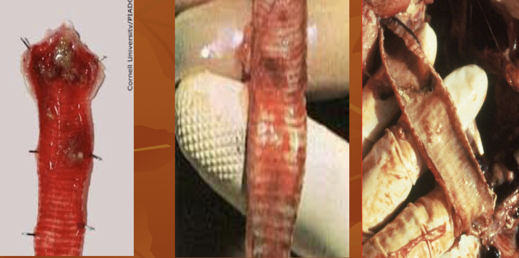
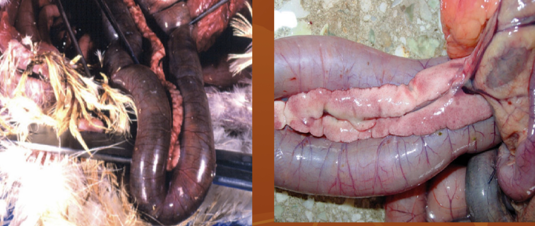
Tracheitis
.
hemorrhagic lesions on the mucous membranes
.
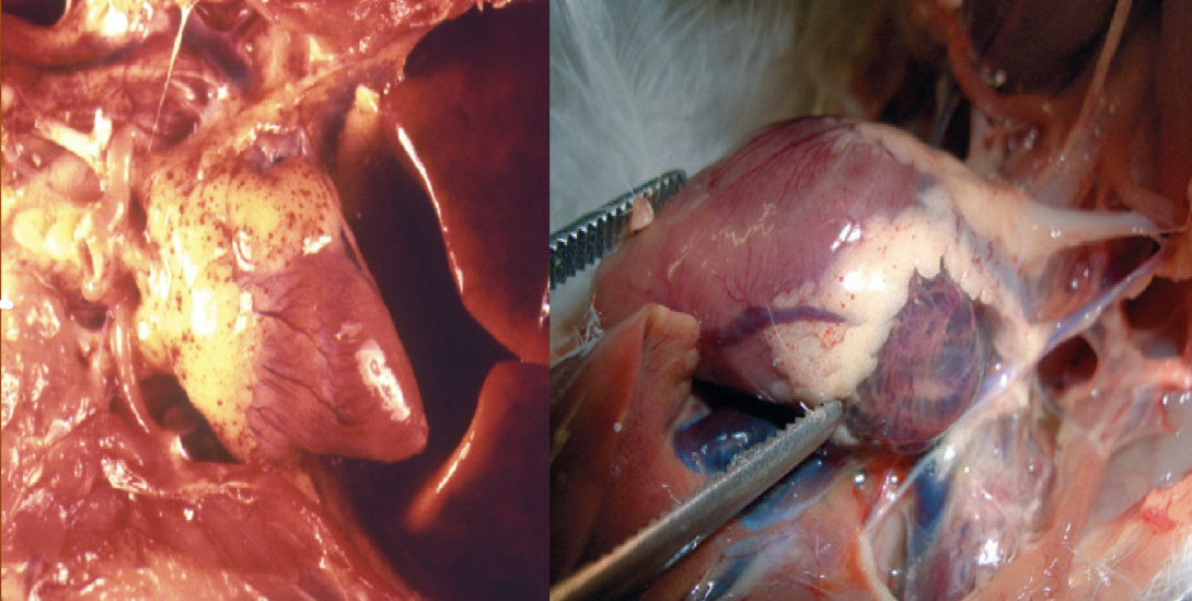
petecchial haemorrhages on pericardial fat
.
Hemorrhages and erosions of the gizzard
.
Proventricular hemorrhages.
.
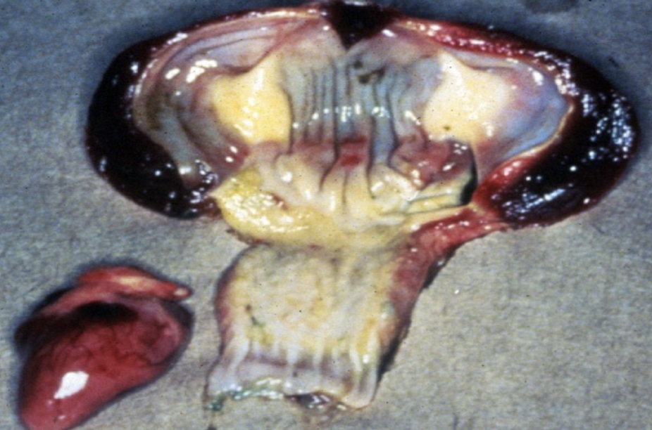
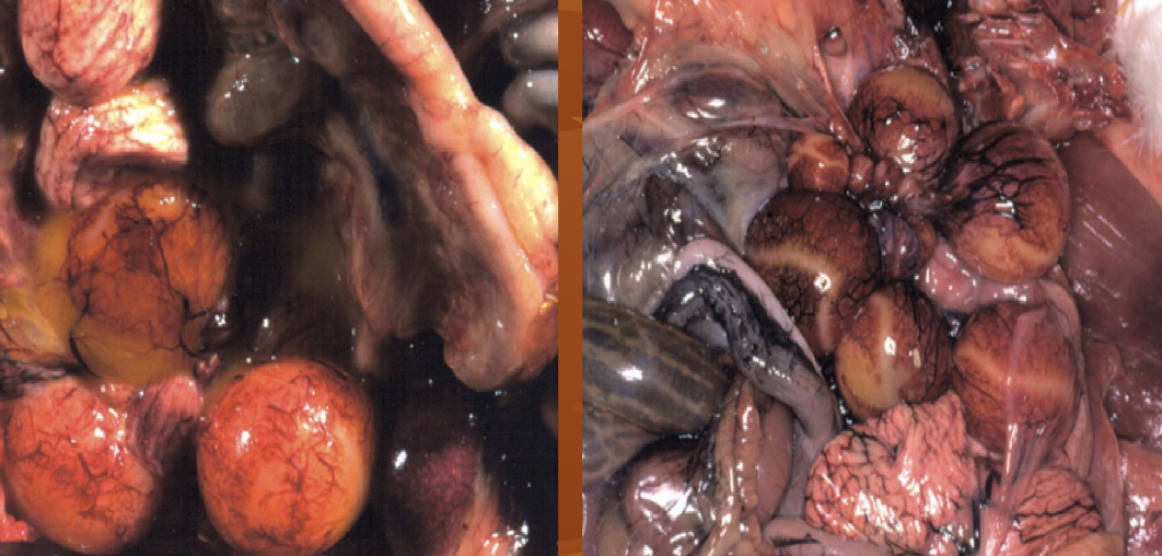
congestion of the ovary and eggyolk peritonitis
haemorrhagic pancreatitis and duodenal distension
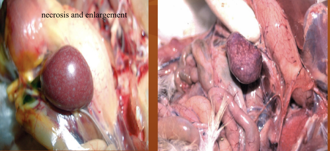
Enlargement, congestion and necrosis of the spleen
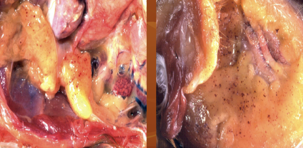
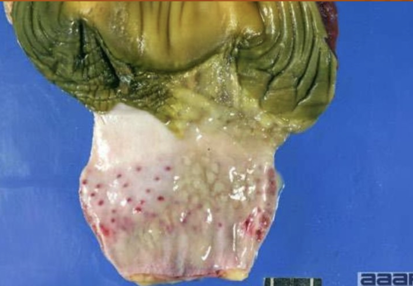
petechial haemorrhages on the abdominal fat
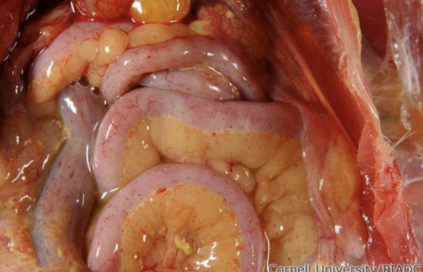
Pinpoint hemorrhages on the serosal surfaces of the intestinal loops and the mesenteric fat
.
petechial haemorrhages on the serosal surfaceof the crop
Diagnosis
1-A presumptive diagnosis may be made on
Clinical signs.
Postmortem lesions.
serology
2-Definitive diagnosis :is by viral isolation and identification
Treatment
• No effective treatment, but good husbandry, nutrition may reduce losses. • Eradication by slaughter is usual in chickens and turkeys.