Equine dentistry 2
 Call Kai
 Learn
Learn Practice Test
 Spaced Repetition
 Match
Match Flashcards
 Knowt Play
1/86

There's no tags or description
Looks like no tags are added yet.
Name | Mastery | Learn | Test | Matching | Spaced | Call with Kai | Chat |
|---|
No analytics yet
Send a link to your students to track their progress
87 Terms
What are the malalignments in equine?
Brachygnathism (congen)
Parrot mouth
Overjet/Overbite
Prognathism
Sow mouth
Campylorrhinus lateralis = wry nose
What is brachyganthism? How do you treat it?
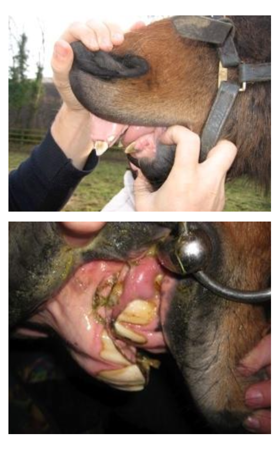
Overly long maxilla compared to mandible —> maxillary incisors lie rostral to mandibular ones
no wearing down of opposing teeth —> problems with overgrowths
cheek teeth not opposing each other → sharp hooks at back of mouth
Severe cases —> option of surgical correction (ethical?)
What are the consequences of brachygnathism?
Ulceration of behind upper incisors
Maxillary rostral 06 overgrowths and mandibular 11 overgrowths will need lifelong attention
What is prognathism? What are the clinical implications?
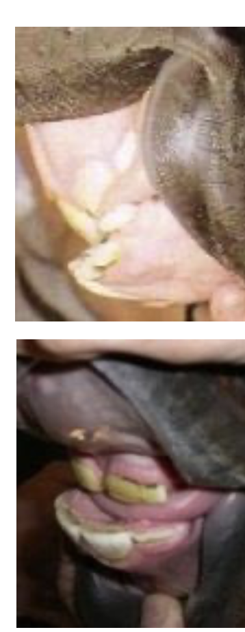
Relative overgrowth of mandible
Miniature breeds over-represented
Clinical Implications
Few incisor problems
Develop lower 06 overgrowths and upper 11 overgrowths
i.e. opposite to normal
Check back of mouth for overgrowths
What is campylorrhinus lateralis (“wry nose”)?
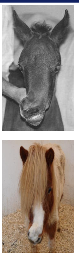
Deviation (& occasional rotation) of entire maxilla involving incisive region, nasal septum & nasal bones
What are the varying degrees of severity of campylorrhinus lateralis?
Minor occlusal problems —> routine dentistry every 6 months
Severe = difficulty nursing & occlusal → breathing problems
Surgical correction can be attempted but complex
What is diagonal bite/slant mouth indicative of?
Eating predominantly on one side
May indicate shear mouth formation of cheek teeth
Which species is smile mouth normal in?
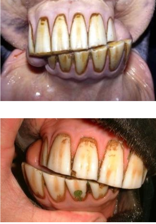
Donkeys
What is being shown here?
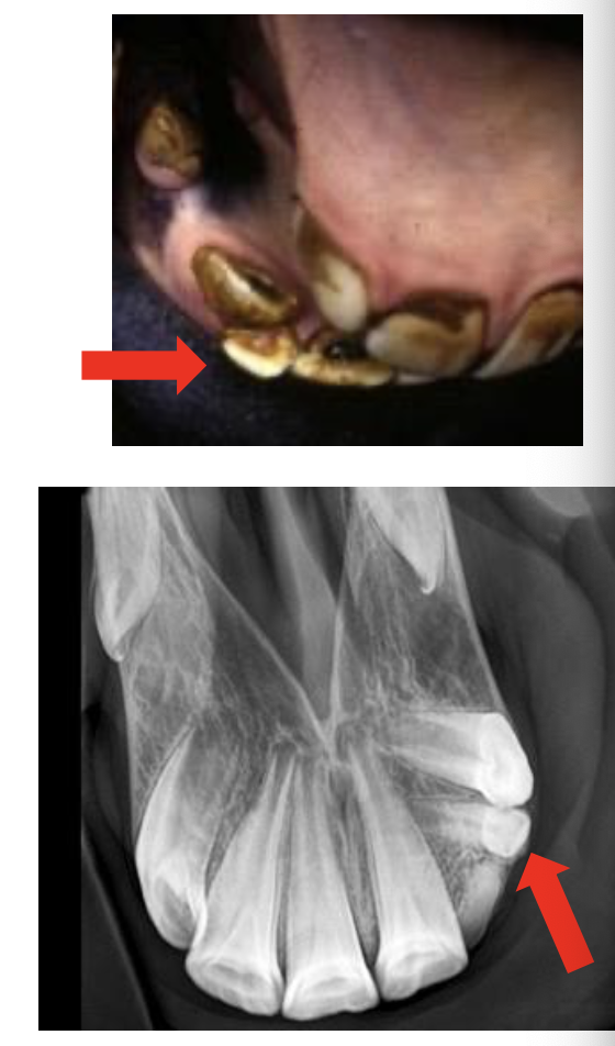
Retained deciduous incisor
Usually rostral & causes caudal displacement of permanent tooth
How are retained deciduous incisors treated?
Loose —> remove with forceps
Firmly attached —> remove with dental elevators
May need to radiograph (easy for incisors & front of mouth)
How do supranumerary incisors present?
Have long crowns (up to 7cm)
Close to reserve crowns and roots of normal permanent incisor
How are supranumerary incisors treated?
Usually cause little problem
Often best not to remove
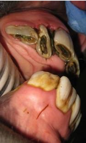
What causes incisor fractures?
Trauma
Kicks
Inquisitive youngstock caught on objects —> e.g. haynets
Crib-biting on fixed objects
What are the two ways that incisors are fractured?
Involve the incisive bone —> may look dramatic but quite easy to reapir, do not remove fragments if repair possible
Incisors themselves —> determine if pulp affected, endodontic repair or extraction
What are incisor diastemata?
Spaces between adjacent teeth
Shouln't be any
May develop with age as incisors taper towards apex
Can lead to periodontal dx
What is valve diastema(ta)?
Narrower at occlusal aspect
Wider at gingival margin
Trap food causing gingivitis, periodontitis & potentially
How do you manage incisor diastemata?
Remove food from spaces
Owner can perform this with a toothbrush x2/week
What is EORTH?
Equine Odontoclastic Tooth Resorption & Hypercementosis
Unknown aetiology —> horses aged >14yrs
Swelling and/or draining tracts over multiple mandibular & maxillary incisors
pain + difficulty prehending food
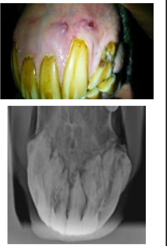
How do you diagnose EOTRH?
Diagnosis —> visual + radiography
Lytic changes / hypercementosis
How do you treat EOTRH?
Extraction of loose incisor curative
Disease progressive in some cases spreads from tooth to tooth
May have to remove all incisors
How are oral neoplasia's classified?
Tissue of origin —> dental, bone, soft tissue
Clinical behaviour + patholoigcal features —> benign/ malignant; invasive/ localise; proliferative / ulcerative
What are the oral neoplasias of dental origin?
Ameloblastoma
Cementoma
Odontoma
Temporal teratoma
What are the features of ameloblastomas?
Most common in:
Older horses
Mandible
Cause a bony swelling
+/-cystic cavity
Benign/locally invasive
Surgical excision —> poss specialist removal of mandible
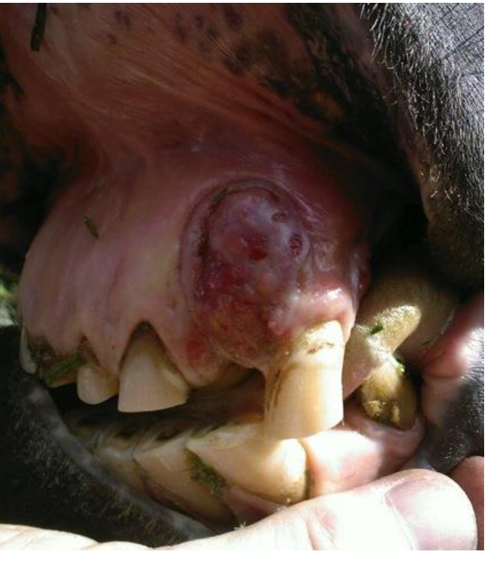
What are the neoplasia of soft tissue origin?
Squamous cell carcinoma (SCC) (most common)
Sarcoid
Epulis
Melanoma
Oral papilloma
Ossifying fibroma
Fibroma
Myxoma/Myxosarcoma
(tx depends on cause, extent & location)
What are the abnormalities of the canine tooth?
Calculus around lower canines most common
Remove with dental forceps
Owner can clean periodontal pockets
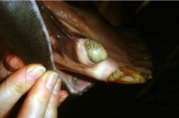
Apical infection/fracture
Endodontic treatment can be considered in some cases
Removal challenging —> long teeth w/ curved roots deeply embedded in bone
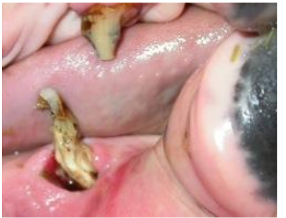
What are the abnormalities of the wolf teeth?
Number, position size variable
Do not cause problems when in normal position and of normal size
(be careful of palatine artery)
What are the indications for wolf tooth removal?
Biting problems/ ulceration
Rostrally displaces, particularly large, mandibular
Loose or displaced
Blindly erupted (painful)
May become molarised (looks like molar) —> always radiograph
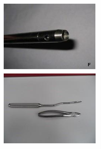
How are wolf teeth removed?
Specialist kit —> incl. apple-core device (Burgess elevator) to go around tooth
Can use a long handled elevator / small animal tooth luxator & pair of SA forceps
What complications can you get with wolf tooth removal?
Fracture of tooth
Fracture of bone
Trauma to the palatine artery → marked haemorrhage
Describe wolf teeth removal
Standing sedation + speculum placed
LA —> infra-orbital / maxillary nerve block → direct infiltration
Use dental elevators circumferentially around tooth to loosen it (be careful of palatal artery)
Remove w/ forceps once loose —> don’t extract until fully loose because risk of breaking tooth
if occurs & small subgingival fragment → try to remove
2 weeks bit rest
What are the two classifications of cheek teeth issues?
Developmental
Acquired
When are cheek teeth first examined?
Ideally briefly in 1st week of life @ same time checking for cleft palate
How often are cheek teeth checked?
Yearly as they begin to erupt
What are eruption cysts?
“3-4 year-old bumps”
Palpable swellings on mandible ventral to apices of Triadan 07/08
Physiological & transient phenomenon
Should not be considered as disorder
What are retained caps?
Remnants of deciduous teeth
Normally shed during eruption of the underlying permanent tooth
Loose / retained caps can cause oral pain / quidding
Usually attached to gingiva in one place, attachement causes pain
How do you treat retained caps?
Easily removed with forceps
If possible contralateral cap should be removed
Do not remove prematurely —> will damage developing tooth
What is the cause of cheek teeth displacements?
Overcrowding during eruption
How do cheek teeth displacements present?
Often bilateral
Usually 09s & 10s
Medial / lateral displacement +/- Rotation
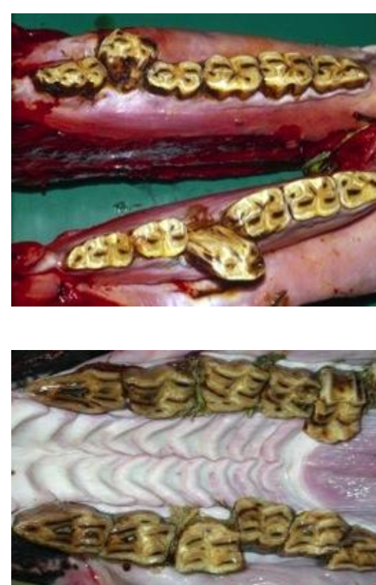
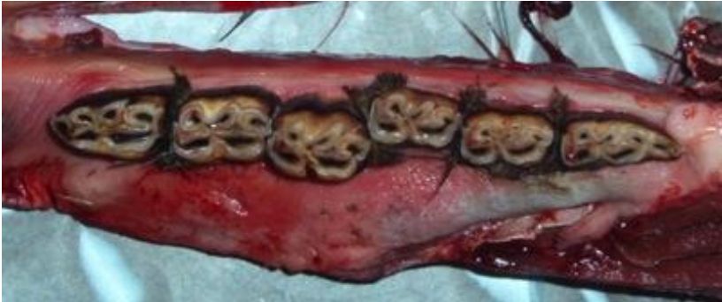
What do cheek teeth displacements lead to?
Buccal/lingual trauma
Diastemata → periodontal disease
What are developmental disatemata?
Cheek teeth occlusal surfaces should be tightly compressed together —> act as one functional grinding surface
If teeth develop too far apart
Spaces develop
Food accumulates
Fermentation
Periodontal disease
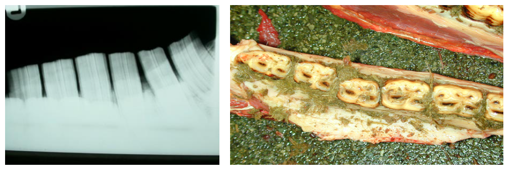
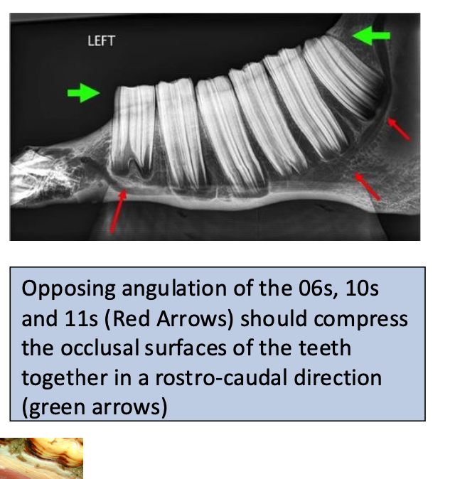
Where do supranumerary CT develop?
Usually at caudal aspect —> so count teeth!
(+ dental dysplasia + oligodontia)
How do you deal with supernumerary CT?
Incidental
Can be left but if not occluded then need to rasp regularly to prevent overgrowth
If cause periodontal disease then extract
Where do enamel overgrowth occur?
Buccal aspect of upper CT
Lingual aspect of lower CT
Due to anisognathism
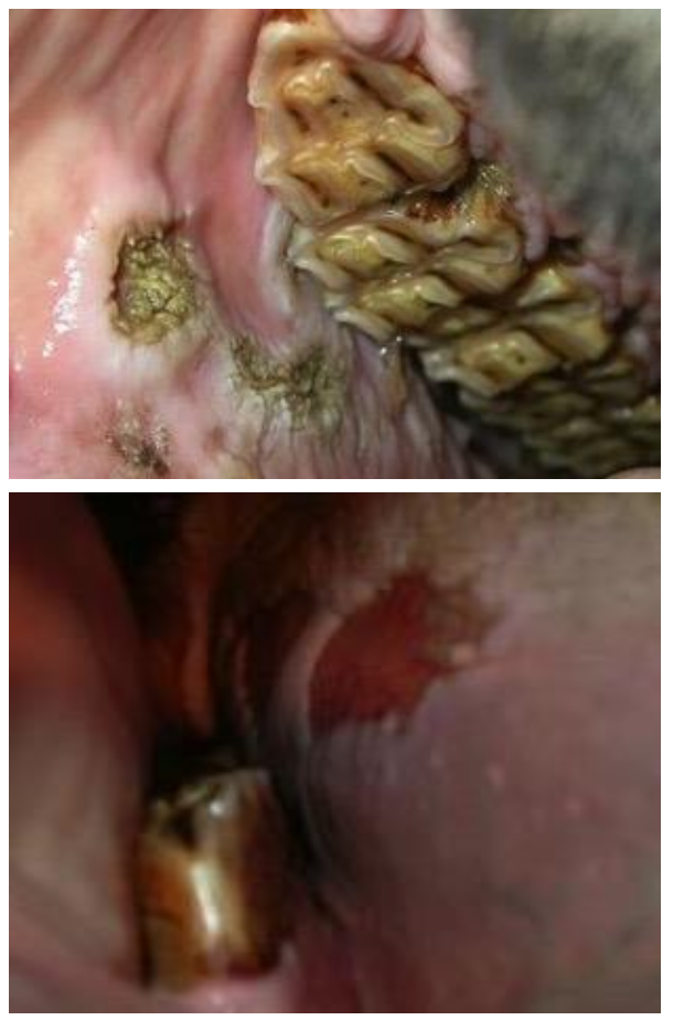
What are the clinical signs of enamel overgrowths?
Quidding, pain when eating, +/- colic
Tack may exarcerbate
More pronounced when horses fed more concentrate bc unnatural masticatory pattern
Tack may exacerbate problem —> mucosal ulceration
How do you treat enamel overgrowths?
Removal during routine dental prophylaxis
What is wavemouth? How do you treat it?
Marked undulation to occusal surface
If mild —> little tx required
Dominant areas can be sequentially reduced every 3 months
What is stepmouth? How do you treat it?
When a focal overgrowth occurs
Usually opposite a missing or extracted tooth
Reduced in stages to prevent pulpar exposure or thermal damage
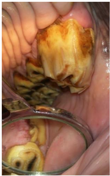
What is shear mouth?
Increase occlusal angle of entire cheek tooth row
Usually 2° to diastemata formation or dental fracture
What should you suspect if shearmouth is bilateral?
Temporomandibular joint arthropathy (joint dx of jaw)
How is shearmouth managed?
Treat 1° problem
Gradual reduction of angle should be performed
Occurs rapidly anyway when horse begins to chew more normally
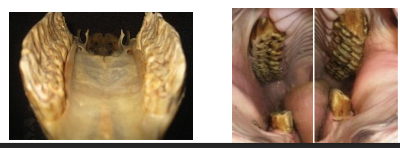
What is being shown here?
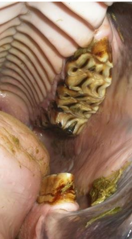
Exaggerated transverse ridges
Do not confuse with normal ridges
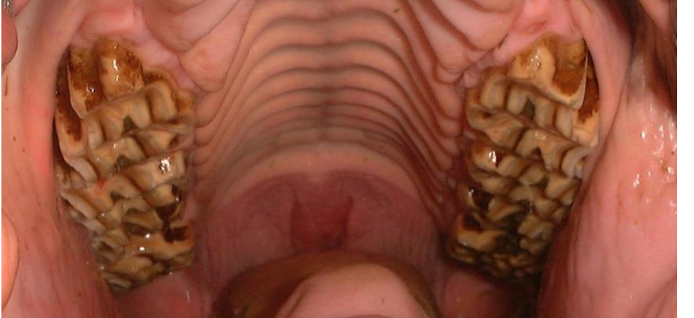
Often opposing diastemata
How do you remove large overgrowths?
Hand or power rasps (careful of thermal energy)
Reduced in stages
Max 3-4 mm q 6 months
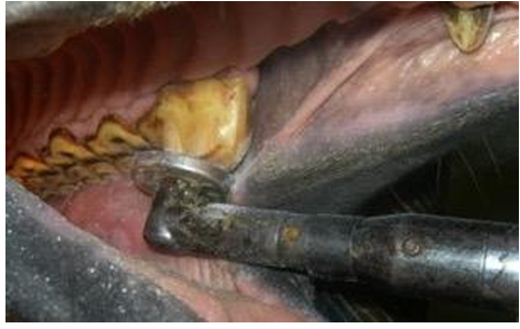
What are the problems with excessive rasping?
Pulp exposure
Thermal damage
Risk apical infection
High risk sites:
Rostral 06s + Caudal 11s due to extra pulp horn
What is bit seating?
Rostral profiling (to prevent bit impingement but misconception —> bit should not sit in this location)
CONTRAINDICATED —> extra pulp horn on 06s
pulp horn exposure
apical infection (18months)
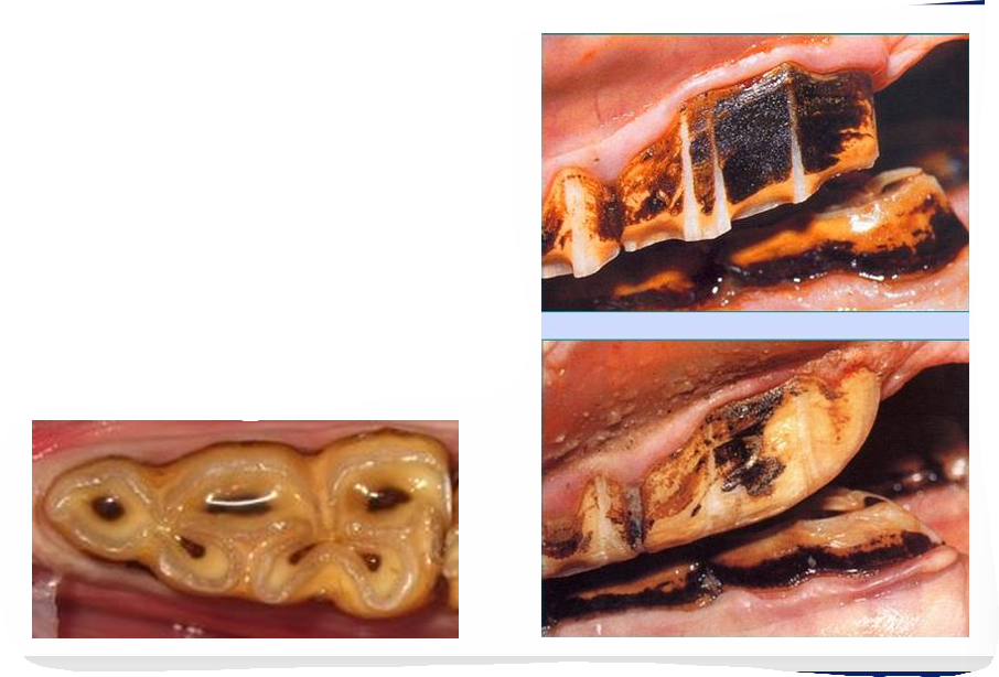
What is smooth mouth?
Senile change
Cheek teeth enamel largely worn away
Sometimes worn down to individual roots
Softer dentine and cementum become smooth
Not very useful for grinding

How do you manage smooth mouth?
Dietary mangement
e.g. feeding chopped forage to help maintain weight
What locations can caries affect?
Peripheral cementum of the maxillary and mandibular cheek teeth
Infundibulae of maxillary cheek teeth
How do dental caries occur?
If food material becomes stagnated in pits of peripheral cementum
Fermentation
Drop in pH of envioronment
Deminerlisation
Pits bigger and blackening of the peripheral cementum
Cementum becomes eroded then may spread into peripheral enamel
What causes infundibular caries?
Developmental predisposition
Cemental hypoplasia
Food accumulates in infundibulum
Fermentation → decay
Young horses may have small central opening = site of former infundibular central artery
(progressive & irreversible)
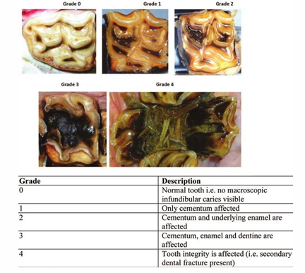
What do infundibular caries predispose a horse to?
Fracture
How are infundibular caries managed?
Infundibular restoration
What are the causes of peripheral caries?
Increase sugar in diet
Haylage
Molasses
(Common)
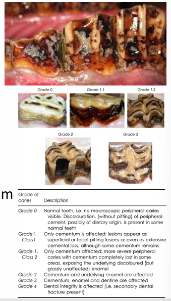
How are peripheral caries managed?
Palliative rasping of roughened cementum
Removal of excess sugars from diet
Oral lavage
What does treatment of diastemata depend on?
Severity of periodontal disease
How do you treat diastemata without periodontal disease?
Must be cleaned out COMPLETELY
Dental Pick / Diastema forceps / High pressure lavage
Remove ETR’s (exaggerated transverse ridges) on opposite arcade and correct other overgrowths that may be contributing to diastema
+/- Pack w/ impression material to prevent refilling
How do you treat diastemata with periodontal disease?
Widen with mechanised burr
Painful —> lidocaine splash block
Remove ETR’s on opposite arcade and correct other overgrowths that may be contributing to diastema
+/- Pack w/ impression material to prevent refilling
Dietary management essential
Short fibre —> grass / ‘short chop’
Avoid long stem hay / haylage
What are the 3 main types of cheek teeth fractures?
Buccal (lateral) slab fracture
Midline sagittal fracture
Occlusal fissure fractures
(v. rarely due to trauma)
How do buccal fractures usually present?
Can be incidental
Quidding
Usually NOT associated with apical infection
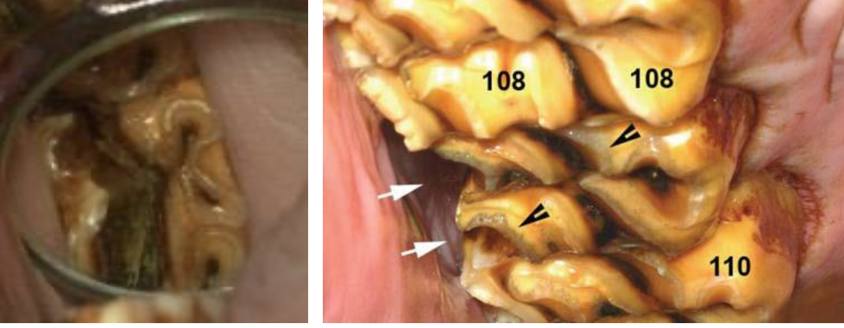
How do you treat buccal fractures (slab fractures)?
Pulp horns seal off —> calcium hydroxide may encourage this
Dental extraction may not be required but remove loose fragment
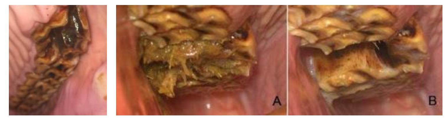
What are the features of midline sagittal fractures?
Most commonly 109 & 209
Pathological fracture through infundibulae
Preceded by infundibular caries
Result in apical infection +/- sinusitis
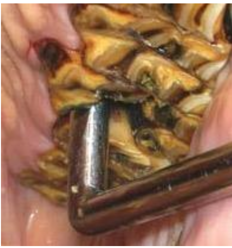
How do you treat midline sagittal fractures?
Extraction
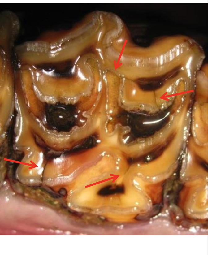
What are the features of occlusal fissue fractures?
Incidental finding, subtle
Require dental mirror/oroscope
Unknown significance
May predispose to atypical fracture
Different configuration
Monitor
What do the clinical signs of an apical infection depend on?
Which cheek teeth are involved
Location in relation to paranasal sinuses
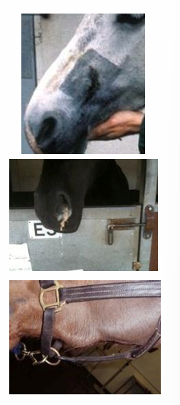
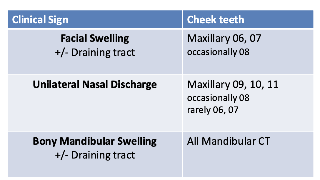
What are the clinical signs of apical infection and which cheek teeth are they related to?
What are the sequelae of pulpitis?
Pulpar oedema → vascular occlusion → necrosis
What are the causes of apical infections?
Anachoresis (= blood borne infection)
Fracture
Periodontal spread (Diastema)
Pulpar exposure
What are the routes of infection of mandib / maxil CT?
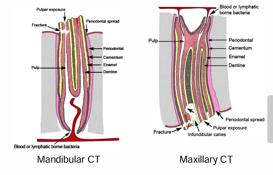
How is apical infection diagnosed?
Clinical signs
Oral exam
Fracture, pulpar exposure
Imaging
Rx, CT (better), Nuclear scintigraphy
What are the options for cheek tooth extraction?
Oral extraction
Modified transbuccal extraction
Lateral buccotomy
Repulsion
(complications poss —> preservation of alveolus important where poss)
What is the sedation of an oral extraction
Regional anaesthesia (nerve + local blocks)
Heavy sedation
NSAID & antimicrobial
How is an oral extraction performed?
(Vet surgeon only)
Gingival elevation and periodontal ligament disruption with picks
Interdental spreading (closed gradually and incrementally in interdental space to stretch periodontal ligament over approx 30 mins)
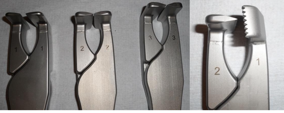
Application of extraction forceps
How are molar forceps used?
Oscillate (gently wiggle to stretch periodontal lig) lateral strain (horizontal rocking) & fulcrum (elevation)
Must be tightly applied to tooth or else risks damage
Many different types depending on tooth shape
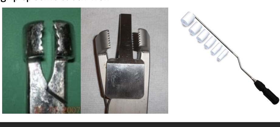
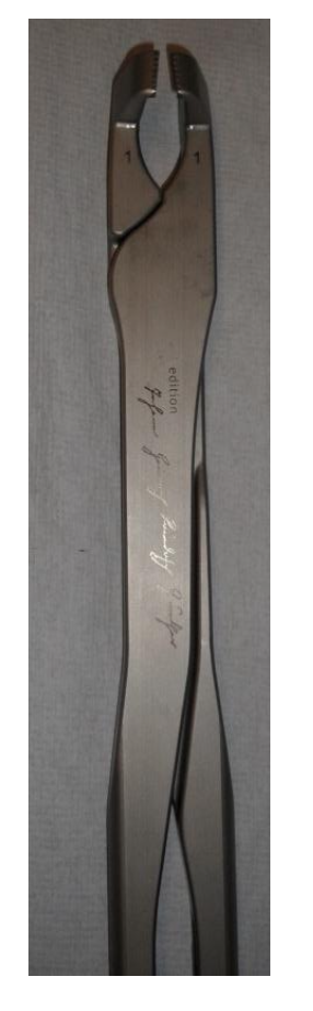
When are Minimally-invasive transbuccal extractions (MTEs) used?
When crown fractured
Oroscopic & radiographic guidance
Preserves alveolar bone
How are MTEs performed?
Chisels to break down ligament
Reserve crown drilled
Hole tapped
Extraction screw inserted
Pulled out orally using mallet
Picks, chisels & fragment forceps
When are repulsion cases used?
Complex cases only
Must be loosened as much as possible first
GA / sedation with LA
Rads guidance imperative
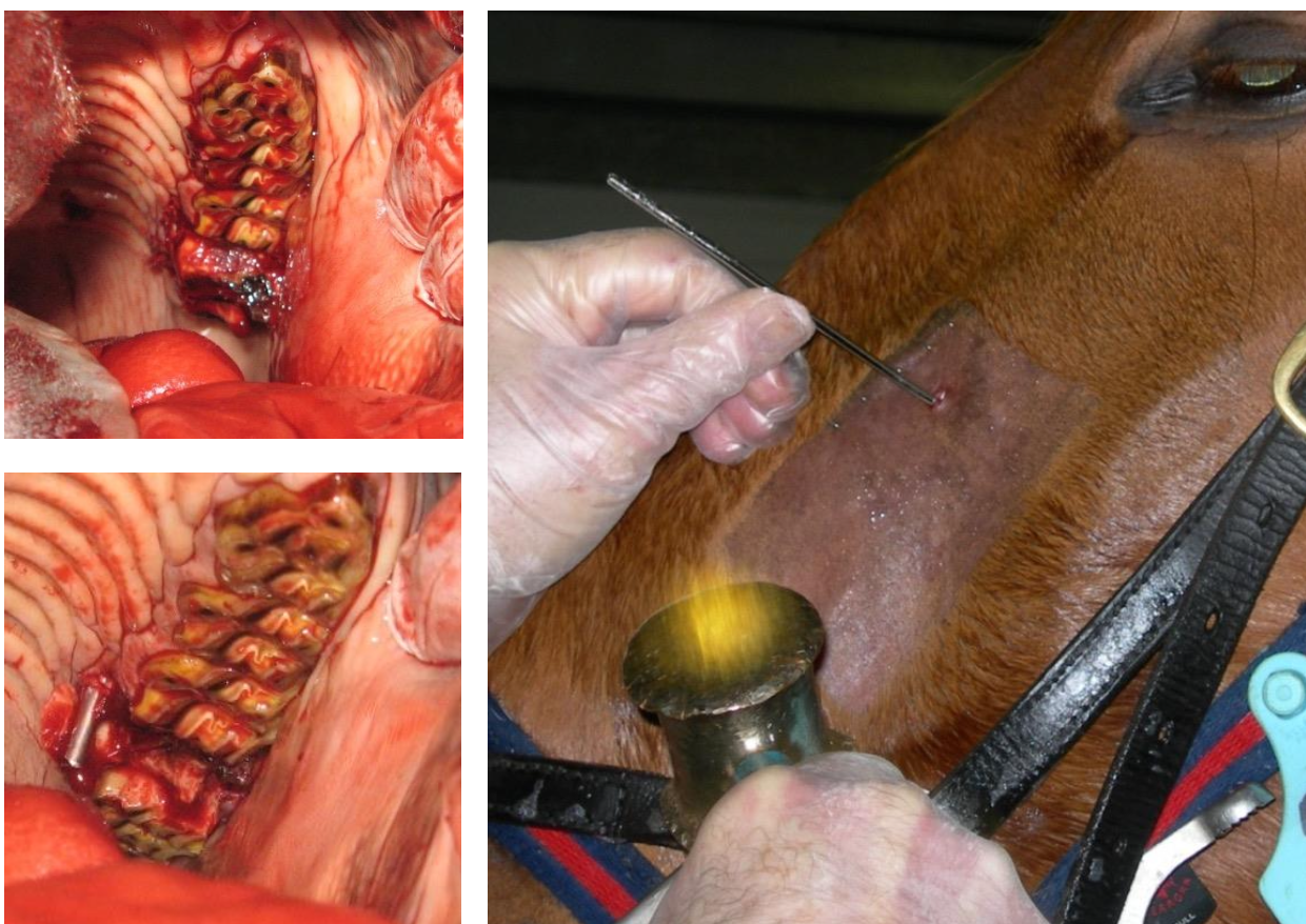
How are repulsions performed?
Trephine over top of tooth
Blunt instrument to drive tooth into mouth
Mechanically inefficient
Alveolar bone damage
Steinnman pin= minimally invasive
Dental punch = very destructive
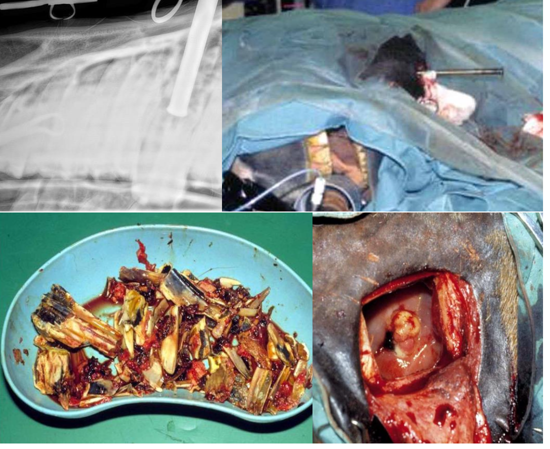
(high potential for complications)
How is a lateral buccotomy performed?
GA often required
Incision through cheek
Removal of lateral alveolar bone
High morbidity (Iatrogenic trauma potential damage to facial nerve & parotid duct + wound breakdown)