Unit 1 440
 Call Kai
 Learn
Learn Practice Test
 Spaced Repetition
 Match
Match Flashcards
 Knowt Play
1/144

There's no tags or description
Looks like no tags are added yet.
Name | Mastery | Learn | Test | Matching | Spaced | Call with Kai | Chat |
|---|
No analytics yet
Send a link to your students to track their progress
145 Terms
List basic tissue from superficial to deep:
skin, subcutaneous tissue, deep fascia, muscle, deep fascia, bone
what is a network of nerves?
nerve plexus
nerve plexus’s branch from what, to innervate the neck, limbs, and body?
ventral rami
What are the 4 plexus’s?
cervical (c1-c4), brachial (c5-c8, t1), lumbar (t12, l1-l4), sacral (l4-l5, s1-s4)
What connects the glands to the dermis in breast tissue?
suspensory retinacula (of cooper)
what carries and stores milk from the glands to the nipple?
lactiferous ducts
Is the lymphatic system a closed circuit?
no
What type of flow is the lymphatic system?
unidirectional flow (towards the heart only)
What are the small, round or bean-shaped aggregates of tissue that are sites of lymphocyte production?
lymph nodes
Lymph nodes filter lymph using what?
macrophages
Where are superficial lymph nodes located?
cervical, axillary, and inguinal regions
Where are the deep lymph nodes located?
abdominal aorta and along iliac arteries
often lymph nodes must be biopsied in malignancies to rule out what?
metastasis
What protects the thoracic viscera as well as helping with limb movement?
muscles of the chest wall
What primarily play a role in forced expiration?
intercostal muscles
What are the 3 types of ribs in the thoracic cage?
true (1-7), false (8-12), floating (11-12)
What are the 3 types of joints in the thoracic cage?
costochondral, costovertebral, sternocostal
What are the 3 primary components of the sternum?
manubrium, body, xiphoid process
What part of the rib is most susceptible to fracture?
area just anterior to the angle
What runs in the costal groove on the inferior aspect of the rib?
neurovascular structures (vein, artery, nerve)
What is a connection between two tubular structure to maintain flow, supplying the same structure?
anastomosis
Intercostal veins will drain the thorax into what?
internal thoracic veins or azygous system
What are the compartments of the thorax?
medial and two lateral
The mediastinum is divided into four additional compartments being?
superior, anterior, middle, posterior
What is contained in the anterior mediastinum?
thymus in children
What is contained in middle mediastinum?
heart, pericardium, roots of the great vessels
What is contained in posterior mediastinum?
esophagus, azygous veins, aorta, trachea, thoracic duct, autonomic nerves
What is the primary muscle of respiration?
diaphragm
The diaphragm is innervated by what?
C3, 4, and 5
What is an immune system gland which involutes and regresses into adipose with age? And almost never identifiable in cadavers
thymus
The thymus is the site of what?
t cell development
The majority of the sternocostal surface is the what?
right ventricle
The left lateral border is mostly the?
left ventricle
The right lateral surface of the heart is mostly what?
right atrium
What makes up a portion of the posterior surface?
left atria
What are the two layers of pericardium the heart is encased in?
serous (visceral and parietal) and fibrous
What circuit is blood vessels that carry blood to and from the lungs?
pulmonary circuit
What is the circuit where blood vessels carry blood to and from the rest of the body?
systemic circuit
Arteries exit the aorta through what?
ostia
What is an opening or orifice between two openings? (sinuses, fallopian tubes, briefly foramen ovale in development)
ostium
The left anterior descending aorta primarily supplies what?
left atrium, left ventricle, and anterior interventricular septum
The right coronary artery primarily supplies what?
right atrium, right ventricle, SA and AV nodes, diaphragmatic surface, and posterior surface
In 70-85% of people, the right coronary artery gives rise to what?
posterior descending artery
What provides oxygenated blood to the posterior surface of the heart?
posterior descending aorta
The right atrium receives blood fro what?
vena cava and coronary sinus
The free wall of the right ventricle is what than the left?
thinner
What typically seals in infancy?
valve of foramen ovale
In some people, the foramen ovale does not properly close
paradoxial emboli
What two ventricles does the interventricular septum separate?
membranous and muscular
What are the five main conduction segments?
sinoatrial node, atrioventricular node, atrioventricular bundle, bundle branches, and purkinje fibers
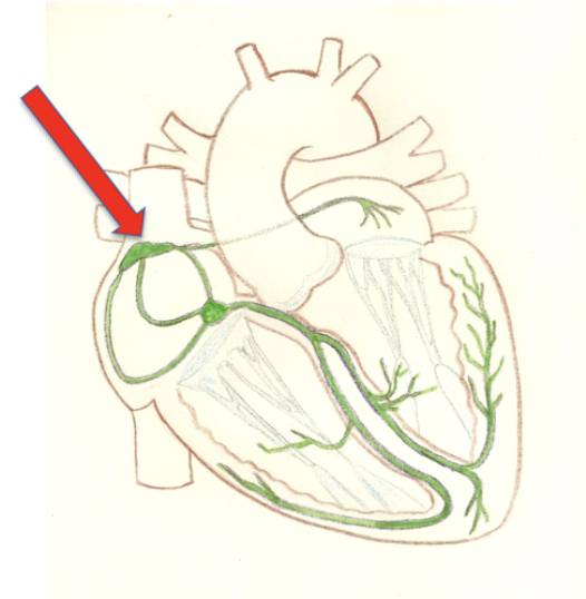
Located in the right atrial wall and is known as the heart’s pacemaker
sinoatrial node
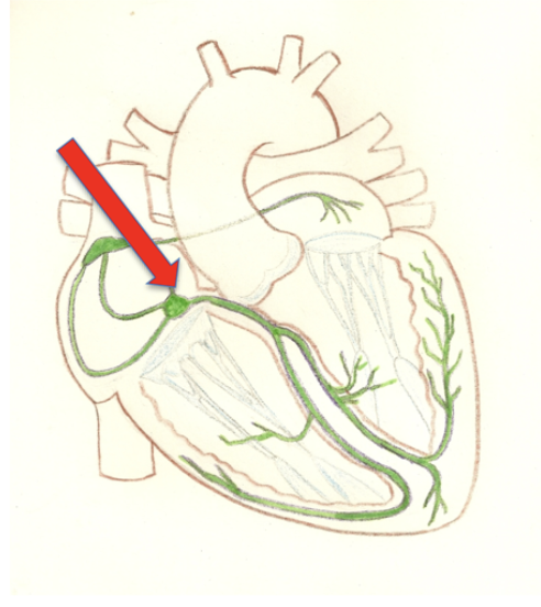
Located in the inferior segment and receives input from SA node. Slow signal due to fewer gap junctions
atrioventricular node
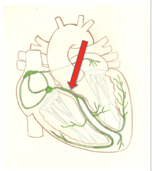
Located in the superior portion and receives signal from the AV node with no gap junctions to pass electrical signals. This is the first segment linking atrial conduction to ventricular conduction
atrioventricular bundle
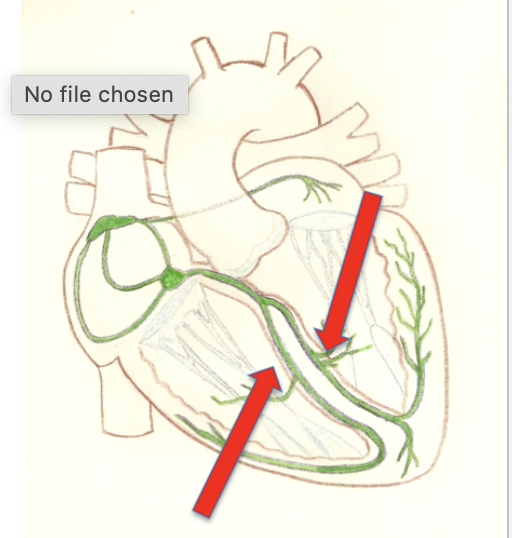
Located along left and right sides and receives signal from the AV bundle and send it down to the apex of the heart. Sends electrical signal to the left and right ventricular walls
bundle branches
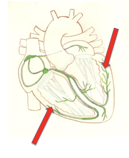
penetrate into the apex and receives signal from the bundle branches and completes the conduction cycle. more elaborate in the left chamber
purkinje fibers
What are the great vessels?
svc, ivc, pulmonary trunk, pulmonary veins, aorta
What vascularizes various structures of the head and neck?
external carotid artery
What does the venous drainage from the superior portions of the body drain through?
subclavians and jugular veins from brachiocephalic veins
the brachiocephalic veins converge to form what?
superior vena cava
What are the drainage sites of lymphatic ducts?
left and right venous angles
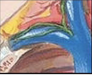
What drains the upper extremity?
right lymphatic duct
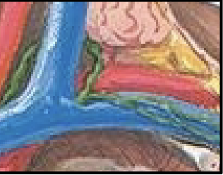
What drains all other aspects of the body and empties into the left venous angle?
thoracic duct
What is air transport and other functions?
conducting
What is gas exchange?
respiratory
Most muscles that control the larynx are controlled by what cranial nerve?
vagus nerve
What is it called when you insert the blade into the vallecula to pull the epiglottis out of the way?
intubation
What is a mediastinal structure which transmits air and has rings of cartilage, connective tissues, and muscle?
trachea
How many times will the bronchial tree divide up to?
23
What extensive surface area increases the efficiency of gas exchange?
alveoli
Alveoli are covered in what?
extensive capillary network
What branch off the aorta to feed the bronchi?
bronchial arteries
Bronchial arteries provide nutrients to what?
pulmonary parenchyma
The pleura of the lungs are composed of what?
visceral and parietal layers
The parietal pleura forms what?
costal, diaphragmatic, mediastinal, and cervical pleura
Pleura reflects in on itself where?
hilum of the lung
This happens when air enters the pleural cavity and can cause the lung to collapse inwards
tension pneumothorax
The lobes of the lung further divide into what?
bronchopulmonary segments
There is extensive lymph drainage of what?
bronchial tree and lungs
What are the openings of the diaphragm that allow passage of structures from the thorax to the abdomen?
caval foramen (T8), esophageal hiatus (T10), aortic hiatus (T12)
shiny surface of the lungs
visceral pleura
shiny surface on the inside of the thoracic cavity
parietal pleura
spaces between the two pleurae, contains lubricating, pleural fluid
pleural cavities
what is a common site of fluid collection in the pleural space?
costodiaphragmatic recess
central receptors of the brainstem sense what?
co2 levels
peripheral receptors in the carotid and aortic bodies sense what?
o2 levels and ph
In a fetus, oxygenated blood comes from what?
placenta
What are the 3 bypasses of a fetus?
2 bypass the fetal lungs (foramen ovale and ductus arteriosis) and 1 bypasses liver (ductus venosus)
the placenta develops from what?
trophoblast cells
What assumes responsibility for hormone production and nutrient/oxygen supply to the fetus?
placenta
What is the umbilical cord composed of?
1 umbilical vein and 2 umbilical arteries
This goes towards the heart and carries O2-rich blood/nutrients from placenta to fetal vessels
umbilical vein
This carries CO2 and waste products from fetus to placenta and arise from internal iliac arteries
umbilical arteries
The placenta detoxifies circulating blood so not all blood needs to pass through what?
liver parenchyma
What shunts some blood away from fetal liver allowing O2/nutrients?
ductus venosus
What is a one way valve from the right atrium to the left atrium?
foramen ovale
Where is the higher pressure in the heart valve?
right atrium
When the foramen ovale closes it becomes what?
foramen ovalis
What is the connection between pulmonary trunk and aorta?
ductus arteriosus
When the ductus arteriosus closes it becomes what?
ligamentum arteriosum
What continues inferior to the pharynx?
proximal esophagus