Antigen and MCH processing
 Call Kai
 Learn
Learn Practice Test
 Spaced Repetition
 Match
Match Flashcards
 Knowt Play
1/19

There's no tags or description
Looks like no tags are added yet.
Name | Mastery | Learn | Test | Matching | Spaced | Call with Kai | Chat |
|---|
No analytics yet
Send a link to your students to track their progress
20 Terms
How does T cells recognize antigens?
T cells do not recognize microbes/antigen but peptides
The peptide needs to be displayed on the surface of the cell
Antigen presenting cells (APCs) degrade microbes and present peptides
APCs display the peptides om MIC-molecules (Major Histocompatibility Complex) - also called HLA in humans
By binding to MHC + peptide with it’s T cell receptor (TCR) the T cell can get activated
What are the two types of MHC molecules, which type of antigens do they present, and which T cells do they activate?
MHC-I molecules
Present peptides from intracellular antigens
Activate CD8+ cytotoxic T cells
MHC-II molecules
Present peptides from extracellular antigens
Activate CD4+ helper T cells
What is the fundamental biological principle behind why different pathogens are directed to either MHC-I/CD8 or MHC-II/CD4 pathways?
Intracellular Threats (Cytosol) = Pathogens multiplying freely in the cytosol (like viruses) require MHC-I to signal CD8+ Cytotoxic T cells to kill the infected cell.
Extracellular/Vesicular Threats = Pathogens outside cells or trapped in vesicles require MHC-II to signal CD4+ Helper T cells to activate cellular help (boosting macrophages or producing antibodies) rather than killing the cell.

How are peptides presented on MHC molecules? What is it that the T cell sees?

What did the Ovalbumin experiments prove about how a cell decides between MHC Class I and MHC Class II presentation?
They proved that antigen location (geography) completely determines the pathway, not the antigen's origin:
Trapped in Vesicles (Endocytosis / Row A) - Binds MHC Class II - Activates CD4+ T cells.
Loose in Cytosol (Synthesis or Artificial Entry / Rows B & C) - Binds MHC Class I - Activates CD8+ T cells.
Key Proof: Forcing an external protein into the cytosol switches its presentation from Class II to Class I.

How are endocytosed antigens processed for MHC-II presentation?
(it is loaded into the vacuole) (expressed on antigen presenting calls)
1. Uptake of extracellular proteins into vesicular compartment of APC
Processing of internalized protein in endosomal/lysosomal vesicles
Biosynthesis and transport of class II MHC molecules to endosomes that is associated with invariant chain (Ii)
After that the the Ii breaks of and the MHC II molecule is left with CLIP that get removed from the binding groove by DM
Expression of peptide-MHC complex on cell surface
MHC II = CD4+
CLIP= CLass II-associated Ii Peptide
(HLA)-DM

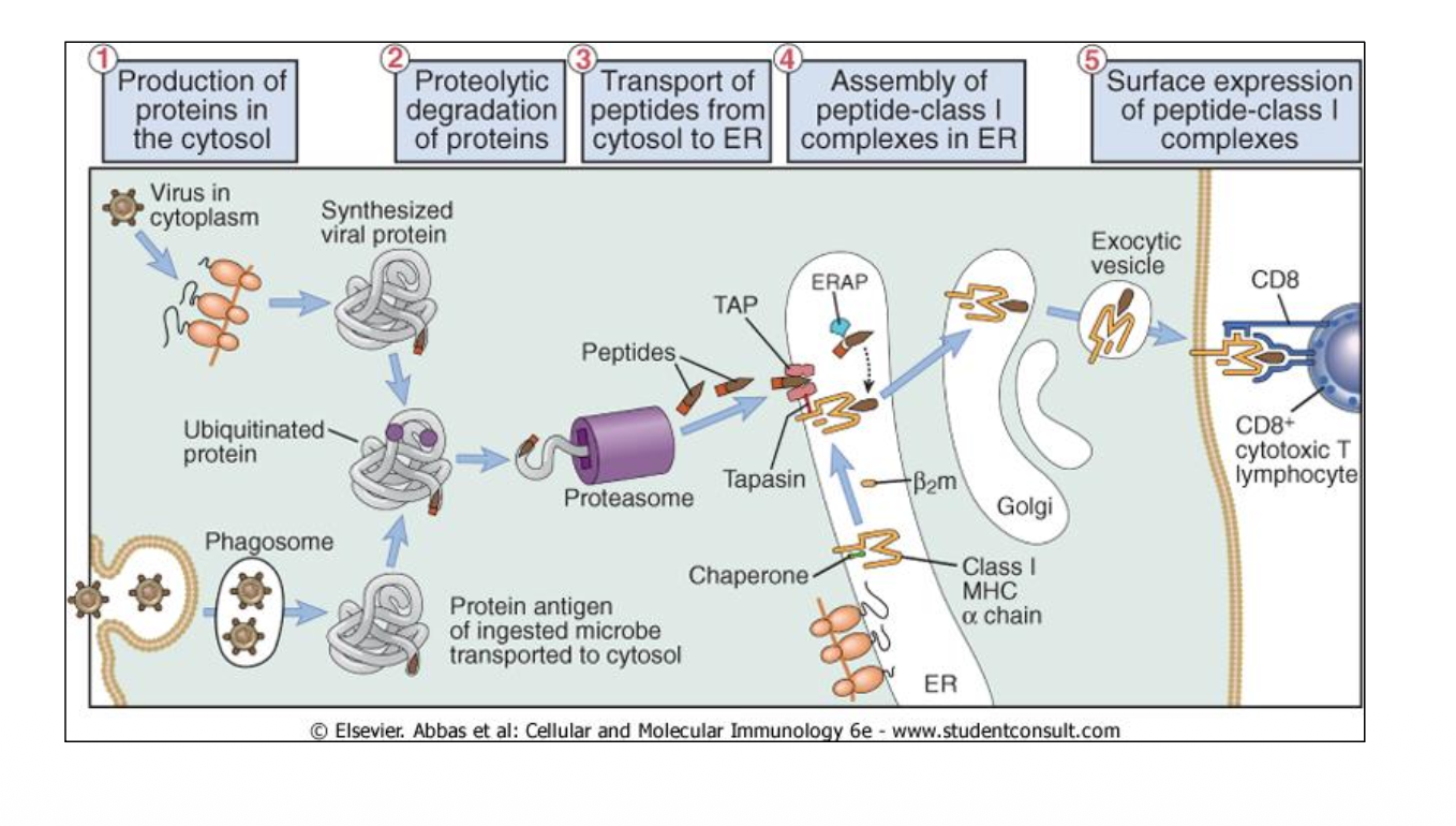
How cytosolic antigens processed for MHC-I presentation?
(It is loaded in the ER) (expressed on all nucleated cells)
Production of proteins in the cytosol
Then the protein get marked by Ubiquitin (Ubiquitination) for the protaesome to recognize it
And then chopped it of into smaller peptide
With the help of the TAP (translocation pore) the peptide is transported into the ER
In the ER there is a link binded to TAP that holds the MHC-I chain (It increases peptide translocation by linking TAP to MHC-I (it acts as protective shield to TAP and shield it from degration) and make the chain more stable)
Before the peptide is bound to the MHC-I chain it get more chopped By ERAP enzyme
Once the MHC Class I-peptide complexes are assembled in the ER, they are transported to the cell surface
MHC-I = CD8+
Cross-presentation is the process where a cell engulfs a microbe into a phagosome (or can be died free particlas of pathogens), transports the ingested microbial proteins into the cytosol, and processes them for display on MHC Class I molecules
Proteasome: It is major non lysosomal degradation system. Large complex 700kDa, 28 subunits, and it can be found in the cytosol.
Transporter associated with antigen processing (TAP): It’s a heterodimer in the ER. Transport peptide into the ER.
In the absence of TAP the MHC-I chain becomes unstable and the peptide become unable to enter the ER and express the antigen to the CD8+ cells.
How do B cells and T cells differ regarding:
Antigen shape recognized?
Types of molecules targeted?
Requirement for antigen processing?
B-cells:
• Recognises a three dimensional structure of the antigen
• Recognises proteins, carbohydrates and lipids.
T-cells:
• Recognises linear peptides
• Conventional T cells only recognises peptides derived from proteins.
• APCs need to degrade the protein and display the peptide to activate the T cell.

How T and B cells generate receptors?
The receptors ar encoded by different gene segment
The gene segments are recombined in the bone marrow (B cells) and Thymus (T cells)
Different chains of the receptors are paired togahter creating one unique receptor that is expressed by each cell

What is that the T cell receptor sees?
It sees the peptide and the MHC molecule. A single MHC molecule cannot bind to all types of peptides.
How does the immune system present so many different microbial peptides?
Polygenic Locus: An individual inherits multiple distinct genes performing the same functional role. This includes three different MHC-I genes (HLA-A, HLA-B, HLA-C) and three different MHC-II genes (HLA-DR, HLA-DQ, HLA-DP).
High Polymorphism: There are thousands of different variants (alleles) of these genes across the human population (e.g., hundreds of variants for HLA-B alone).
Co-Dominant Expression: Both copies of the HLA locus inherited from the parents are expressed simultaneously on the cell surface. This maximizes the total number of individual MHC molecules available to present antigens in a single person.
How does the enormous diversity of MHC molecules affect antigen presentation versus organ transplantation?
Antigen Presentation (Benefit): It ensures an individual can present a vast number of unique foreign peptides to fight diverse infections.
Organ Transplantation (Drawback): It complicates donor matching because a recipient's immune system easily recognizes a donor's unique MHC molecules as foreign, leading to graft rejection.
What is a syngeneic strain of mice, and how did it lead to the discovery of the MHC?
Syngeneic strain: A group of laboratory mice that are genetically identical to one another (essentially clones). (homozygous at every locus)
The mice have zero genetic variation, which allowed scientists to discover the MHC locus through skin graft transplantation experiments (grafts between syngeneic mice are accepted, while grafts between different strains are rejected).

What is the concept of MHC Restriction demonstrated by this experiment?
MHC Restriction is the principle that a T cell receptor (TCR) cannot recognize an antigen on its own.
To trigger an immune response, the T cell must simultaneously recognize both:
The specific foreign peptide
The correct "self" MHC molecule hosting that peptide
If either the peptide is wrong (uninfected cell) or the MHC molecule is from a different genetic strain (allogeneic MHC), the T cell remains blind to the threat and will not activate.

Summery of MHC I and II:
They are also heterodimer

What are the three professional Antigen-Presenting Cells (APCs), and what are their primary mechanisms for antigen uptake?

What are the key characteristics of Dendritic Cells (DCs) regarding their location, movement, and primary role in the immune system?
Primary Role: They are the main cells that initiate primary T cell responses.
Location & Anatomy: They have a large surface area and are stationed as scouts in all peripheral tissues (where they are phenotypically immature).
Migration: After capturing an antigen, they migrate to the draining lymph node.
Final Destination: Once inside lymphoid tissues, phenotypically mature DCs home directly to T cell areas to present antigens and activate naive T cells.
(DC originate in the bone marrow, travel through the blood into peripheral tissues to scout for antigens, and then migrate via lymphatic vessels to settle inside a lymph node).
What four major changes occur during dendritic cell (DC) activation after encountering a microbe?
Upregulated CCR7: Increases migration to regional lymph nodes
Downregulated antigen processing: Halts further pathogen uptake
Upregulated surface MHC: Optimizes the physical interaction with T cells
Upregulated co-stimulatory molecules: Delivers the "second signal" to inform T cells that a microbe was encountered

What three signals are required to activate a naive T cell, and what is the immediate cellular result?
Three Required Signals:
Signal 1: Antigen-specific recognition via MHC-TCR binding
Signal 2: Co-stimulatory
Signal 3: Cytokine secretion
Resulting Downstream Steps:
IL-2 secretion
The IL-2 binds back to the T cell.
Proliferation: The T cell begins rapid cell division.