ANAT 403 Back and Spinal Cord
 Call Kai
 Learn
Learn Practice Test
 Spaced Repetition
 Match
Match Flashcards
 Knowt Play
1/49

There's no tags or description
Looks like no tags are added yet.
Name | Mastery | Learn | Test | Matching | Spaced | Call with Kai | Chat |
|---|
No analytics yet
Send a link to your students to track their progress
50 Terms
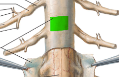
Spinal cord dura mater
First layer, protective. Ends at S2 level.
Spinal cord arachnoid mater
Second layer, beneath which lies the subarachnoid space filled with CSF.
Spinal cord pia mater
Innermost layer, intimate to the spinal cord itself (impossible to seperate).

Dorsal root ganglion
Where the pseudounipolar sensory neuron cell bodies lie.

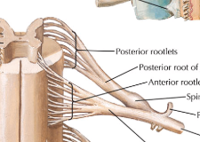
Typical mixed spinal nerve
Contains a motor and sensory portion, emerging from both the dorsal and ventral roots of the spinal cord
Conus Medullaris
Portion of the spinal cord in between L1-2 that is cone shaped and marks the end of the main portion

Cauda equina
Outgrowth of spinal nerves which hang down past the conus medullaris to continue on to their exit points (sort of look like a horses tail). Formed because the vertebral column grows faster and larger than the main spinal cord ever does.
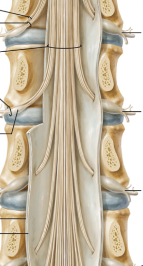
Filum terminale
String (outgrowth of the pia mater surrounding the spinal cord to S2), then outgrowth of dura mater tethers the coccyx to anchor the spinal cord.

Denticulate ligaments
Tether the chord laterally to the pia mater
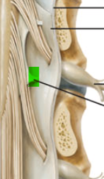
Ligamentum flavum
Holds the lamina, yellow due to elastic property
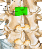
Posterior longitudinal ligaments
Inside the vertebral canal, against the bodies.

Anterior lognitudinal ligament
Most anteiror against the vertebral canal, travels from the skull to the sacrum.
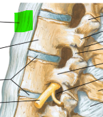
Interspinous ligaments
Holds the spinous processes together, second most posterior

Supraspinous ligaments
Most posterior, runs along the spinous process

Inner portion of spinal cord
Grey matter (reverse from brain - made of all the unmyelinated cell bodies and dendrites)

Dorsal primary ramus
Relaying information to/from erector spinous muscles and skin of back (do not have to travel very far/do much work at all).

Ventral primary ramus
Relaying information to/from many other places in the body, projecting all the way to anterior surfaces (lots more work being done).

Midsection dermatomes
Responsible for the feeling of reffered pain found in visceral structures (entire dermatome hurts). T1 = starts at clavicles, T4 = nipples, T7 = xiphoid process (tip of sternum), T10 = umbillicus and T12/L1 = fold at hip joint.

Primary vertebral curves
Project backwards (thoracic and sacral) - red
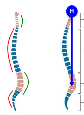
Secondary curves
Project forwards (lumbar and cervical) - green
Why does the spine curve?
Lines the head up with the center of gravity; a curved spine is also able to withstand compression much better than a straight spine, seen in infants.
Vertebral body
1
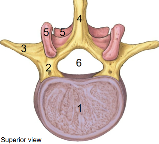
Pedicle
2
Transverse process
3
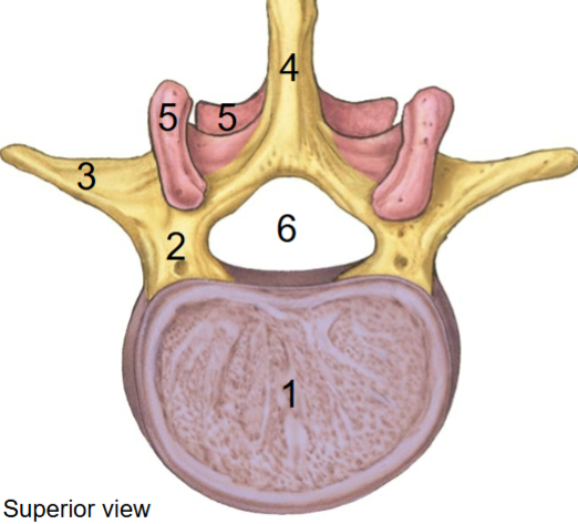
Spinous process
4 (the ones we can feel)
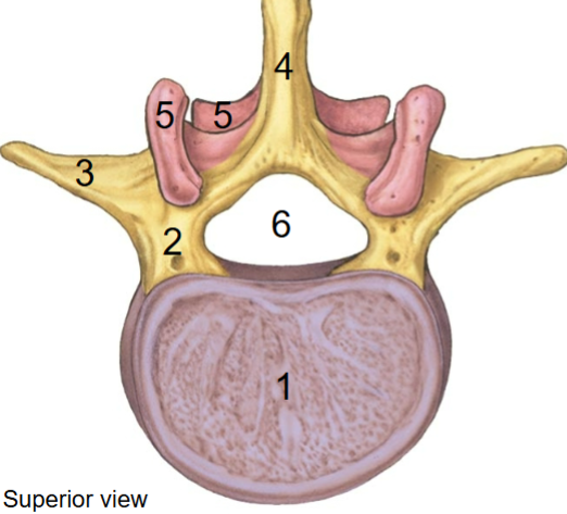
Articular process
5
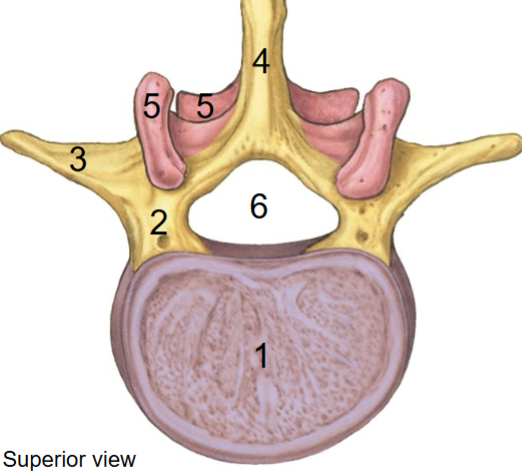
Vertebral joints (2)
Intervertebral joint (discs): between adjacent vertebrae bodies (secondary cartilaginous joint)
Zygapophyseal joint: Between two adjacent vertebral processes (synovial joint)
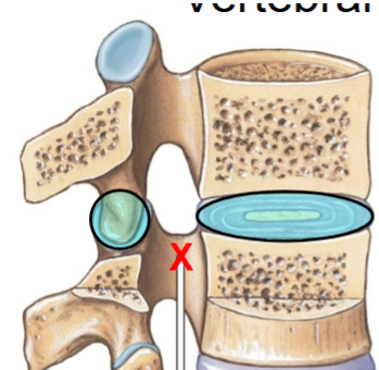
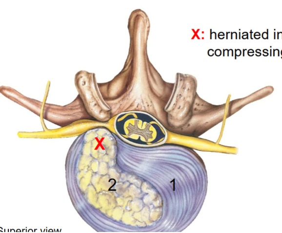
Annulus fibrosa
Cartilage portion of the body in vertebra (1)
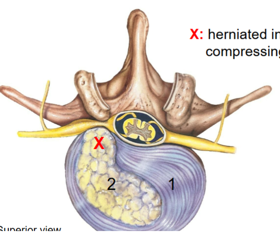
Nucleus pulposus
Gelatinous, cushy material in the body of the vertebra (2). Formed from the notochord.
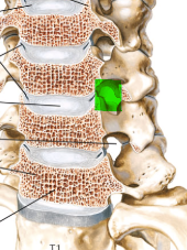
Intervertebral foreamen
Space between the pedicles of two adjacent vertebrae, to allow for spinal nerve passage.
Vertebral foreamen
The central passage formed by the vertebral canal which allows for the spinal cord to pass through

Cervical vertebrae
7 total. Contain a transverse foramen for vertebral arteries to travel through up towards the brain; bifid spinous process (except for C7/C1/C2). Coronal plane zygopophysis (articular facet). Increased ability for motion, but this also means an increased liklihood of injury via movement in multiple planes. Contain a larger vertebral foreamen since the spinal cord increases in size (because of brain needs and plexus emerging here which contain control of the hands = fine motor skills and thus large amounts of nerves)
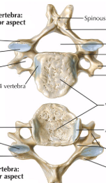
Atlas
C1. Wonky, allows for flexion/extension of the head, as well as rotation (to a lesser extent). Body is replaced by two arches, contains a massive zygopophysis joint to hold the skull (forms the atlanto-occipital joint).

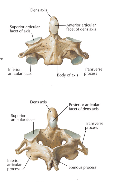
Axis
C2. Wonky, supports atlas to allow for rotation (through the atlanto-axial synovial joint). Contains the Dens/Odontoid process (fits through the anterior arch of the atlas).
Thoracic vertebrae
12 total. Contain two more synovial joints (costovertebral joint) for ribs to attach to (allows them to move slightly when breathing). Possess coronal plane zygopophysis (articular facets), which allows for rotation, but this is restricted by the ribs until floating ribs (T11 and T12). Some movement of rotation but no bending of back (no flexion here due to ribs limiting movement)

T12
Transitional vertebrae. Thoracic top half, lumbar bottom half. Contains facet for ribs.

Lumbar vertebrae
5 total. Very thick bodies, look like a moose head laterally. Articular facets (zygopophysis) oriented in sagital plane, which allows for flexion/extension but not rotation/twist.

Sacrum
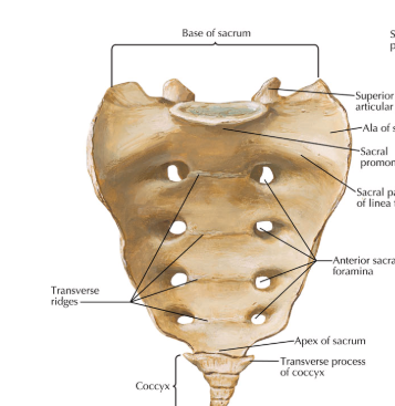
S1 - S5 combine together to form this one structure. Contain anterior and posterior foreamen (the holes that look like they were drilled into the bone) - these are for the lower dorsal and ventral rami, which turn into the lumbrosacral plexus and the sciatic nerve. Triangular vertebral foreamen
Coccyx
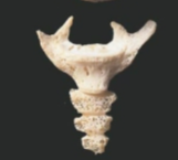
Can be up to 4. Leftover remains of tails. Largely useless.
Scapular spine
7
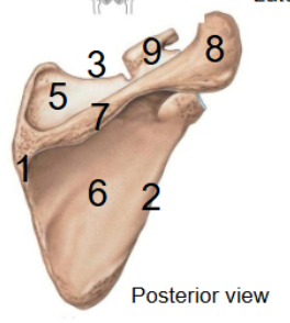
Acromion
8
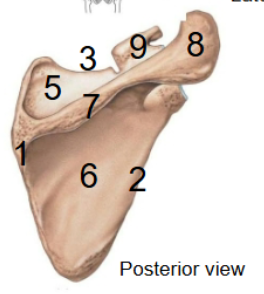
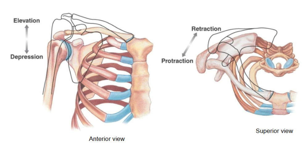
What’s special about the scapula
Has the scapulothoracic “joint”, which is not really a joint but rather is the term used to describe how the scap is able to glide along the rib cage. Can do both elevation/depression (shrugs) and retraction/protraction (reaching forward)
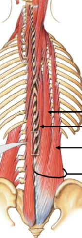
Erector Spinae
Spinalis, Longissiums and Iliocostalis muscles
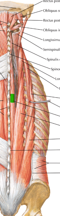
Spinalis
Extension and lateral flexion of the vertebral column. Epaxial (dorsal root of spinal nerves). Most medial of the three (nearest the vertebral column), best for sagital plane movement
Capitis branch (head): spinous process of C7-T1 → occipital bone (proturburance)
Colli branch (neck): Spinous process of C7-T2 → spinous process of C2-C4
Thoracic branch: Spinous process of T11-L2 → Spinous process of T2-T8
Longissimus
Extension and lateral flexion of the vertebral column. Epaxial (dorsal root of spinal nerves). In the centre, does all purpose movement.
Capitis branch (head): Transverse process C4-T4 → mastoid process
Colli branch (neck): Transverse process T1-T5 → Transverse process C2-C6
Thoracic branch: Spinous process L1-L5 and posterior sacrum → T1-T12, vertebrae ribs 5-12, accessory and transverse process of L1-L5 vertebrae

Iliocostalis
Extension and lateral flexion of the vertebral column. Epaxial (dorsal root of spinal nerves). Most lateral, best for lateral movement (coronal plane).
Colli branch (neck): Ribs 3-6 → posterior turbercles C4-C6 vertebrae
Thoracic branch: Ribs 7-12 → ribs 1-6
Lumborum: Sacrum and illiac crest → Ribs 4-12, transverse processes of L1-L4 vertebrae

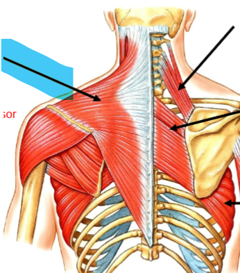
Trapezius
Pectoral girdle muscle - elevator/depressor/retractor of the scapula (at scapular-thoracic joint). Convergent from spinous process’ C7-T12 → scapular spine and first third of the clavicle. CN XI. (Hypaxial)
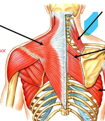
Levator scapulae
Elevates the scapula (at scapular-thoracic joint). Attached at C1/C2 transverse process, C3/C4 posterior turbucle → medial border of scapula (superior to the spine). Ventral rami of C3/C4 Spinal nerve (Hypaxial)
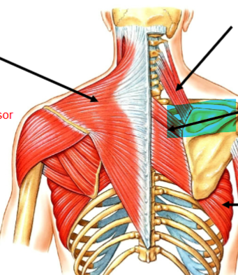
Rhomboids (major and minor)
Retracts the scapula (at scapular-thoracic joint). Attached at Spinous process C7-T5 → medial border of scapula (inferior to spine). Dorsal scapular nerve (hypaxial)
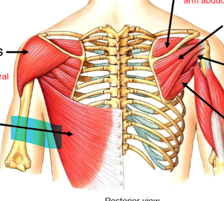
Latissimus dorsi
Humerous extension, adductor and medial rotation (at shoulder joint). Attached at spinous process of T7-T12, posterior layer of thoracolumbar fascia (L1-L5 and iliac crest, ribs 10-12 → intertubucular sulcus of humerus. Thoracodorsal nerve (Hypaxial)