Nitrogen Metabolism I: Biochem MT2
 Call Kai
 Learn
Learn Practice Test
 Spaced Repetition
 Match
Match Flashcards
 Knowt Play
1/60

There's no tags or description
Looks like no tags are added yet.
Name | Mastery | Learn | Test | Matching | Spaced | Call with Kai | Chat |
|---|
No analytics yet
Send a link to your students to track their progress
61 Terms
Excess AA ____ be stored->_____ Turnover: _____
~46-56 gm degraded/day need to replace
can't
degraded
400 gm/day
AA degradation links to what?
TCA cycle
acetoacetate
pyruvate
fumarate
aKG
Prop CoA
OAA
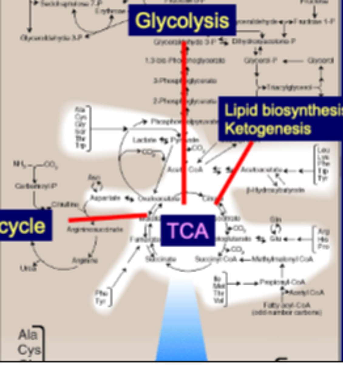
Glucogenic AAs
Glucogenic AAs can be converted to "Glucose/Glycogen"
-generates Pyruvate or an intermediate of TCA cycle
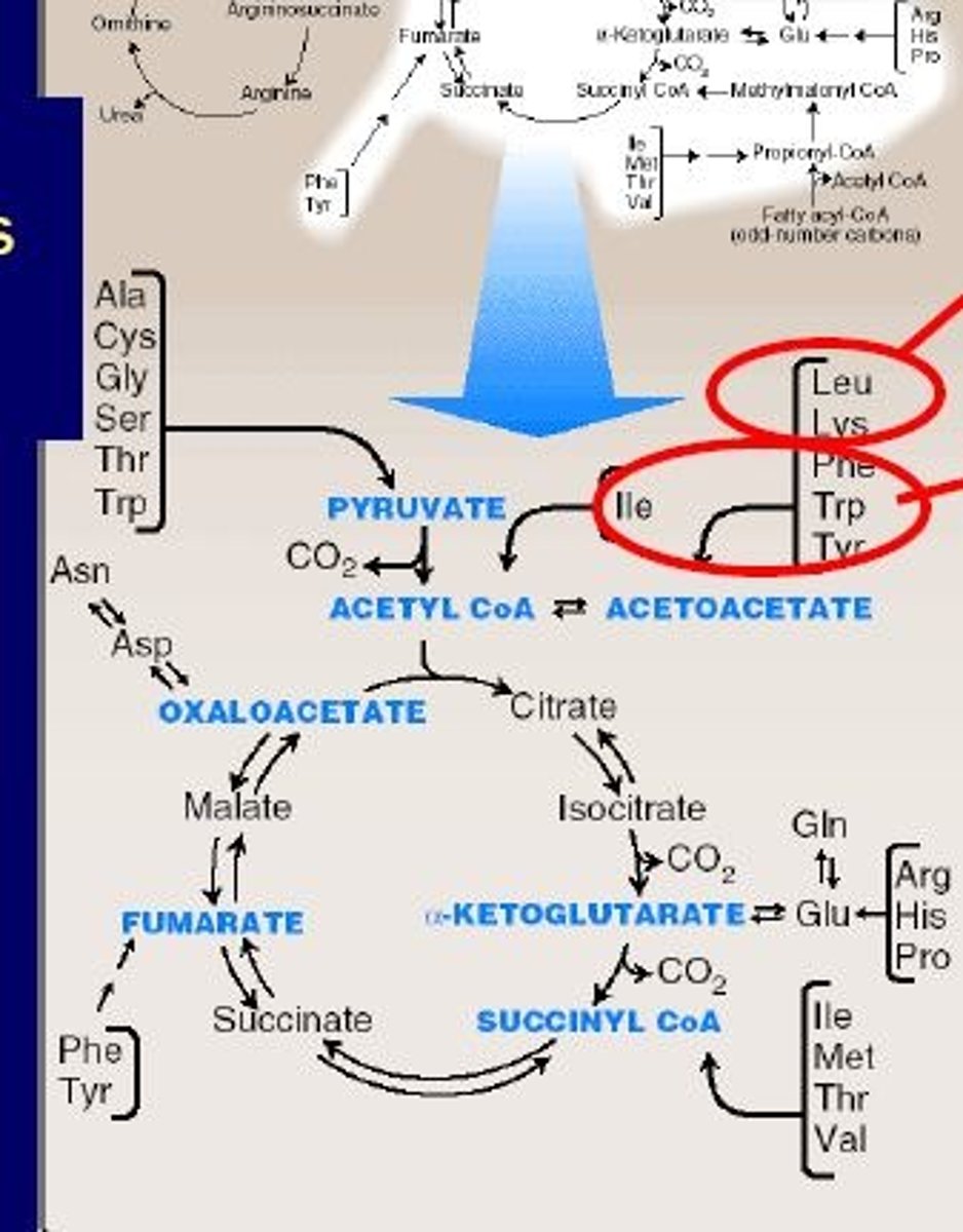
Ketogenic AAs
Ketogenic can be converted into ketone bodies
-directly generates Ketone bodies (acetoacetate, acetyl CoA or acetoacetyl CoA)
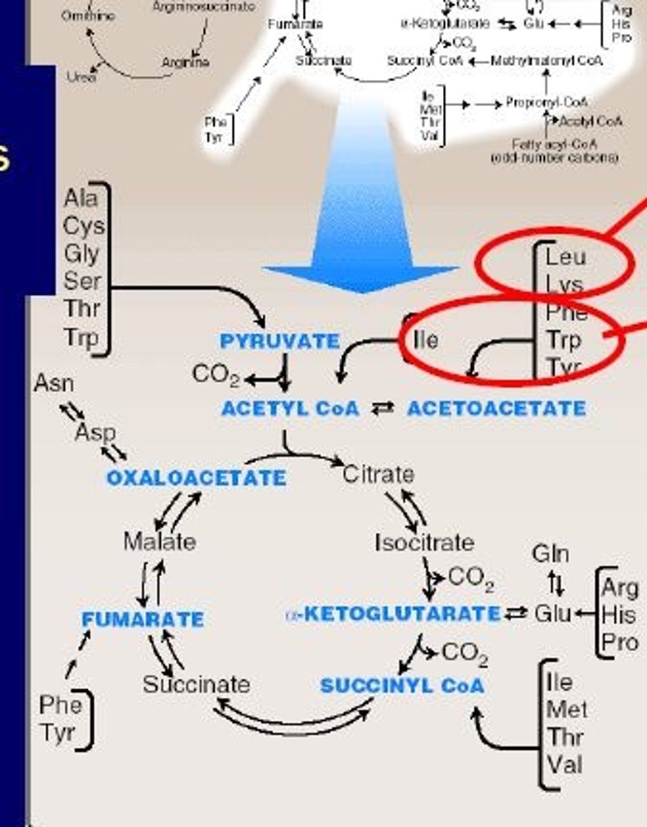
Ketogenic AA examples
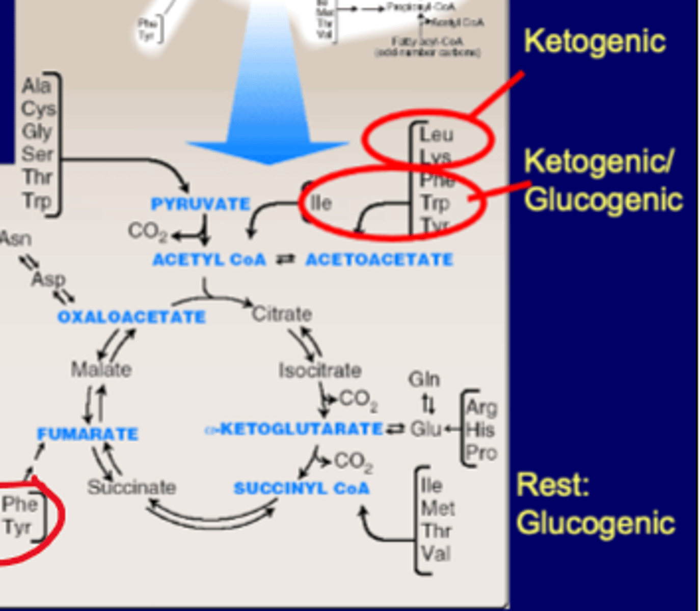
Leu, Lys->Acetoacetate
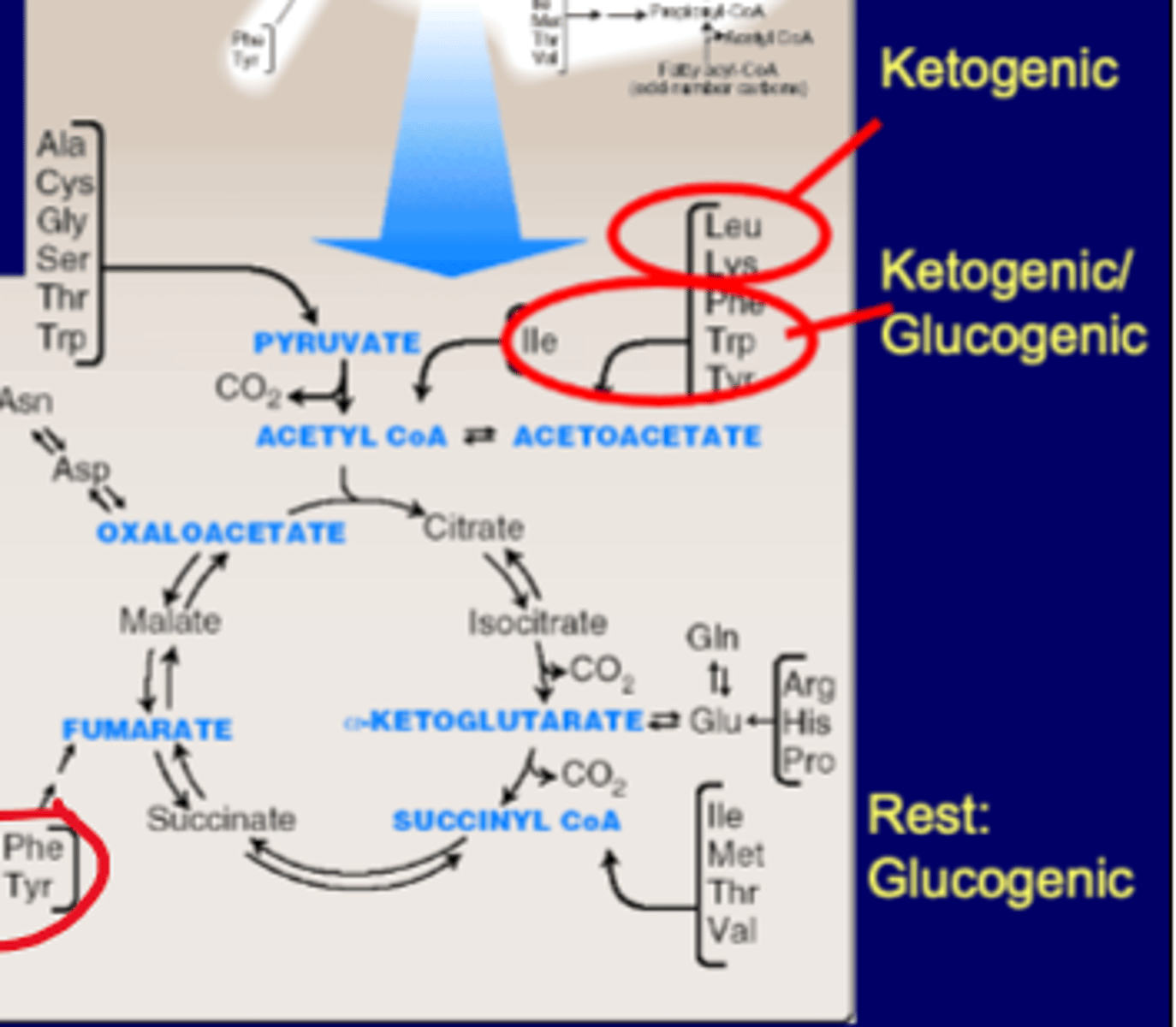
Ketogenic/Glucogenic examples
Phe, Trp, Tyr->Acetoacetate (Phe and Tyr also gluco for fumarate and Trp gluco for pyruvate)
Ile->ACoA (also gluco for Succ CoA)
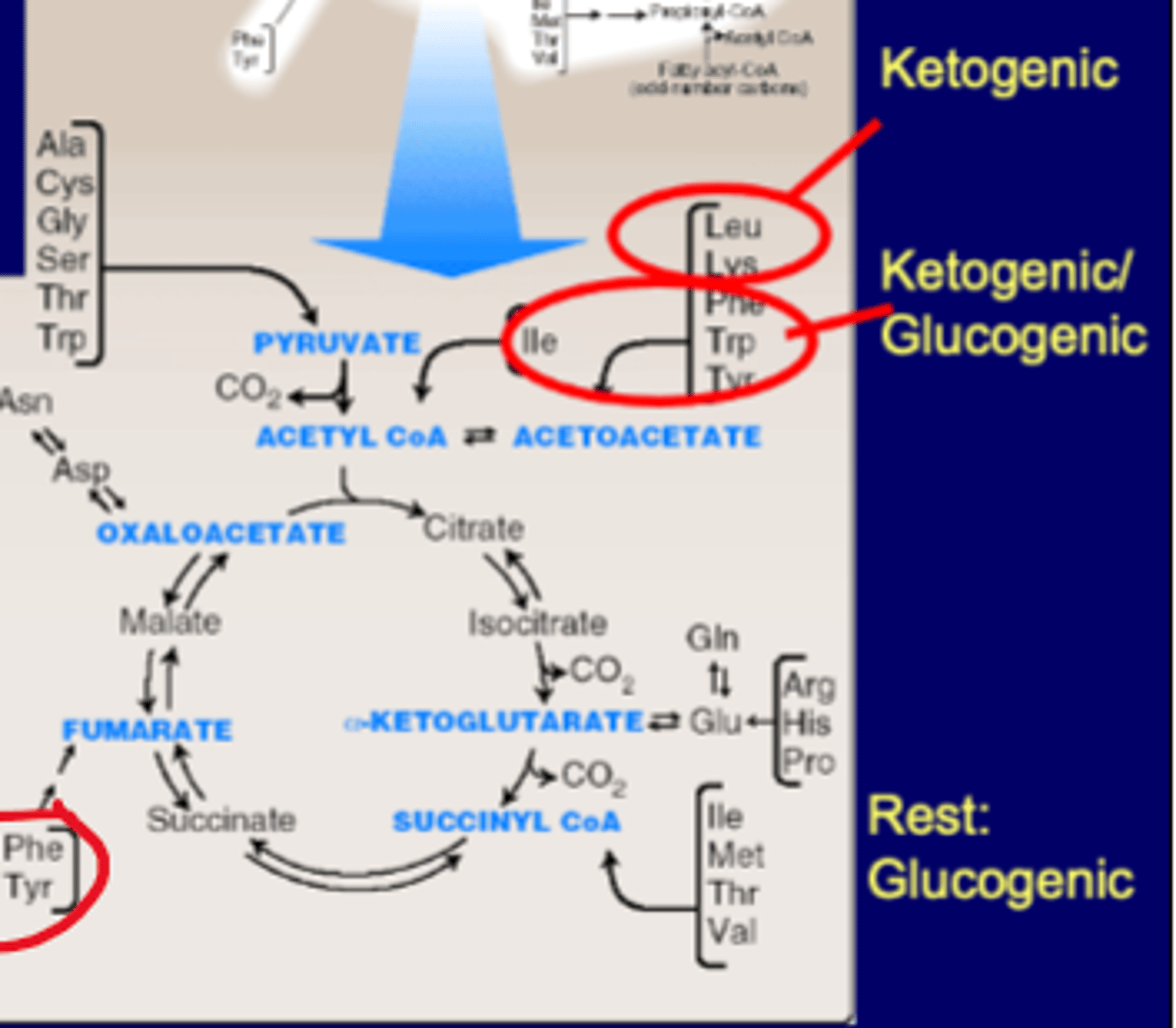
Glucogenic (the rest) examples
Phe, Tyr->Fumarate (Phe and Tyr also keto for acetoacetate)
Arg, His, Pro->Glu->aKG
Glu-><-Gln
Ile, Met, Thr, Val->Succ CoA (Ile also keto for ACoA)
Ala, Cys, Gly, Ser, Thr, Trp->Pyruvate (Trp also keto for acetoacetate)
Degradation of branched-chain amino acids (BCAAs)
-Universal fuels (~40% of total amino acids)
-metabolized by thes peripheral tissue (particularly muscle)
-defects cause accumulation of a-keto acids (Maple syrup urine disease)-a-keto acids have same co factors as a
KG (lipoic acid etc)
Maple Syrup Urine Disease
-caused by defect in a keto acid dehydrogenase
-causes brown color, sweet smelling urine
Catabolism of AAs: the Amino nitrogen portion of an AA turns into what and the carbon skeleton to what?
-Amino Nitrogen to urea and glutamine
-the carbon skeleton to intermediates metabolism
Three coenzymes used in AA metabolism
-Pyriodxal phosphate (PLP/B6)
-Tetrahydrofolate (FH4) (folic acid, B vitamin)
-Tetrahydrobiopterin (BH4)
Catabolism of amino acids: Two major reactions
-transamination
-oxidative deamination
Transamination reaction
-transamination is followed by deamination
-transports NH2 from aspartate to a KG to form glutamate
-glutamate can lose NH3 (NH2 plus an H) to the urea cycle
Aspartate Transaminase (AST)
Aspartate + a-keto glutarate w/AST -> OAA + glutamate (can also go other way)
transfers NH2 group from Asp to aKG
same pathway as GOT, can use either one
most transaminases use aKG
Glutamate Oxaloacetate Transaminase (GOT)
Aspartate + a-keto glutarate w/AST -> OAA + glutamate
transfers NH2 group from Asp to aKG
same pathway as AST, can use either one
most transaminases use aKG
Transaminases or Amino transferases require
Pyridoxal phosphate (PLP/B6)
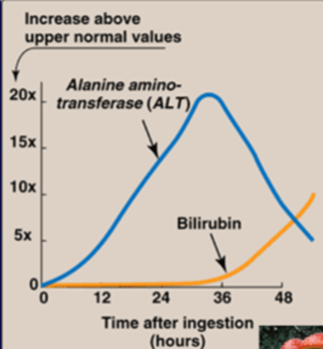
Transaminase (ex. ALT-ALanine amino-transferase) is a marker of ?
liver damage
-more effective than bilirubin as a marker (slow)
-amanita is toxic, can cause liver damage from mushrooms->causes ALT to leak out of the liver cell membrane
Serum transaminase: ALT (GPT) is more specific for
for liver cytoplasmic
AST (GOT) is specific for
mitochondrial & cytoplasmic liver disease
serum [AST]=2x [ALT] in?
alcoholic liver disease
ALT transfers
alanine to alpha-keto glutarate, resulting in the formation of pyruvate and glutamate
Role of glutamate in urea cycle
-1st step is AA w/ TA->glutamate using aKG
-2nd step is moving nitrogen from glutamate to make free ammonium using GDH (oxidative deamination)
-glutamate w/ TA + OAA -> Aspartate (keeps nitrogen, also uses aKG)
-glutamine shuttle from brain (protects brain), can also be from muscle
-alanine shuttle from muscle only
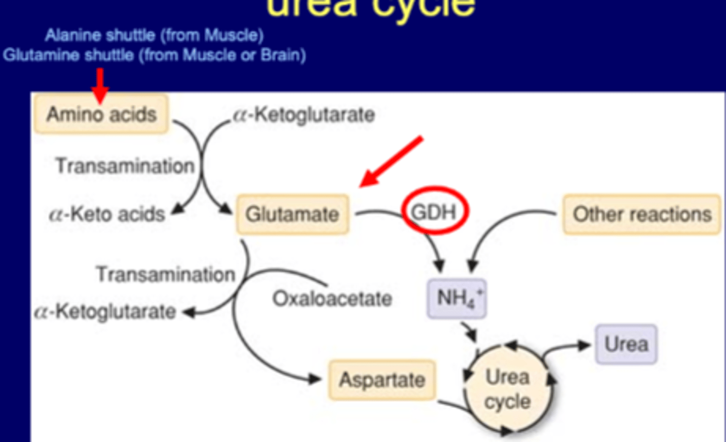
Glutamate dehydrogenase (oxidative deamination)
the deamination of glutamate by glutamate dehydrogenase (GDH) (coenzyme is NADH or NADPH) to form alpha keto-glutarate and NH3
Fate of AA and role of urea cycle
AA-> Carbon path or Nitrogen path
Nitrogen moved by TAs
-make free ammonium or ASP for urea cycle
-when urea cycle knocked out cause hyperammonemia and increased AAs
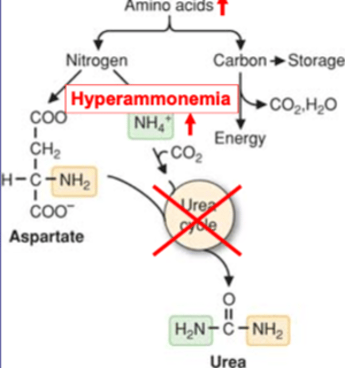
Urea cycle disorders (UCD) broadly
-hyperammonemia
-urea cycle disorders (UCD)
-UCDs, except for arginase deficiency, present in newborn who are initially well and become hyperammonemia after a period of protein feeding.
Hyperammonemia
-acquired: common: liver disease
-congenital: UCD (type of inborn errors of metabolism, IEM)
Urea cycle disorders (UCD) specific
deficiency of any of the urea cycle enzymes
-Ornithine transcarbamylase (OTC) deficiency (most common; X-linked; other UCDs are AR)
-Citrullinemia (ASS1/enzyme and SLC25A13/transporter mutations)
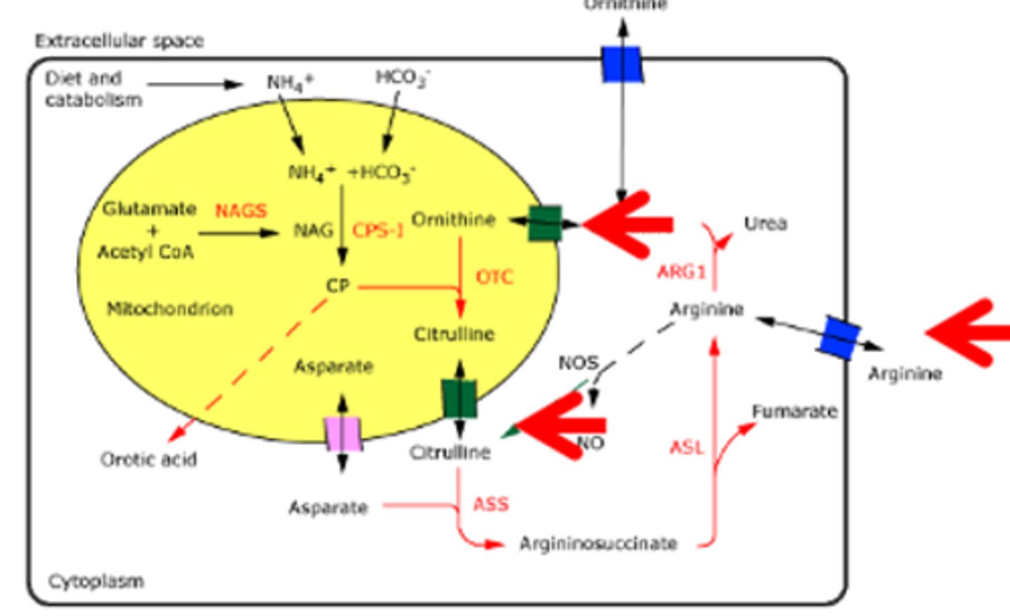
Urea Cycle Overview
-CPS1: rate limiting
-OTC, ASS: disorders
-Arginine: directly to urea cycle
-Enzymes: ARG1, ASL, ASS, CPS1, NAG, NAGS, OTC
most occurs in mito, citrulline has to be transported out, and orthenine has to be transported in
Urea Cycle Disfunction
OTC most common, X-linked (Orthenine to Citrulline)
all others are autosomal recessive
OTC deficient->accumulate CP, ammonium
ASS mutation affects citrulline transporter (to leave mito), will increase citrulline
ASS deficient/citrulline transport issue->accumulate citrulline, ammonium
all mutations except arginase will increase ammonium (NH4)
even if knock out arginase, can still release arginine, liver release
Essential AA
Carbon skeleton cannot be synthesized
Non-essential AA
can be synthesized
How many essential AAs? How many conditionally essential?
10 essential
6 conditional
Non-essential AA examples
non-essential AA Precursor
Glutamate, Glutamine aKG (common)
Aspartate, Asparagine OAA (TCA)
Serine 3-phosphoglycerate
Glycine serine
Tyrosine phenylalanine (important)
Proline glutamate
Alanine pyruvic acid (pyruvate, glycolysis)
Cysteine serine, methionine (met most)
Essential AA Examples
10 AAs
Phenylalanine 6.6 (most abundant)
Valine 4.4
Tryptophan 1.0 (less abundant)
Threonine 3.2
Isoleucine 3.8
Methionine 2.3
Histidine* 1.5
Arginine*
Leucine 6.4
Lysine 4.6
ideal proportion=ovalbumin
Arginine can be made, still need to eat-conditionally non-essential, Histidine essential for infants
Synthesis of AA
-synthesis by amidation
-synthesis from aKG
-proline synthesis from glutamate
-derived from glycolysis intermediates
-from essential AA
Conditionally essential AAs
6 AAs
Arginine
Cysteine
Glycine
Glutamine
Proline
Tyrosine
essential under catabolic disease (PKU) and other conditions
AAs synthesized by amidation
glutamine (from glutamate)
asparagine (amide donor is glutamine)
Amidation for glutamine
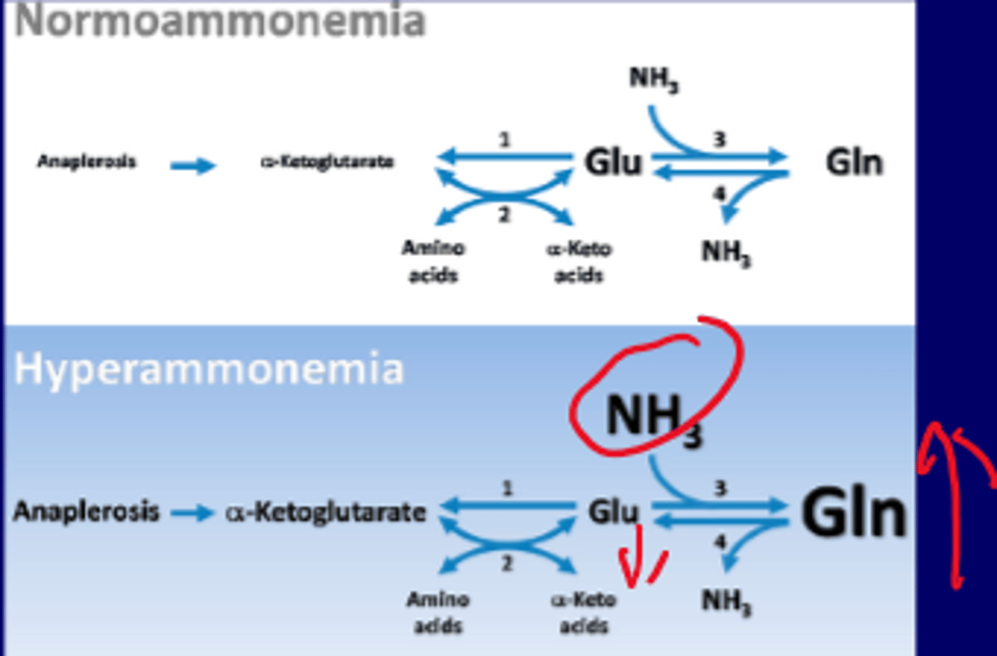
prevention of hyperammonemia
-detoxification of ammonia in brain and liver
-nontoxic storage for NH3
-uses aKG to make Glu, glutamine synthetase for Gln
-Glutamate (Glu) down; Glutamine (Gln) up, NH3 up (during hyperammonemia)
(treatment: can be removed by phenylbutyrate (prodrug) to phenylacetate to phenylacetylglutamine)
glutamine synthetase
an enzyme that plays an essential role in the metabolism of nitrogen by catalyzing the condensation of glutamate and ammonia to form glutamine
important in the brain
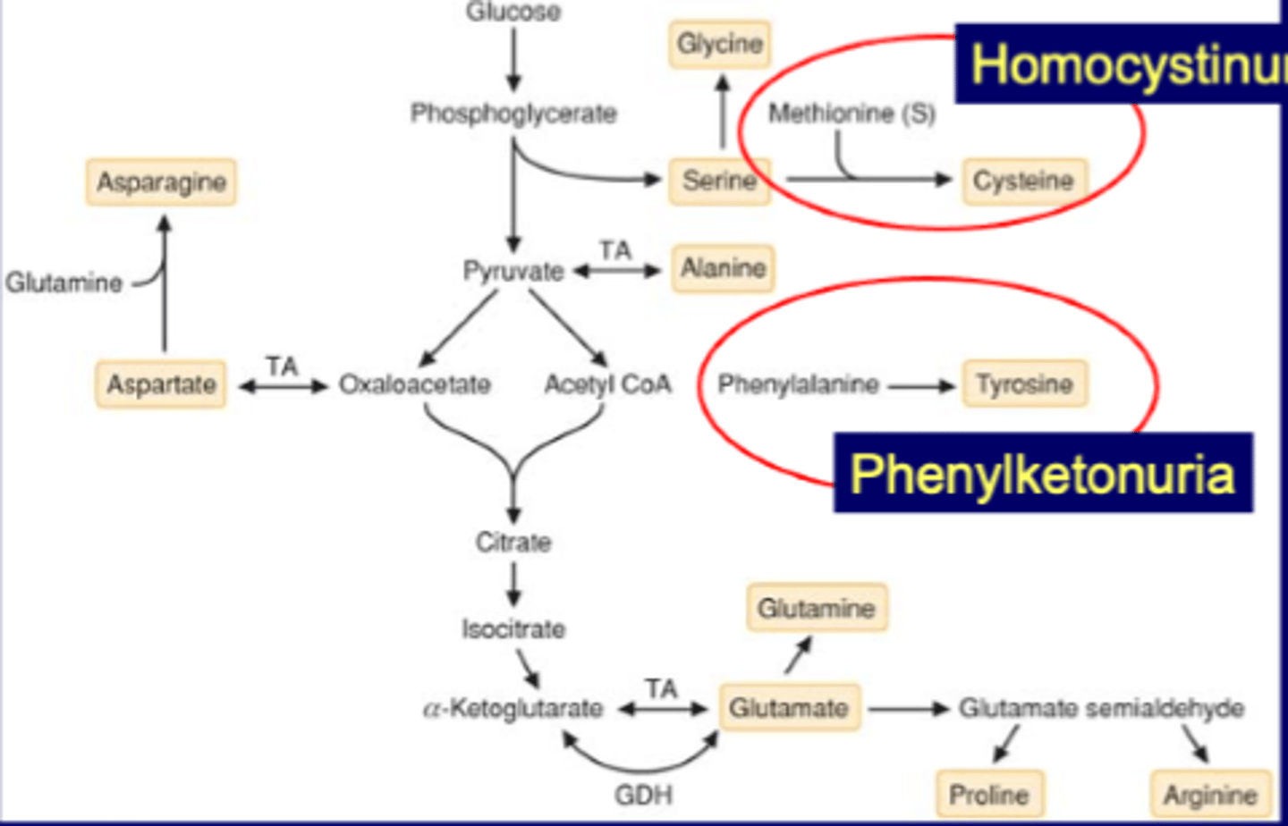
Overview: synthesis of non-essential AA
4 from glycolysis
Serine->Cysteine
Methionine (S)->Cysteine (deficiency is homocystinuria)
Phe->Tyrosine (deficiency is phenylketonuria (PKU))
Cysteine synthesis
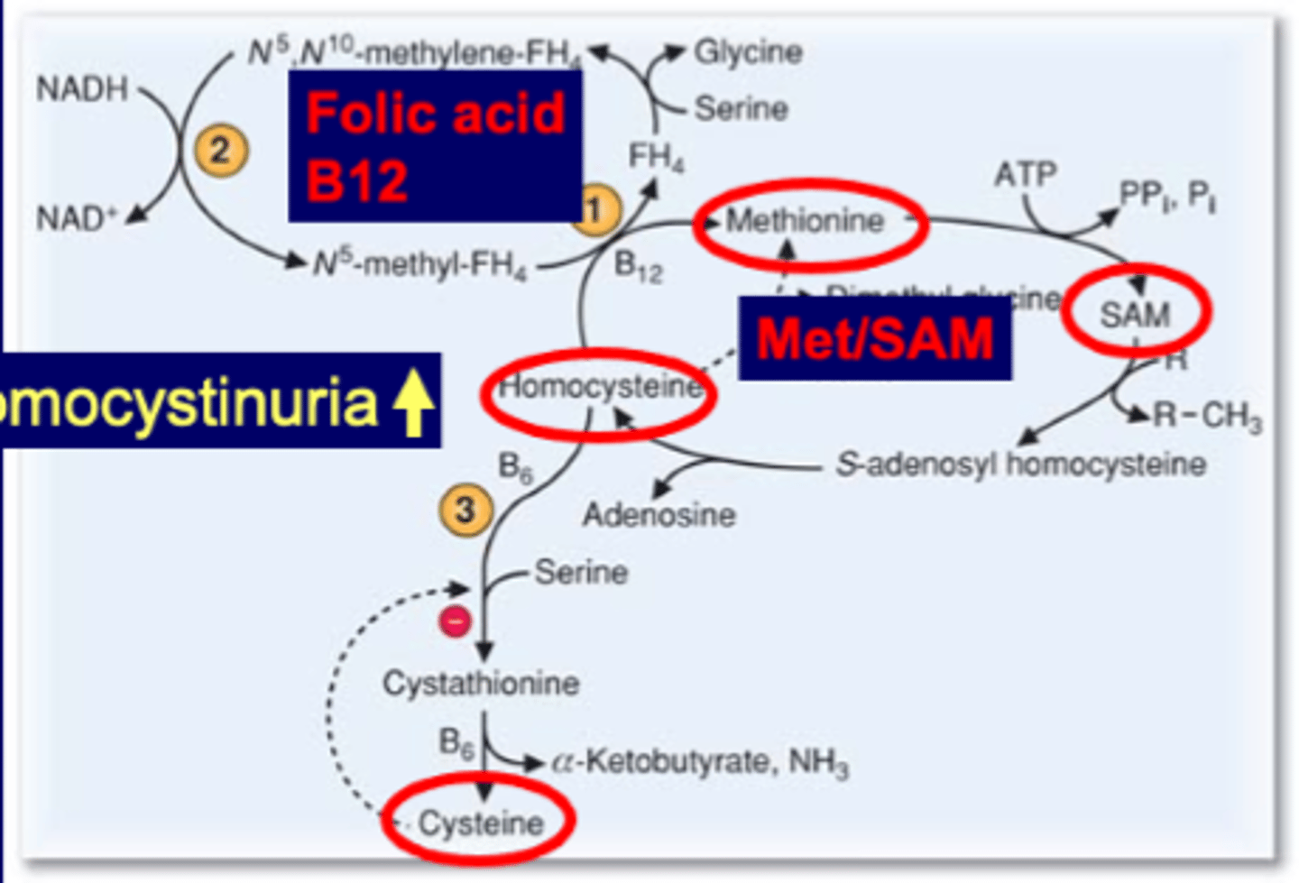
from methionine (and serine)
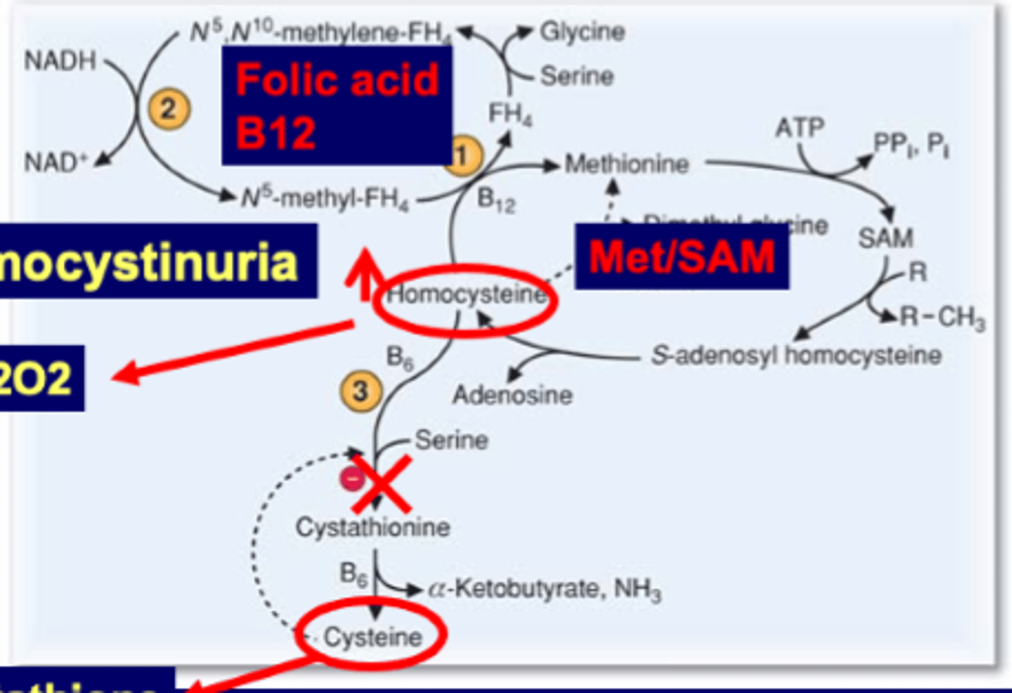
Met cycle->SAM->SAH->homocysteine->cysteine
Folic acid (FH4) coupled with B12 can use methionine for other stuff (B12 convert homocystein->met). When knock out met goes only to homocysteine->cysteine. Overloads cysteine->negative feedback, blocks pathway and accumulates homocysteine
B6 two points: homocysteine->cystathionine and cystathionine->cysteine
If knock out accumulate homocysteine
The coenzymes in methionine metabolism
-pyriodoxal phosphate (PLP/B6)
-tetrahydrofolate (FH4)/B12
Amino acidopathy
-inability to metabolize (urea deficiency)
-inability to detoxify ammonia through urea cycle (AA problem)
Inherited diseases of AA metabolism
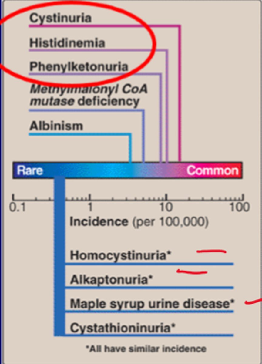
(Common)
Cystinuria
Histidinemia
Phenylketonuria
(Rare)
Homocystinuria
Alkaptonuria
Maple syrup urine disease
Aminoacidureas
Cystinuria
Histidinemia
Phenylketonuria
MSUD
Histidinemia
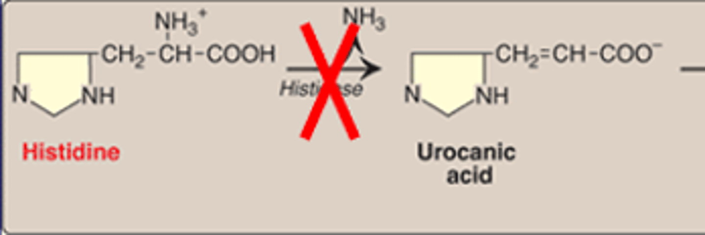
Genetic metabolic disorder of histidine
-histidine can't degrade to glutamine
-enzyme problem with histidase
Cysienuria
genetic disorder of renal AA transport. Cysteine metabolism defect
-AA->COAL (cysteine, ornithine, arginine and lysine; hexagonal kidney or bladder stones)
Phenylketornuria
Genetic metabolic disorder. detected by blood screening
Accumulation of phenylalanine and low tyrosine
CNS problems. Hypopigmentation
Aminoaciduria cont.
Methylmalonyl CoA mutase deficiency (similar to B12 deficiency)
Albinism (missing melanin due to mutations in several genes, including tyrosinase gene)
Alkaptonuria (mutations in homogenistic acid oxidase gene in the tyrosine degradation pathway. Urine turn black when exposed to air. Ochronosis, a buildup of dark pigment in connective tissues such as cartilage and skin)
Hartnup disease (mutations in the neutral AA transporter gene. Shows ataxia and also is associated with niacin deficiency)
Methylmalonyl CoA mutase deficiency
similar to B12 deficiency
Albinism
missing melanin due to mutations in several genes, including tyrosinase gene
Alkaptonuria
mutations in homogenistic acid oxidase gene in the tyrosine degradation pathway. Urine turn black when exposed to air.
Ochronosis, a buildup of dark pigment in connective tissues such as cartilage and skin
Hartnup disease
mutations in the neutral AA transporter gene.
shows ataxia and also is associated with niacin deficiency
Deficiencies in Phe to Tyr (PKU)
common inborn error (prevalence 1:15,000)
two major causes
-PAH deficiency (classic)
-BH4 deficiency (non-classic)
Tyr->fumarate (glucogenic)
Tyr->acetoacetate (ketogenic)
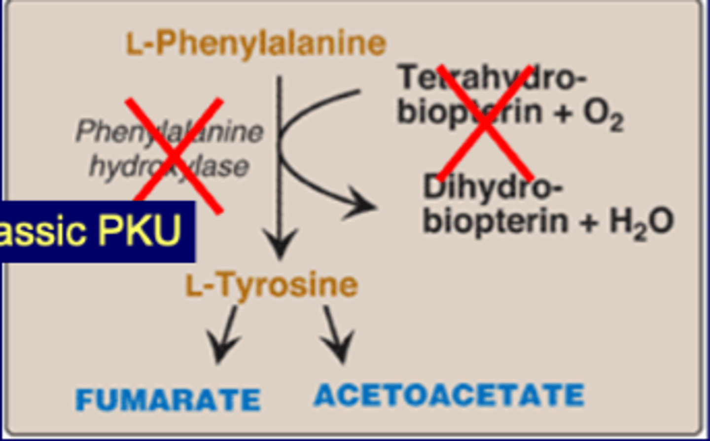
Classic PKU
-IEM
-PAH deficiency (phenylalanine hydroxylase)
-autosomal recessive
-1 in 13,500 to 19,00 births
-CNS deficiencies
--inhibition of LNAAT (large neutral AA transporter)
--elevated Phe (LNAA)->inhibit LNAA transporter (may saturate LNAAT)
--low LNAA is thought to cause intellectual disability
--phenylketones are high, clogged system or high ketones
-hypopigmentation (high phe inhibits tyr for melanin)
-Guthrie test for early diagnosis
PKU cont.
>400 mutations known in PAH gene
lifelong dietary restrict Phe
neonatal screening after 24-48 hours (not immediate)
diagnosis
-elevated serum concentration of phe
-useful lab method->tandem-mass spec (MS-MS), can identify IEM in a single sample
-confirm with 2nd plasma AA analysis
-enzyme analysis not performed to confirm diagnosis, since PAH expressed only in liver (baby liver too small)
Non-classic PKU
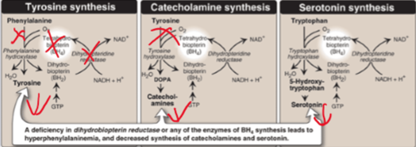
BH4 (tetrahydrobiopterin) deficiency, pleiotropic
Biopterin reductase mutation
Phe up, tyr down
impacts Tyr->DOPA and other catecholamines
Classic-tyrosine
Non-classic-serotonin or catecholines cause of biopterin
Homocystinuria
cystathionine beta synthase deficiency.
Low cysteine (low glutathione). High homocysteine (High H2O2, oxid stress).
Homocystinuria symptoms
-1:330,000 autosomal recessive
interferes with collagen cross linking
-dislocated lens
-elongated, thin, arachnodactyly
-scoliosis, osteoporosis
oxidative stress (ROS generated)
intellectual disability
similar but NOT marfan's (not tall and no aortic aneurysm)
Summary: homocysteinuria
high homocysteine
multiple causes:
-cystathionine beta synthase deficiency (most common)
-B12 or folic acid deficiencies lead to high cysteines, leading to feedback inhibition
increased oxidative stress:
-high homocysteine levels increase oxid stress
-low cysteine levels reduce ROS scavenging ability
interferes with collagen cross linking
increased risk of atherosclerosis and dementia
Recommended Uniform Screening Panel (RUSP)
List of conditions that all newborn screening programs should include. Established by United States Secretary of Health and Human Services.
Panel includes (not limited to):
-sickle cell disease
-critical congenital heart disease (most common)
-cystic fibrosis
-classical galactosemia
-severe combined immunodeficiencies
-phenylketonuria (AA problems)
-maple syrup urine disease