Normal GYN
 Call Kai
 Learn
Learn Practice Test
 Spaced Repetition
 Match
Match Flashcards
 Knowt Play
1/92

There's no tags or description
Looks like no tags are added yet.
Name | Mastery | Learn | Test | Matching | Spaced | Call with Kai | Chat |
|---|
No analytics yet
Send a link to your students to track their progress
93 Terms
What is the collection of the Pelvic bone called?
Pelvic Girdle
What is the Pelvic Girdle made up of?
True/Minor pelvis
False/Major pelvis
What does the True pelvis contain?
Contain the bladder and pelvic organ
What is contained in the False pelvis?
Bowel and extend to the Umbilicus
What are the 5 muscles in the pelvis?
Levator Ani
Oburator Internus
Iliopsoas
Coccygeus
Piriformis
What does the Levator Ani muscles form?
Form the Pelvic flood
Inf to vagina
Considered to be Hammock shaped
Describe the Oburator Internus muscle?
Triangle shaped
Contains the Ant and Lat pelvic sidewalls
What is the iliopsoas muscle?
Continous to the Psoas Muscle
Large strap muscles
Best seen in Long
Most lateral landmark of the pelvis
Describe the Coccygeus muscle?
Is lateral and Post to cervix
Forms the Post Pelvic floor
If muscle fails it can cause uterine prolapse
Seperate the pelvis from peritoneum
Describe the Piriformis muscle?
Is lat and Post to uterine body
How can we visualize the Obturator Internus?
Tranabdominal or Transvaginally
Hypoechoic
Ovoid
Lateral to the Bladder
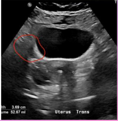
What muscle is this showing?
OBTURATOR INTERNUS
What is the Levator Ani muscle?
post and inf to Obturator Internus
Best seen in Transabdominal
Has a curved comma shape
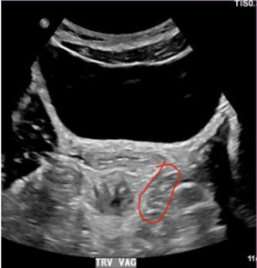
What muscle is this?
Levator ANI
Describe the Broad Ligament?
-Not a true ligament
-Looks like a peritoneal fold on either side of Uterus
-Surrounds FT,Ovaries,Ovarian and UT vessels
Describe the Round Ligament?
-BEgins at UT horn
-goes through the Inguinal canal ends at mons pubis,labia majora
-helps hold UT in forward position
Describe the Ovarian Ligament?
-Attach to the Inf Med part of ovary
-Lateral wall of outer uterus
Describe the Uterosacral Ligment?
-Support UT at base
-Determine if UT is Anteflexed or Retroflexed
Describe the Cardinal Ligament?
-Support UT at cervix
Extend from side wall of Pelvis
When are most ligaments seen?
Not well seen Unless there is Ascites or Free Fluid
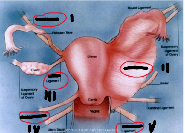
Lable this image
1-Round
2-Ovarian
3-Broad
4-Cardinal
5-Uterosacral
Describe the Urinary bladder?
-Landmark as a window
-Holds 500mL of urine
-Uses the Uterus away to see ovaries more clearly(Full)
Describe the anatomy of Bladder?
Post to rectus abdominus and Pubic bone
Ant to Uterus,cervix,vagina,rectum
How does the Uterus Run?
Anterior to Internal Ilaic artery and Posterior to Ovaries
Describe the Uterus?
Not visible on Pelvic U/S
We see bladder jets filling bladder
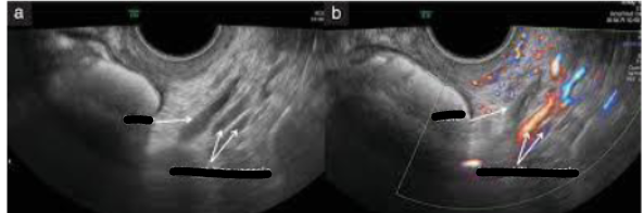
Lable this image?
Uterus
Internal Iliac vessels
What are the Pelvic spaces?
Anterior cul de sac
Posterior Cul de sac
Space of REtzius
Describe the Anterior Cul de Sac?
-Called Uterovesical or Vesicouterine pouch
-Seperate the Ut and Bladder
-Normally empty but can have small fluid
Describe the Posterior Cul De Sac?
-Called pouch of douglas and Rectouterine pouch
-Seperate posterior UT and REctum
-very common place for fluid to collect
Describe the Space of Retzius?
-Between the Ant Bladder and Post Pubic Symphasis
-Uncommon for fluid to collect here
Describe the Pelvis Arterial Vasculature?
Aorta→Iliac→External & Internal Iliac Arteries
-Uterine arteries branch off Internal ILiac Arteries
Where does the Ovarian arteries originate?
From the aorta then anastomoses with Uterine Arteries at Ut Cornua
-Ut artery then branch into and becomes Arcuate Artery
Describe the Venous Pelvic Vascular Flow?
Internal and External Iliac veins→IVC
-Internal drains→Ut and Pelvic muscles
-External drains→legs
Where does the Ovarian veins drain?
Rt Ov vein- drains directly into IVC
Lt OV vein- drains into LRV
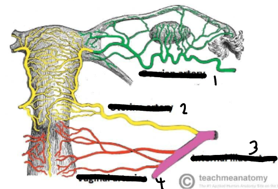
Lable this image?
1-Ov Arteries
2-UT arteries
3-Internal Iliac Arteries
4-Vaginal Arteries
Describe the Embryology timeline?
Week 5→2 gestational ridges develop form gonadals
Early Week 6→Primitive germ cells becomes egg and sperm travel to yolk sac. Ridges develop into sex cords
Second Half week 6→Y chrosomes activated,female mechanism develop
Week 7→Grow more and cell differenciates
Week 8→Sex cords degenerate into either Mularian or Wolfian Ducts
Describe the Mullerian Ducts
-Called Paramesonephric Duct
-Leads toFemale Reproductive organ
-Female(originate Kidney)
-degenerate in Men
-fuse formin Vagina and UT
-Unfused part become Fallopian Tube
Describe the Wolffian Duct?
-Called Mesenteric Duct
-Found in men(originate kidney)
-Degrade in women
-Remnats in Women(Gartner duct cyst, Hydatoid Cyst,Paraovarian Cyst)
What are the parts of the Uterus?
Vagina
Cervix
Uterus
Endometium
Fallopian Tube
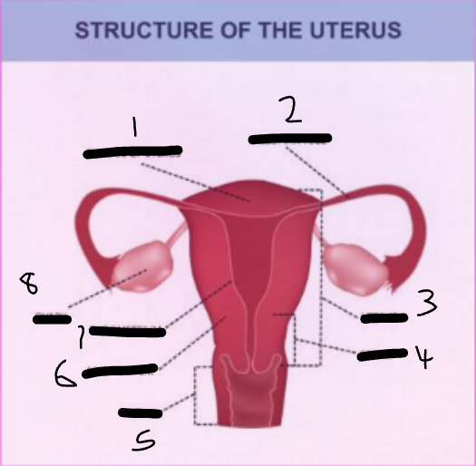
Lable this image
1-FT
2-UT
3-CV
4-Vag
5-Myometrium
6-Endometrium
7-Ovary
Describe the Vagina?
-Opening extend from vulva to Cervix
-Fibromuscular tube measure 9cm
-Vag Cervix located in upper part of vag at external os of cervix
-Fornices called pelvic spaces
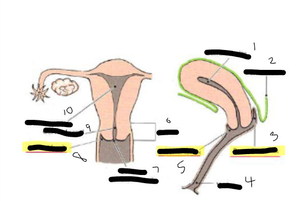
Lable this image?
1-Uterine Cavity
2-Peritoneum
4-Posterior Fornix
5-Vagina
6-Anterior Fornix
7-Cervix
9-External OS
10-Lateral Fornix
11-Cervical canal
12-Uterine Cavity
Describe the Cervix?
-Narrowing of the lower end of Uterus
-Opening of vag=external os
-Opening in the Uterus=internal os
-Also called birthing canal
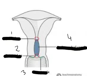
Lable this Cervical image?
1-Internal Os
2-External OS
3-Ectocervix
4-Endocervical Canal
Describe the Uterus?
-Is muscular organ
-Sole purpose is for gestatation
-divided into 4 segemnts[Cervix,Insthmus,Ut Body,Fundus]
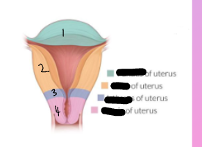
Lable this Uterine image?
1-Fundus
2-Uterus
3-Isthmus
4-Cervix
Describe the Uterus based on age?
-largest organ in the pelvis
-Length=6-8 cm
-Ap=3-5
-Ut volume considered enlarge=>120ml
What are the 3 layers of the Uterus?
Serosa/Peri,etrium=outer
myometrium=middle
Endometriusm=Inner
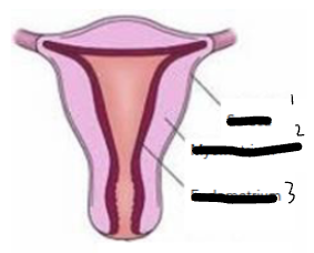
Lable the Uterine Layers?
1-serosa
2-Myometrium
3-Endometrium
Describe the Endometrial layer
-Innermost layer
-Has 2 layers(Zona Funtionalis,Basalis)
What is the Zona Functionalis?
Superficial layer
Made of gland and stroma tissue
Stretches with menses
What is the Zona Basalis?
Thinner deeper layer
Connect Endometrial canal to myometrium
Regenerates
Describe the Vasculature of Endometrium?
-Arcuate Artery branch into myometrium then become radial artery
-Radial Artery branch into straight artery supply zona basalis
-Becomes larger and supply zona basalis
-Blood from spiral arteries shed in menses
What are the positions of the Uterus?
Anteverted
Anteflexed
Retroverted
Retroflexed
Describe Anteverted or Anteflexed?
most common
bend foward
Describe retroverted or Retroflexed?
normal
Ut bend post to rectum
What does it mean the Uterus is Dextroflexed or levorted?
Bent Lt or RT
What is version refer to to as?
anteverted-angled anteriorly
Retroverted-angled posteriorly
What is Flexion?
Bend of the Fundus
Anteflexed-Fundus bent anteriorly
Retroflexed-Fundus folds posteriorly
What are the Fallopian tubes called?
Oviducts
Describe the Fallopian tubes?
-Muscular tube that connects to the UT at cornus
-Open at the peritoneal cavity and curve over ovary
What is the length of the Fallopian tube?
10-12 cm long
1-4 cm diameter
How does the Fallopian tube lies?
lies superior to utero-ovarian and round ligament
Are the Fallopian tubes be seen on the scan?
Not easily visualized but can use color to distinguish
What are the segments of the Fallopian tubes?
Infundibulum
Ampulla
Isthmus
Interstitial
Describe the Infundibulum?
-Lateral part of tube
-Funnel shape over ovaries
-Finger like structures called Fimbriae
Describe the Ampulla?
-Middle
-Longest part of the Tube
-Where fertilization of egg occurs
Describe the Isthmus of FT?
Medial part of tube
Narrows and lies anterior to the Uterus
Describe the Interstitial portion of the FT?
-Narrowest part of tube
-connects and passes through the Uterine Cornua
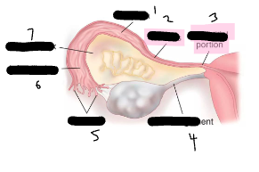
Lable the portions of the Fallopian Tube?
1-Ampulla
2-Isthmus
3-Interstitial portion
4-Ovarian ligament
5-Fimbriae
6-Infundibulum
7-Mesosalpinx
Describe the Ovaries?
-Almond shaped
-Lateral and posterior to Uterus near cornua
-Volume over 10ml is abnormal
Where are the Ovarian ligaments suspended?
suspended by the posterior aspect of broad ligament
-medially supported by the Ovarian ligament
-laterally supported by the suspensory or infundibulumpelvic ligament
What is the primary role of the ovaries?
-produce the female reproductive cells
Ovum,estrogen(secreted by the follicles) and progesterone(secreted by the corpus luteum)
What are the layers of the Ovaries?
Cortex(outer)
Central Medulla(Medulla)
Describe the Cortex of the Ovaries?
-Outer portion
-Surrounds the medulla
-Consist of follicles
-Cortex is covered by 2 tissues (Tunica Albuginea,Germinal Epithelium)
describe the Medulla?
-Central part of the ovary
-Compuses of connective tissue
-Contains blood vessels,lymphatic vessels,smooth vessels
Describe the Menstrual Cycle?
-Typically begins at age 11-13
-likely end at age 50
Describe the 3 menstrual status?
Premenarche -prepuberty before menses
Menarche- Menses occur on a regular basis
Menopause- endo of the women Menstrual cycle
Describe the menstural cycle?
-Last 28 days
-Day 1 =bleeding
What is the Menstural cycle regulated by?
regulated by the Hypothalamus in the brain
releases estrogen and progesterone from the ovaries
What is the Ovulation cycle?
-Release of an egg each month
-Ovulation occurs mid cycle day 14
-Woman born with over 200,000 eggs
Write on white board
Slide 50
Describe the Follicular phase of the Ovarian cycle?
-Secretion of the FSH
-1st phase of menstural cycle
-continue until day 14 of cycle
-Follicles grow and fill with fluid and increase amount of estrogen
Describe the LH Surge & Ovulation?
5-8 follicles but only 1 reaches maturity
Mature follicles called Grafian Follicle
LH surges peak until the 10-12 hour before ovulation
Describe the Luteal Phase?
2nd phase of Menstural cycle 14 days after ovulation
Rupture follicle turn into Corpus Luteum and secrete Progesterone
Decline in progesterone causes cycle to begin
Describe the Menstral Cycle?
Endometrium changes through Menstral cycle
What are the 3 stages of the Endometrium during the menstrual cycle?
Menstrual
Proliferative
Secretory
Describe the Menstrual phase of the Menstural cycle
-Day 1-5 bleeding occurs
-Progesterone levels decline
-low levels of progesterone causes the spiral artery to constrict result in shedding of the Zona Functionalis of endometrium
-In association with the Early Follicular phase
-We can see fluid sometimes
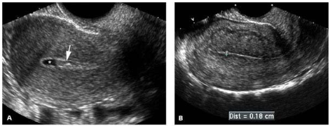
What phase is this?
Menstural Phase
Describe the Proliferative phase?
-When the follicles grow the ovaries secrete estrogen
-Regeneration of the regrowth occurs in the Proliferative phase
Day 5-14
-endometrim usually measures 4-8mm (three line patterns)
-the endometriumis 6-10mm before ovulation
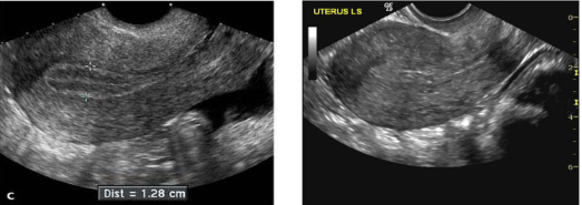
What part of the Menstral Cycle this patient is in?
Proliferative phase
Describe the Secretory phase?
-Right after ovulation
-Secrete progesterone form the corpus luteum
-Causes spiral arteries to swell
-Occurs day 15-28
-Normally in association with the Luteal phase
-thickest and most echogenic aroud 14mm
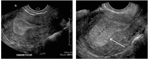
What phase of the cycle is this patient in?
Secretory phase